. 2012 Jan-Feb;20(1):61-73.
doi: 10.1111/j.1524-475X.2011.00757.x.

Topical androgen antagonism promotes cutaneous wound healing without systemic androgen deprivation by blocking β-catenin nuclear translocation and cross-talk with TGF-β signaling in keratinocytes

Affiliations

Topical androgen antagonism promotes cutaneous wound healing without systemic androgen deprivation by blocking β-catenin nuclear translocation and cross-talk with TGF-β signaling in keratinocytes

Gianluca Toraldo et al. Wound Repair Regen. 2012 Jan-Feb.

Abstract

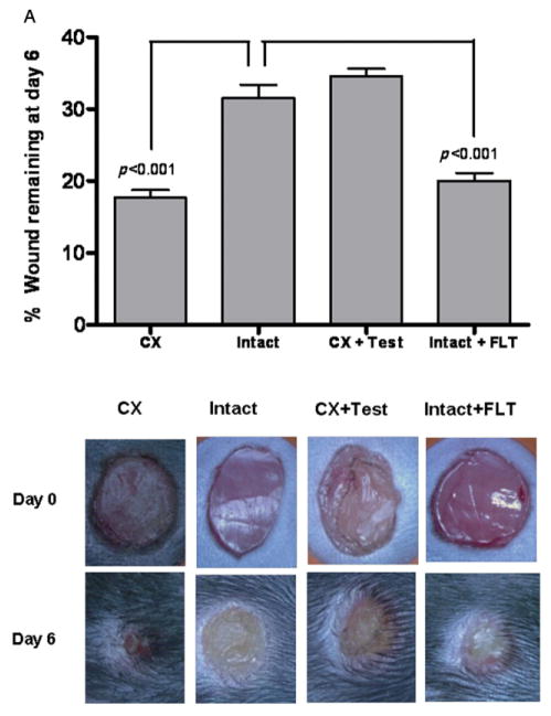
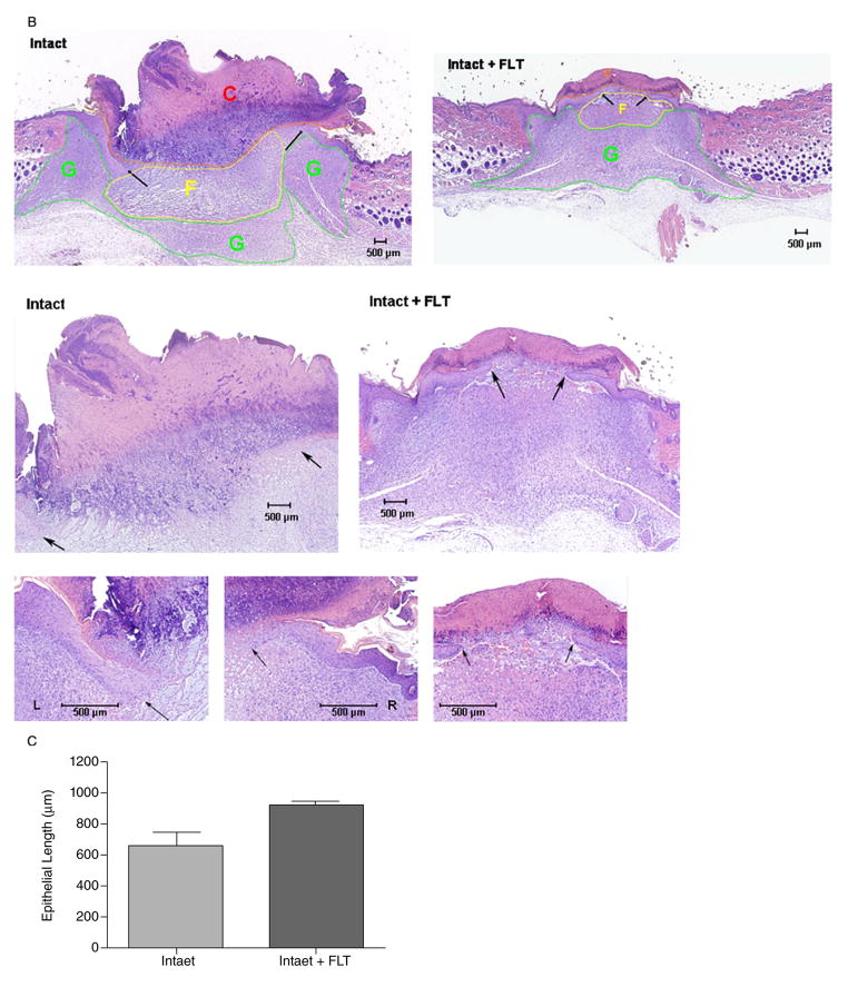
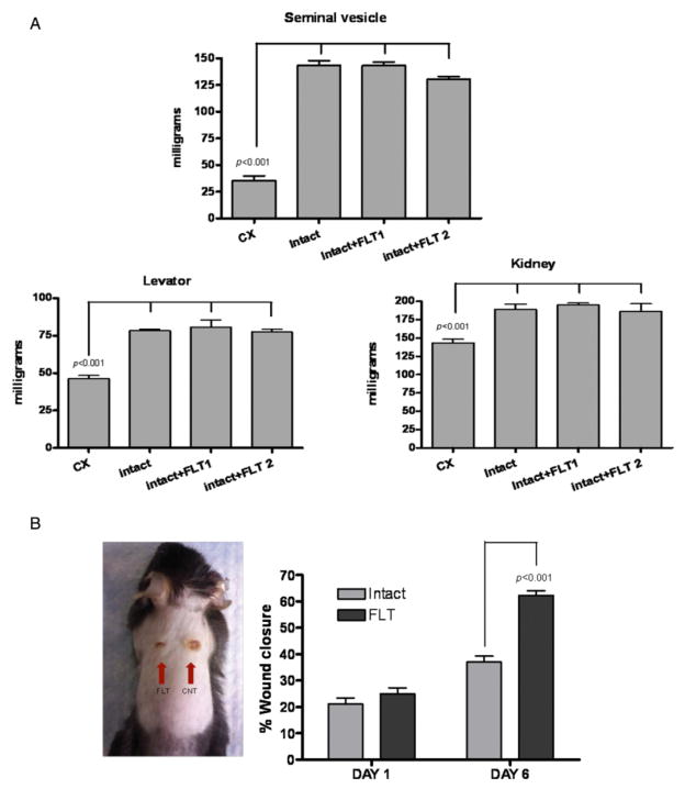
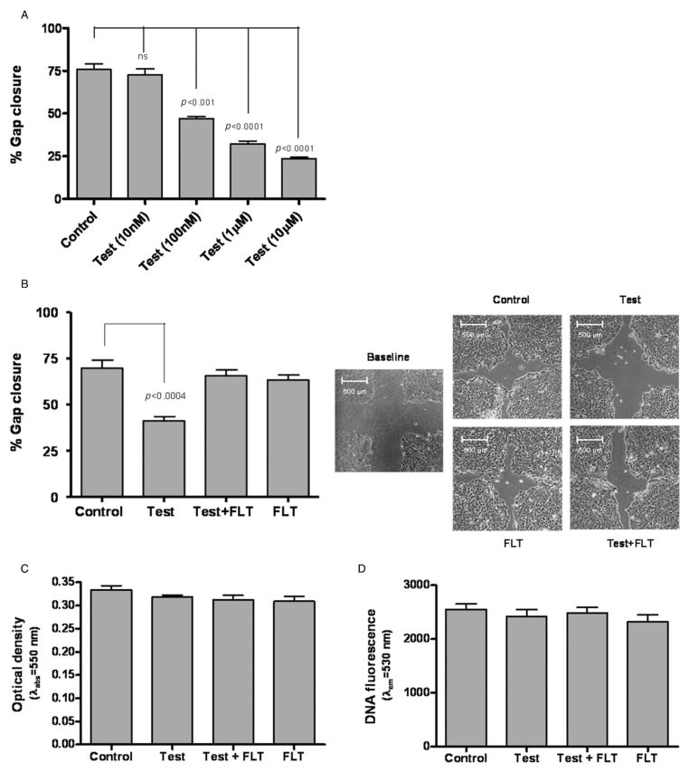
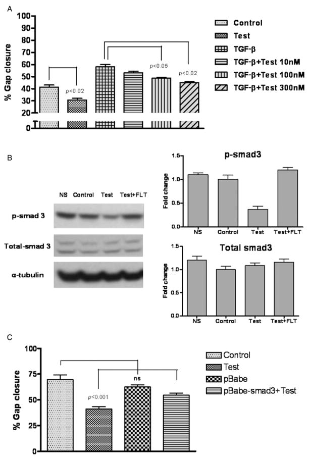
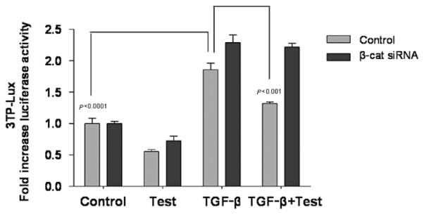
Orchidectomy in rodents and lower testosterone levels in men are associated with improved cutaneous wound healing. However, due to the adverse effects on skeletal and sexual tissues, systemic androgen blockade is not a viable therapeutic intervention. Accordingly, we tested the hypothesis that topical application of an androgen antagonist would elicit accelerated wound healing without systemic androgen antagonism. Full-thickness cutaneous wounds were created on adult C57BL6/J mice. Daily topical application of androgen receptor antagonist, flutamide, resulted in improved gap closure similar to orchiectomized controls and faster than orchidectomized mice treated with topical testosterone. In vivo data showed that the effects of androgen antagonism on wound closure primarily accelerate keratinocytes migration without effecting wound contraction. Consequently, mechanisms of testosterone action on reepithelialization were investigated in vitro by scratch wounding assays in confluent keratinocytes. Testosterone inhibited keratinocyte migration and this effect was in part mediated through promotion of nuclear translocation of β-catenin and by attenuating transforming growth factor-β (TGF-β) signaling through β-catenin. The link between Wnt and TGF beta signaling was confirmed by blocking β-catenin and by following TGF-β-induced transcription of a luciferase reporter gene. Together, these data show that blockade of β-catenin can, as a potential target for novel therapeutic interventions, accelerate cutaneous wound healing.


Figures

Figure 1
Figure 1
Effect of topical blockade of androgen receptor on wound healing. (A) Mean ± SEM unhealed wound area after 6 days in vehicle-treated castrated (Cx), sham-operated (Intact), testosterone-treated castrated (CX + Test), and flutamide-treated sham-operated (Intact + FLT). n = 12/group. Illustrative photographs of the wounds on Days 0 and 6 from each treatment group are shown in the lower panel. (B) Histological analysis of wounds. Pictures were taken at 2×, 4×, and 10× magnifications. Intact: Scale crust (C) overlying the ulcer bed (between arrows). The base of the ulcer shows fibrin (F) and necrotic debris. A large epithelial gap is apparent with substantially less reepithelialization than in the flutamide-treated wound, underlying granulation tissue (G). Flutamide: Scale crust overlying the ulcer bed (between arrows). The base of the ulcer shows minimal fibrinous exudate (F) and marked granulation tissue. (C) Morphometric analysis of the length of the wound epithelium.
Figure 1
Figure 1
Effect of topical blockade of androgen receptor on wound healing. (A) Mean ± SEM unhealed wound area after 6 days in vehicle-treated castrated (Cx), sham-operated (Intact), testosterone-treated castrated (CX + Test), and flutamide-treated sham-operated (Intact + FLT). n = 12/group. Illustrative photographs of the wounds on Days 0 and 6 from each treatment group are shown in the lower panel. (B) Histological analysis of wounds. Pictures were taken at 2×, 4×, and 10× magnifications. Intact: Scale crust (C) overlying the ulcer bed (between arrows). The base of the ulcer shows fibrin (F) and necrotic debris. A large epithelial gap is apparent with substantially less reepithelialization than in the flutamide-treated wound, underlying granulation tissue (G). Flutamide: Scale crust overlying the ulcer bed (between arrows). The base of the ulcer shows minimal fibrinous exudate (F) and marked granulation tissue. (C) Morphometric analysis of the length of the wound epithelium.
Figure 2
Figure 2
Topical application of androgen antagonist is not associated with systemic androgen blockade and its effects are confined to the site of application. (A) Topical application of flutamide does not induce a state of systemic androgen antagonism. Two doses of flutamide (Intact + FLT1: 30 μg; Intact + FLT2: 300 μg) were applied topically on the wounds of intact animals. The weights of the androgen-sensitive tissues (seminal vesicles, kidneys, and levator ani muscle) were similar among intact mice treated with the vehicle and those treated with either of the two doses of flutamide. (B) Two wounds were created on the back of intact mice. One side was treated with placebo (Intact = CNT) and the other with flutamide (FLT, 30 μg). Topical flutamide significantly accelerates wound healing. Data are mean ± SEM; n = 8/group. CX, castrated.
Figure 3
Figure 3
Topical application of androgen antagonist is not associated with wound contraction. (A) Schematic of the Contraction area (C) and Wound area (W). A plastic Template (T) containing a circular window was used to briefly hold the mice. (B) Representative pictures of the wounds on Days 0 and 6 of control and flutamide (FLT)-treated intact animals (n = 6/group). (C) Effect of topical flutamide on wound healing and wound contraction. Data were expressed as a ratio between treated and not-treated wounds (n = 6/group).
Figure 4
Figure 4
Effects of testosterone in an in vitro scratch assay. (A) HaCaT monolayers were scratch-wounded and treated with increasing concentrations of testosterone (10 nM–10 μM); gap closure was measured after 24 hours. (B) Flutamide (FLT) blocks the effects of testosterone in the in vitro scratch-wound assay. Testosterone treatment (100 nM) delayed gap closure compared with control. Co-incubation with flutamide (1 μM) blocked the inhibitory effects of testosterone. The right panel shows illustrative photographs of the HaCaT cell monolayers at baseline, and after 24 hours of incubation in respective treatments. (C, D) Testosterone does not affect HaCaT cell proliferation. HaCaT cells were treated for 48 hours with testosterone (100 nM) with or without flutamide (1 μM), and cell growth was determined using the MTT (C) or CyQUANT® Cell Proliferation Assay (D).
Figure 5
Figure 5
Testosterone increases nuclear β-catenin translocation. (A) HaCaT cells were treated for 1 hour with testosterone (100 nM) with or without flutamide (1 μM), and analyzed for β-catenin expression. Densitometric analysis shows that cells incubated with testosterone had higher level of nuclear β-catenin. (B) HaCaT cell infected with type 5 adenovirus encoding β-catenin with eGPF were scratch-wounded and treated with medium, testosterone (100 nM), or lithium chloride LiCl (2 mM). Extent of β-catenin nuclear translocation was monitored after 1 hour; 4′,6-diamidino-2-phenylindole (DAPI) counterstain was used to localize the nuclei. Three independent experiments were performed, and representative photographs are shown. (C) β-catenin small interfering RNA siRNA decreases the expression levels of β-catenin. HaCaT cells were transiently transfected with specific β-catenin siRNA (βcat-siRNA) and representative densitometric data of one of multiple independent experiments are shown. (D) βcat-siRNA attenuates testosterone’s effect on gap closure. HaCaT cells, transfected with either nonspecific oligonucleotide (c-siRNA) or specific βcat-siRNA were incubated with or without testosterone (100 nM).
Figure 6
Figure 6
(A) Testosterone inhibits the effects of transforming growth factor-β TGF-β on keratinocyte migration in a dose-dependent manner. HaCaT cell monolayers were scratch-wounded and treated with TGF-β (10 ng/mL) with our without testosterone (10 nM to 300 nM). Gap closure was measured 16 hours after treatment. No significant difference was observed between control and cells treated with TGF-β + 300 nM of testosterone. (B) The effects of testosterone on p-Smad3 expression in HaCaT cell monolayer. HaCaT cells were treated with testosterone (100 nM) with or without flutamide (1 μM) for 1 hour after which the cells were analyzed for p-Smad3 and total-Smad3 expression. NS = nonscratched control. Respective densitometric analyses are shown in the right panels. (C) Smad3 hyperexpression partially blocks the effect of testosterone on keratinocyte migration in HaCaT cell scratch assay.
Figure 7
Figure 7
β-catenin small interfering RNA siRNA attenuates the effects of testosterone on transforming growth factor-β TGF-β-induced luciferase reporter activity HaCaT cell were co-transfected with 3TP-Lux and β-catenin siRNA. After 48 hours of β-catenin silencing, cells were scratched and incubated with testosterone (100 nM), TGF-β (10 ng/mL), or both. Mean ± SD luciferase activity from two separate experiments conducted in triplicates is shown.

Similar articles

Cited by

References

    1. Branski LK, Gauglitz GG, Herndon DN, Jeschke MG. A review of gene and stem cell therapy in cutaneous wound healing. Burns. 2009;35:171–80. - PMC - PubMed
    1. Sen CK, Gordillo GM, Roy S, Kirsner R, Lambert L, Hunt TK, Gottrup F, Gurtner GC, Longaker MT. Human skin wounds: a major and snowballing threat to public health and the economy. Wound Rep Regen. 2009;17:763–71. - PMC - PubMed
    1. Ashcroft GS, Dodsworth J, van Boxtel E, Tarnuzzer RW, Horan MA, Schultz GS, Ferguson MW. Estrogen accelerates cutaneous wound healing associated with an increase in TGF-beta1 levels. Nat Med. 1997;3:1209–15. - PubMed
    1. Ashcroft GS, Mills SJ, Lei K, Gibbons L, Jeong MJ, Taniguchi M, Burow M, Horan MA, Wahl SM, Nakayama T. Estrogen modulates cutaneous wound healing by downregulating macrophage migration inhibitory factor. J Clin Invest. 2003;111:1309–18. - PMC - PubMed
    1. Gilliver SC, Ashworth JJ, Mills SJ, Hardman MJ, Ashcroft GS. Androgens modulate the inflammatory response during acute wound healing. J Cell Sci. 2006;119 (Pt 4):722–32. - PubMed

Publication types

MeSH terms