

Effect of the HIV nucleoside reverse transcriptase inhibitor zidovudine on the growth and differentiation of primary gingival epithelium
- PMID: 22276657
- PMCID: PMC3319519
- DOI: 10.1111/j.1468-1293.2011.00973.x
Effect of the HIV nucleoside reverse transcriptase inhibitor zidovudine on the growth and differentiation of primary gingival epithelium
Abstract
Objectives: Oral complications associated with HIV infection and with the antiretroviral drugs used to treat it are of increasing concern in HIV-infected patients. Protease inhibitors have been shown to change the proliferation and differentiation state of oral tissues but the effect of nucleoside reverse transcriptase inhibitors is currently unknown. This study examined the effect of zidovudine on the growth and differentiation of the gingival epithelium.
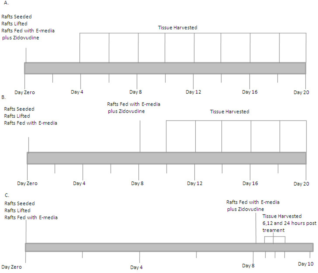
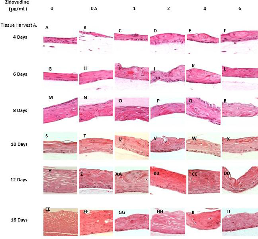
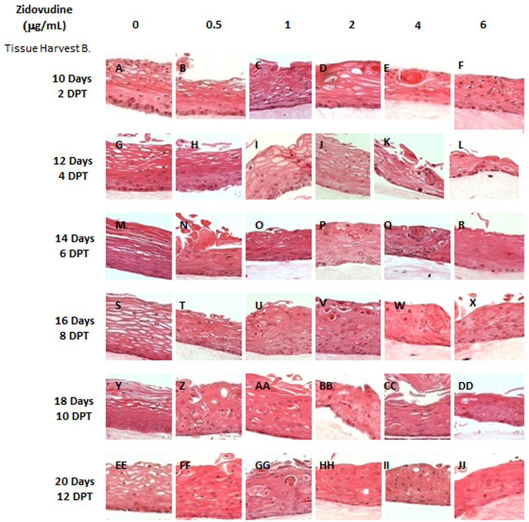
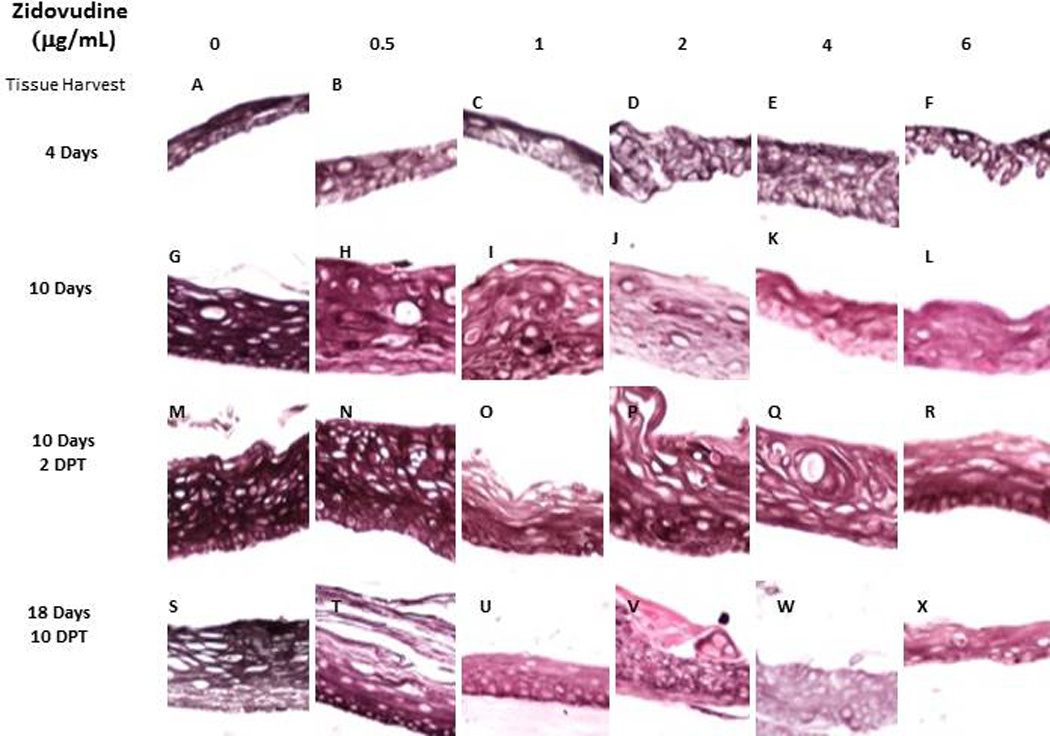
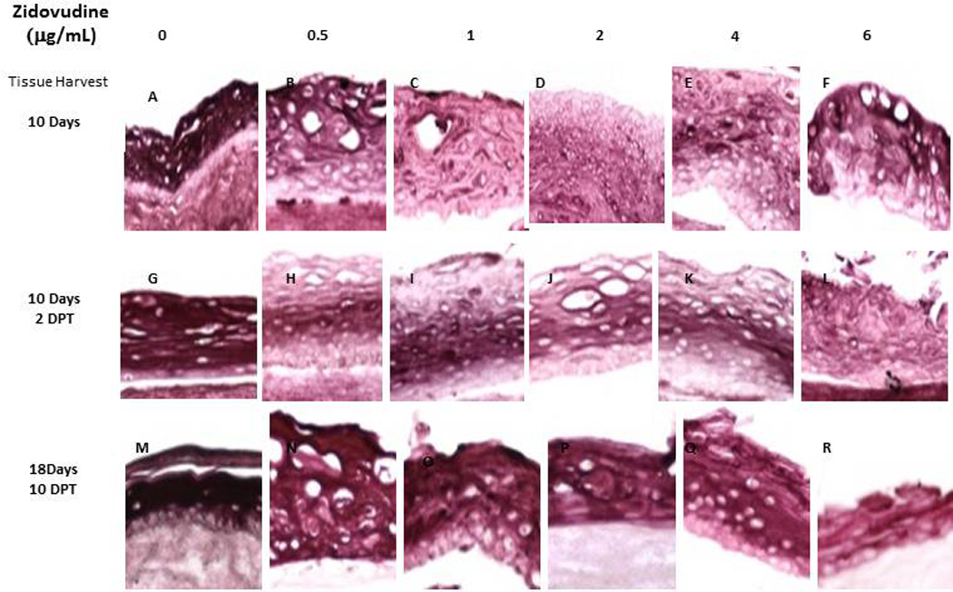
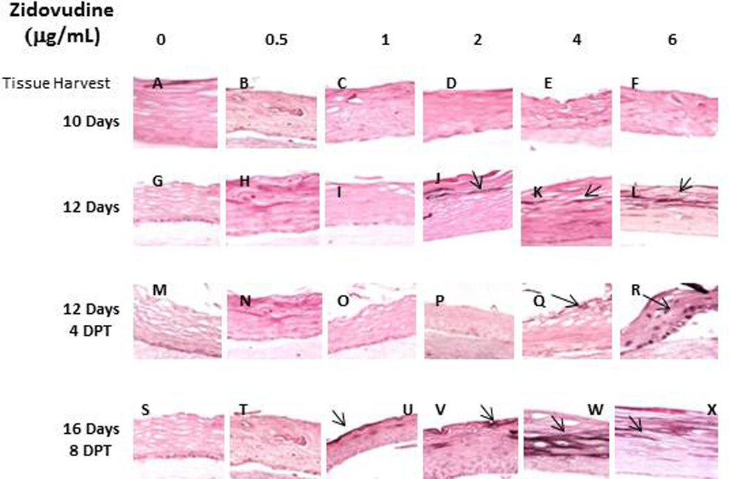
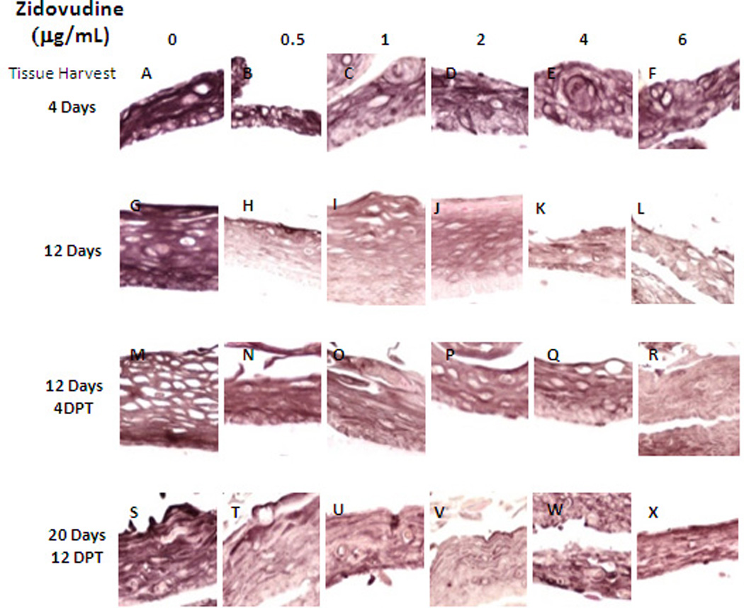
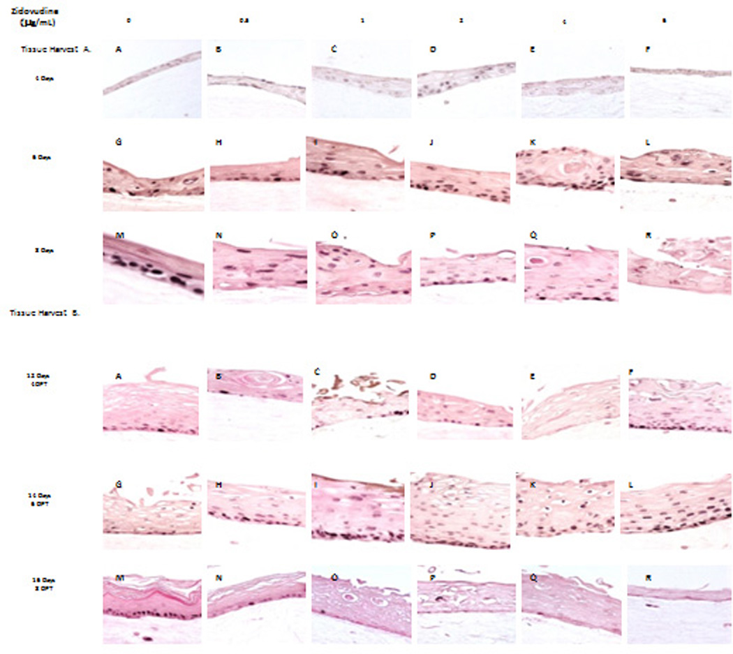
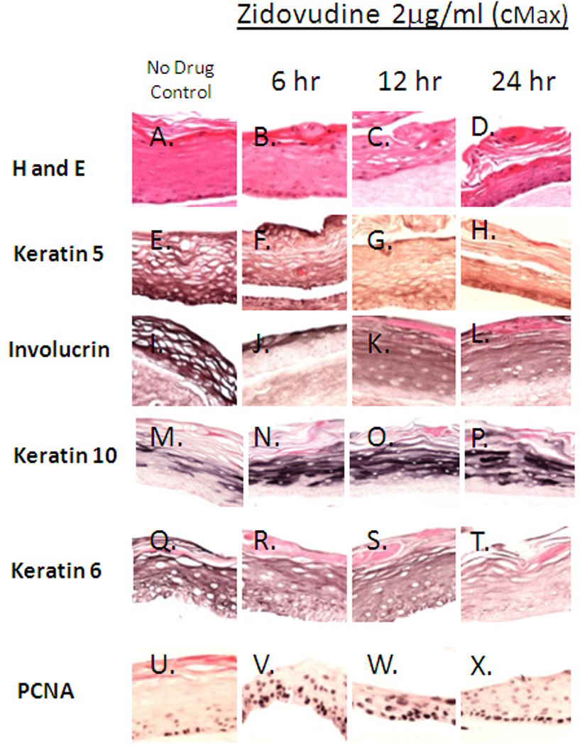
Methods: Gingival keratinocyte organotypic (raft) cultures were established. The raft cultures were treated with a range of zidovudine concentrations. Haematoxylin and eosin staining was performed to examine the effect of zidovudine on gingival epithelium growth and stratification. Raft cultures were immunohistochemically analysed to determine the effect of this drug on the expression of key differentiation and proliferation markers, including cytokeratins and proliferating cell nuclear antigen (PCNA).
Results: Zidovudine dramatically changed the proliferation and differentiation state of gingival tissues both when it was present throughout the growth period of the tissue and when it was added to established tissue at day 8. Zidovudine treatment increased the expression of cytokeratin 10, PCNA and cyclin A. Conversely, cytokeratin 5, involucrin and cytokeratin 6 expression was decreased. The tissue exhibited characteristics of increased proliferation in the suprabasal layers as well as an increased fragility and an inability to heal itself.
Conclusions: Zidovudine treatment, even when applied at low concentrations for short periods of time, deregulated the cell cycle/proliferation and differentiation pathways, resulting in abnormal epithelial repair and proliferation. Our system could potentially be developed as a model for studying the effects of HIV and highly active antiretroviral therapy in vitro.
© 2012 British HIV Association.
Figures
Similar articles
-
HIV nucleoside reverse transcriptase inhibitors efavirenz and tenofovir change the growth and differentiation of primary gingival epithelium.HIV Med. 2014 Apr;15(4):196-202. doi: 10.1111/hiv.12100. Epub 2013 Oct 31. HIV Med. 2014. PMID: 24580719 Free PMC article.
-
Effect of the HIV protease inhibitor amprenavir on the growth and differentiation of primary gingival epithelium.Antivir Ther. 2010;15(2):253-65. doi: 10.3851/IMP1512. Antivir Ther. 2010. PMID: 20386081 Free PMC article.
-
The HIV protease inhibitor lopinavir/ritonavir (Kaletra) alters the growth, differentiation and proliferation of primary gingival epithelium.HIV Med. 2011 Mar;12(3):145-56. doi: 10.1111/j.1468-1293.2010.00863.x. Epub 2010 Aug 15. HIV Med. 2011. PMID: 20722750 Free PMC article.
-
Zidovudine: a review of its use in the management of vertically-acquired pediatric HIV infection.Paediatr Drugs. 2002;4(8):515-53. doi: 10.2165/00128072-200204080-00004. Paediatr Drugs. 2002. PMID: 12126455 Review.
-
Delavirdine: a review of its use in HIV infection.Drugs. 2000 Dec;60(6):1411-44. doi: 10.2165/00003495-200060060-00013. Drugs. 2000. PMID: 11152019 Review.
Cited by
-
Comparisons of VLP-Based ELISA, Neutralization Assays with Native HPV, and Neutralization Assays with PsV in Detecting HPV Antibody Responses in HIV-Infected Women.J AIDS Clin Res. 2015 Mar;6(3):433. doi: 10.4172/2155-6113.1000433. J AIDS Clin Res. 2015. PMID: 26085957 Free PMC article.
-
The Epstein-Barr Virus Glycoprotein BDLF2 Is Essential for Efficient Viral Spread in Stratified Epithelium.J Virol. 2023 Feb 28;97(2):e0152822. doi: 10.1128/jvi.01528-22. Epub 2023 Jan 23. J Virol. 2023. PMID: 36688650 Free PMC article.
-
Oral lichen planus: a microbiologist point of view.Int Microbiol. 2021 Aug;24(3):275-289. doi: 10.1007/s10123-021-00168-y. Epub 2021 Mar 10. Int Microbiol. 2021. PMID: 33751292 Free PMC article. Review.
-
HIV nucleoside reverse transcriptase inhibitors efavirenz and tenofovir change the growth and differentiation of primary gingival epithelium.HIV Med. 2014 Apr;15(4):196-202. doi: 10.1111/hiv.12100. Epub 2013 Oct 31. HIV Med. 2014. PMID: 24580719 Free PMC article.
-
Three-dimensional cell culture models for investigating human viruses.Virol Sin. 2016 Oct;31(5):363-379. doi: 10.1007/s12250-016-3889-z. Epub 2016 Oct 27. Virol Sin. 2016. PMID: 27822716 Free PMC article. Review.
References
-
- Global summary of the AIDS epidemic. The Joint United Nations Programe on HIV AIDS. 2007 http://data.unaids.org/pub/EPISlides/2007/2007_epiupdate_en.pdf.
-
- Greenspan D, Canchola AJ, MacPhail LA, Cheikh B, Greenspan JS. Effect of highly active antiretroviral therapy on frequency of oral warts. Lancet. 2001 May 5;357(9266):1411–1412. - PubMed
-
- Greenspan JS, Greenspan D. The epidemiology of the oral lesions of HIV infection in the developed world. Oral Dis. 2002;8 Suppl 2:34–39. - PubMed
-
- Patton LL, McKaig R, Strauss R, Rogers D, Eron JJ., Jr Changing prevalence of oral manifestations of human immuno-deficiency virus in the era of protease inhibitor therapy. Oral Surg Oral Med Oral Pathol Oral Radiol Endod. 2000 Mar;89(3):299–304. - PubMed
-
- Hodgson TA, Greenspan D, Greenspan JS. Oral lesions of HIV disease and HAART in industrialized countries. Adv Dent Res. 2006;19(1):57–62. - PubMed
Publication types
MeSH terms
Substances
Grants and funding
LinkOut - more resources
Full Text Sources
Medical
Research Materials
Miscellaneous
