

Histological alterations in the liver of rats induced by different gold nanoparticle sizes, doses and exposure duration
- PMID: 22276919
- PMCID: PMC3281779
- DOI: 10.1186/1477-3155-10-5
Histological alterations in the liver of rats induced by different gold nanoparticle sizes, doses and exposure duration
Abstract
Background: Nanoparticles (NPs) can potentially cause adverse effects on organ, tissue, cellular, subcellular and protein levels due to their unusual physicochemical properties. Advances in nanotechnology have identified promising candidates for many biological and biomedical applications. Since the properties of NPs differ from that of their bulk materials, they are being increasingly exploited for medical uses and other industrial applications. The aim of the present study was to investigate the particle-size effect of gold nanoparticles (GNPs) on the hepatic tissue in an attempt to cover and understand the toxicity and the potential threat of their therapeutic and diagnostic use.
Methods: To investigate particle-size effect of GNPs on the hepatic tissue, a total of 70 healthy male Wistar-Kyoto rats were exposed to GNPs received 50 or 100 ul of GNPs infusion of size (10, 20 and 50 nm for 3 or 7 days).
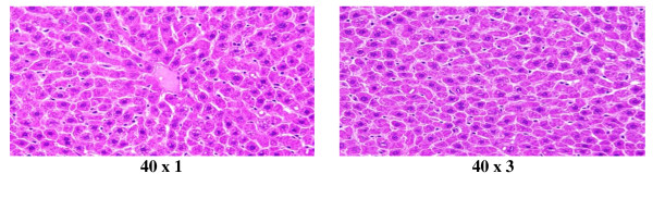
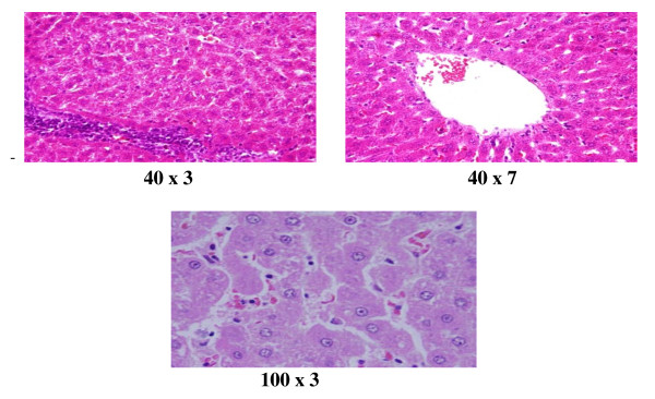
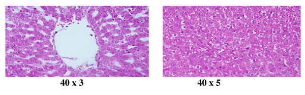
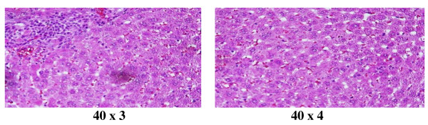
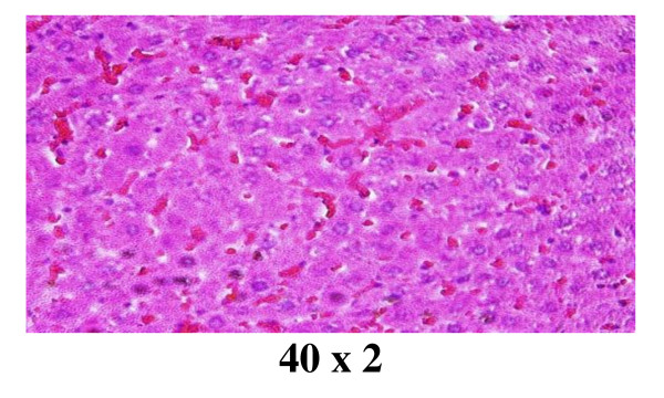
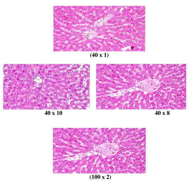
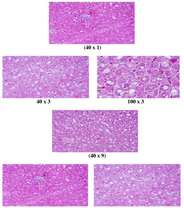
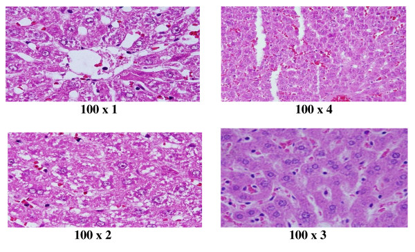
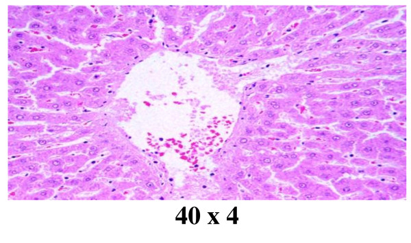
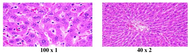
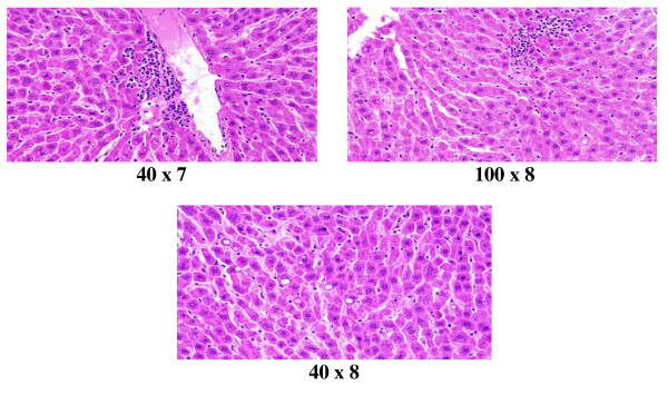
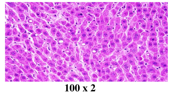
Results: In comparison with respective control rats, exposure to GNPs doses has produced alterations in the hepatocytes, portal triads and the sinusoids. The alterations in the hepatocytes were mainly summarized as hydropic degeneration, cloudy swelling, fatty degeneration, portal and lobular infiltrate by chronic inflammatory cells and congestive dilated central veins.
Conclusions: The induced histological alterations might be an indication of injured hepatocytes due to GNPs toxicity that became unable to deal with the accumulated residues resulting from metabolic and structural disturbances caused by these NPs. These alterations were size-dependent with smaller ones induced the most effects and related with time exposure of GNPs. The appearance of hepatocytes cytoplasmic degeneration and nuclear destruction may suggest that GNPs interact with proteins and enzymes of the hepatic tissue interfering with the antioxidant defense mechanism and leading to reactive oxygen species (ROS) generation which in turn may induce stress in the hepatocytes to undergo atrophy and necrosis. More histomorphologcal, histochemical and ultrastrucural investigations are needed in relation of the application of GNPs with their potential threat as a therapeutic and diagnostic tool.
Figures
References
-
- Yu LE, Yung L-YL, Balasubramaniam KS, Hartono D. et al.Translocation and effects of gold nanoparticles after inhalation exposure in rats. Nanotoxicology. 2007;1(3):235–42. doi: 10.1080/17435390701763108. - DOI
Publication types
MeSH terms
Substances
LinkOut - more resources
Full Text Sources
