. 2012 Jan 26:5:5.
doi: 10.1186/1757-2215-5-5.

The role of melatonin as an antioxidant in the follicle

Affiliations

The role of melatonin as an antioxidant in the follicle

Hiroshi Tamura et al. J Ovarian Res. .

Abstract

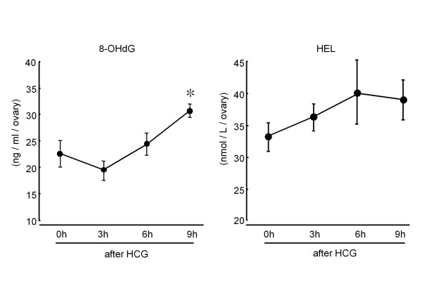
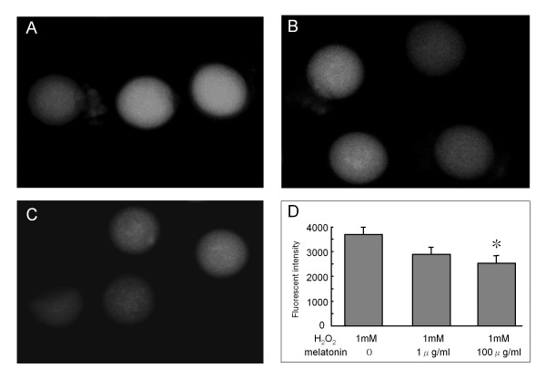
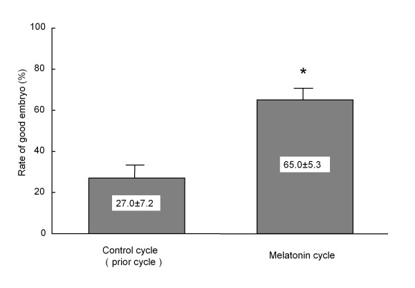
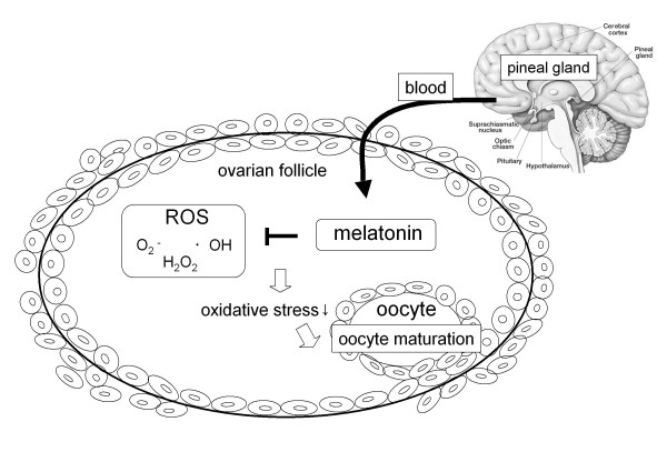
Melatonin (N-acetyl-5-methoxytryptamine) is secreted during the dark hours at night by pineal gland, and it regulates a variety of important central and peripheral actions related to circadian rhythms and reproduction. It has been believed that melatonin regulates ovarian function by the regulation of gonadotropin release in the hypothalamus-pituitary gland axis via its specific receptors. In addition to the receptor mediated action, the discovery of melatonin as a direct free radical scavenger has greatly broadened the understanding of melatonin's mechanisms which benefit reproductive physiology. Higher concentrations of melatonin have been found in human preovulatory follicular fluid compared to serum, and there is growing evidence of the direct effects of melatonin on ovarian function especially oocyte maturation and embryo development. Many scientists have focused on the direct role of melatonin on oocyte maturation and embryo development as an anti-oxidant to reduce oxidative stress induced by reactive oxygen species, which are produced during ovulation process. The beneficial effects of melatonin administration on oocyte maturation and embryo development have been confirmed by in vitro and in vivo experiments in animals. This review also discusses the first application of melatonin to the clinical treatment of infertile women and confirms that melatonin administration reduces intrafollicular oxidative damage and increase fertilization rates. This review summarizes our recent works and new findings related to the reported beneficial effects of melatonin on reproductive physiology in its role as a reducer of oxidative stress, especially on oocyte maturation and embryo development.


Figures

Figure 1
Figure 1
Changes in intrafollicular concentrations of 8-hydroxy-2'-deoxyguanosine (8-OHdG) and hexanoyl-lysine adduct (HEL) during ovulation process. Immature (3 wks) rats received a subcutaneous injection of 20 units of pregnant mare serum gonadotropin (PMSG) to stimulate the multiple follicles. 48 hrs after PMSG injection, human chorionic gonadotropin (HCG) injection was performed to induce ovulation. The ovaries were quickly removed 0, 3, 6 and 9 hr after HCG injection and were transferred to the 500 μl of 0.01 M PBS buffer. Follicular fluids were collected by puncturing ovarian follicles with a 26-gauge needle under dissecting microscope and centrifuged at 300 g for 10 min, and the supernatants were collected and stored at -80C until assay. 8-OHdG and HEL concentrations were measured using ELISA kit. Data are shown as the mean ± SEM for 4 rats. *: p < 0.05 versus 0 h, 3 h.
Figure 2
Figure 2
The effect of melatonin on intracellular ROS production. Immature (3 wks) ICR mice were given an injection of 20 units of pregnant mare serum gonadotropin (PMSG) to stimulate the development of multiple follicles. Oocytes were collected by puncturing ovarian follicles after 48 hrs PMSG injection, and then the surrounding cumulus cells were removed. Oocytes were incubated with 1 mM H2O2 for 10 min in the presence of melatonin (0, 1 μg/ml, 100 μg/ml). Intracellular ROS were detected using an intracellular dye (dichlorofuorescin:DCF-DA). The nonfluorescent DCF-DA is oxidized by intracellular ROS to form the highly fluorescent DCF, intracellular ROS formation was visualized by fluorescence image and fluorescence intensity was analyzed using MetaMorph software. (A) H2O2 (1 mM); (B) H2O2 (1 mM)+ melatonin (1 μg/ml); (C) H2O2 (1 mM)+ melatonin (100 μg/ml); (D) Fluorescence intensity in the oocytes. Data are shown as the mean ± SEM for (6-9 oocytes). *: p < 0.05 versus H2O2 (1 mM).
Figure 3
Figure 3
Effect of melatonin treatment on embryo development in patients who underwent IVF-ET program. Nine patients undergoing IVF-ET who failed to become pregnant in the prior IVF-ET cycle were given a 3 mg tablet of melatonin (KAL, Park City, UT, USA) orally at 22:00 hr from the fifth day of the previous menstrual cycle until the day of oocyte retrieval. Embryo quality was assessed according to Veeck criteria at 2 days after insemination, and Veeck I or II embryo was defined as a good embryo. Rate of good embryos was calculated; divide the number of good embryos (VeeckI or II) by the number of oocytes retrieved. Data are shown as the mean ± SEM. *: p < 0.05 versus control cycle.
Figure 4
Figure 4
Schematic representation of the presumed roles of melatonin in ovarian antral follicle. Melatonin, secreted by pineal gland, is taken up into the follicular fluid from the blood. ROS produced within the follicles, especially ovulation process, were scavenged by melatonin, and reduced oxidative stress may be involved in oocyte maturation and embryo development.

Similar articles

Cited by

References

    1. Brigelius-Flohe R, Banning A, Kny M, Bol GF. Redox events in interleukin-1 signaling. Arch Biochem Biophys. 2004;423(1):66–73. doi: 10.1016/j.abb.2003.12.008. - DOI - PubMed
    1. Forman HJ, Torres M. Reactive oxygen species and cell signaling: respiratory burst in macrophage signaling. Am J Respir Crit Care Med. 2002;166(12 Pt 2):S4–8. - PubMed
    1. Li Q, Sanlioglu S, Li S, Ritchie T, Oberley L, Engelhardt JF. GPx-1 gene delivery modulates NFkappaB activation following diverse environmental injuries through a specific subunit of the IKK complex. Antioxid Redox Signal. 2001;3(3):415–432. doi: 10.1089/15230860152409068. - DOI - PubMed
    1. Agarwal A, Gupta S, Sharma RK. Role of oxidative stress in female reproduction. Reprod Biol Endocrinol. 2005;3:28. doi: 10.1186/1477-7827-3-28. - DOI - PMC - PubMed
    1. Goud AP, Goud PT, Diamond MP, Gonik B, Abu-Soud HM. Reactive oxygen species and oocyte aging: role of superoxide, hydrogen peroxide, and hypochlorous acid. Free Radic Biol Med. 2008;44(7):1295–1304. doi: 10.1016/j.freeradbiomed.2007.11.014. - DOI - PMC - PubMed