. 2012 Apr;129(4):1000-10.e3.
doi: 10.1016/j.jaci.2011.12.965. Epub 2012 Jan 24.

Activin A and TGF-β promote T(H)9 cell-mediated pulmonary allergic pathology

Activin A and TGF-β promote T(H)9 cell-mediated pulmonary allergic pathology

Carla P Jones et al. J Allergy Clin Immunol. 2012 Apr.

Abstract

Background: IL-9-secreting (T(H)9) T cells are thought to represent a distinct T-cell subset. However, evidence for their functionality in disease is uncertain.

Objective: To define a functional phenotype for T(H)9-driven pathology in vivo.

Methods: We used fluorescence-activated cell sorting to identify circulating T(H)9 cells in atopic and nonatopic subjects. In mice we utilized a model of allergic airways disease induced by house dust mite to determine T(H)9 cell function in vivo and the role of activin A in T(H)9 generation.

Results: Allergic patients have elevated T(H)9 cell numbers in comparison to nonatopic donors, which correlates with elevated IgE levels. In a murine model, allergen challenge with house dust mite leads to rapid T(H)9 differentiation and proliferation, with much faster kinetics than for T(H)2 cell differentiation, resulting in the specific recruitment and activation of mast cells. The TGF-β superfamily member activin A replicates the function of TGF-β1 in driving the in vitro generation of T(H)9 cells. Importantly, the in vivo inhibition of T(H)9 differentiation induced by allergen was achieved only when activin A and TGF-β were blocked in conjunction but not alone, resulting in reduced airway hyperreactivity and collagen deposition. Conversely, adoptive transfer of T(H)9 cells results in enhanced pathology.

Conclusion: Our data identify a distinct functional role for T(H)9 cells and outline a novel pathway for their generation in vitro and in vivo. Functionally, T(H)9 cells promote allergic responses resulting in enhanced pathology mediated by the specific recruitment and activation of mast cells in the lungs.


Figures

FIG 1
FIG 1
Identification of in vivo generated TH9 cells. A, Number of TH9 cells in PBMCs from nonallergic and allergic donors. B, Correlation between the number of circulating TH9 cells and plasma IgE levels in humans. C, Percentage of freshly isolated human TH9 cells expressing PU.1. D, TH9 and TH2 cells in the lungs of mice challenged with PBS or HDM for up to 3 weeks. E, Phenotype of murine in vivo generated TH9 cells after 1 week of HDM challenge. F, Allergen specificity of in vivo generated pulmonary T cells. G, Percentage of TH9 cells expressing PU.1 following 1-week HDM challenge in mice. Data shown represent means ± SEMs. *P < .05.
FIG 2
FIG 2
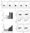
Induction of TH9 differentiation in vitro by activin A. A, TH9 cells generated in the presence of IL-4 alone or in association with TGF-β1, activin A, and/or IL-25. B, Percentage of TH9 cells plotted in a histogram. C, Phenotype of TH9 cells differentiated in the presence of IL-4 with either TGF-β1 or activin A. D, IL-9 secretion from activin A–derived TH9 cells after 4 days in culture in the presence of 0.1 ng/mL of TGF-β1. Data shown represent means ± SEMs.
FIG 3
FIG 3
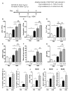
TH9 Adoptive transfer induces mast cell recruitment to the lungs. A, Schematic of experimental protocol. B, Total cells recovered from the lung 1 week after TH9 cell transfer and challenge with either PBS or HDM. Eosinophils (Siglec F+) (C) and TH2 cells (D) recovered from the lung. Serum mMCP-1 levels (E) and intraepithelial mast cell numbers (F) scored in lung sections expressed as total number of mouse tryptase beta 1 positive cells per lung section. G, Serum IgE levels. IL-13 (H) and IL-9 (I) levels in supernatants from HDM-stimulated LN cell cultures. J, Total cells recovered from the lungs of SCID mice adoptively transferred with TH9 cells and treated with either PBS or HDM. Serum mMCP-1 levels (K) and intraepithelial mast cells numbers (L) in SCID mice. Data shown represent means ± SEMs. *P < .05. LN, Lymph node; mMCP-1, mouse mast cell protease-1; NS, not significant; SCID, severe combined immunodeficiency.
FIG 4
FIG 4
Acute blockade of TGF-β and activin A inhibits TH9 differentiation. A, Schematic of experimental protocol. B, TH9 cells recovered from the lung after treatment with either anti–TGF-β and/or anti–activin A in mice challenged with either PBS or HDM. Total cells (C), lung eosinophils (D), and TH2 cells (E) recovered from the lung. Serum mMCP-1 levels (F) and intraepithelial mast cells (G) per lung section. IL-9 (H) and IL-13 (I) levels in supernatants from lymph node cell cultures. J, IL-25 levels in the lungs. Data shown represent means ± SEMs. *P < .05. mMCP-1, Mouse mast cell protease-1.
FIG 5
FIG 5
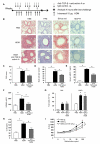
Chronic blockade of TGF-β and activin A reduces airway remodeling. A, Schematic of experimental protocol. B, Peribronchial and perivascular cellular infiltrate (H&E), purple-colored mucin-containing cells in the epithelium (PAS), and perivascular and peribronchiolar collagen (sirrius red) and brown stained intraepithelial mast cells. Original magnification ×40. Scale bar = 50 μm. C, Quantification of mucus positive cells. D, Intraepithelial mast cells per lung section. E, Serum mMCP-1 levels. F, Total lung collagen. G, Total cells recovered from BAL and lung. H, IL-25 levels in the lung. I, Airway resistance (RI) following 3-week HDM challenge. Data shown represent means ± SEMs. *P < .05. BAL, Bronchoalveolar lavage; MCPT7, mouse tryptase beta 1; mMCP-1, mouse mast cell protease-1; PAS, periodic acid-Schiff.

Comment in

  • Reply: To PMID 22277204.
    Korošec P, Silar M, Zidarn M, Košnik M. Korošec P, et al. J Allergy Clin Immunol. 2012 Sep;130(3):818-9. doi: 10.1016/j.jaci.2012.05.048. Epub 2012 Jul 11. J Allergy Clin Immunol. 2012. PMID: 22795372 No abstract available.

References

    1. Nicolaides NC, Holroyd KJ, Ewart SL, Eleff SM, Kiser MB, Dragwa CR, et al. Interleukin 9: a candidate gene for asthma. Proc Natl Acad Sci U S A. 1997;94:13175–80. - PMC - PubMed
    1. Doull IJ, Lawrence S, Watson M, Begishvili T, Beasley RW, Lampe F, et al. Allelic association of gene markers on chromosomes 5q and 11q with atopy and bronchial hyperresponsiveness. Am J Respir Crit Care Med. 1996;153:1280–4. - PubMed
    1. Jager A, Dardalhon V, Sobel RA, Bettelli E, Kuchroo VK. Th1, Th17, and Th9 effector cells induce experimental autoimmune encephalomyelitis with different pathological phenotypes. J Immunol. 2009;183:7169–77. - PMC - PubMed
    1. Kearley J, Erjefalt JS, Andersson C, Benjamin E, Jones CP, Robichaud A, et al. IL-9 governs allergen-induced mast cell numbers in the lung and chronic remodeling of the airways. Am J Respir Crit Care Med. 2011;183:865–75. - PMC - PubMed
    1. Temann UA, Geba GP, Rankin JA, Flavell RA. Expression of interleukin 9 in the lungs of transgenic mice causes airway inflammation, mast cell hyperplasia, and bronchial hyperresponsiveness. J Exp Med. 1998;188:1307–20. - PMC - PubMed

Publication types

MeSH terms