. 2012 Jan 25:7:8.
doi: 10.1186/1750-1172-7-8.

Development and application of a next-generation-sequencing (NGS) approach to detect known and novel gene defects underlying retinal diseases

Affiliations

Development and application of a next-generation-sequencing (NGS) approach to detect known and novel gene defects underlying retinal diseases

Isabelle Audo et al. Orphanet J Rare Dis. .

Abstract

Background: Inherited retinal disorders are clinically and genetically heterogeneous with more than 150 gene defects accounting for the diversity of disease phenotypes. So far, mutation detection was mainly performed by APEX technology and direct Sanger sequencing of known genes. However, these methods are time consuming, expensive and unable to provide a result if the patient carries a new gene mutation. In addition, multiplicity of phenotypes associated with the same gene defect may be overlooked.

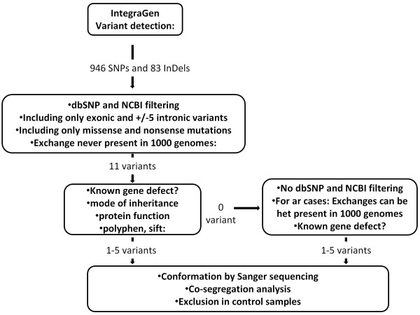
Methods: To overcome these challenges, we designed an exon sequencing array to target 254 known and candidate genes using Agilent capture. Subsequently, 20 DNA samples from 17 different families, including four patients with known mutations were sequenced using Illumina Genome Analyzer IIx next-generation-sequencing (NGS) platform. Different filtering approaches were applied to identify the genetic defect. The most likely disease causing variants were analyzed by Sanger sequencing. Co-segregation and sequencing analysis of control samples validated the pathogenicity of the observed variants.

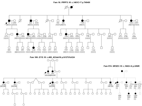
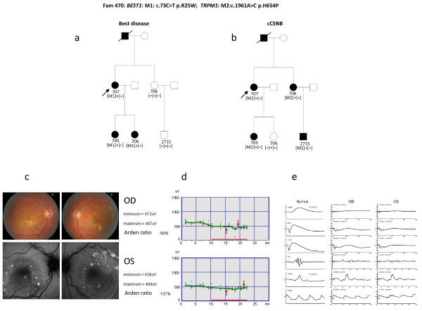
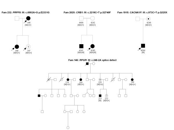
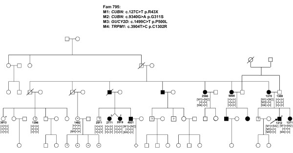
Results: The phenotype of the patients included retinitis pigmentosa, congenital stationary night blindness, Best disease, early-onset cone dystrophy and Stargardt disease. In three of four control samples with known genotypes NGS detected the expected mutations. Three known and five novel mutations were identified in NR2E3, PRPF3, EYS, PRPF8, CRB1, TRPM1 and CACNA1F. One of the control samples with a known genotype belongs to a family with two clinical phenotypes (Best and CSNB), where a novel mutation was identified for CSNB. In six families the disease associated mutations were not found, indicating that novel gene defects remain to be identified.

Conclusions: In summary, this unbiased and time-efficient NGS approach allowed mutation detection in 75% of control cases and in 57% of test cases. Furthermore, it has the possibility of associating known gene defects with novel phenotypes and mode of inheritance.


Figures

Figure 1
Figure 1
Flow chart of variant analysis. IntegraGen provided the results in form of excel tables. For each sample on average 946 SNPs and 83 inDels were detected, of which 11 represent missense, nonsense or putative splice site mutations, which were absent in dbSNB, NCBI and 1000 genome databases. Of those 1-5 variants were predicted to be pathogenic. In case where none of the variants were predicted to be pathogenic, dbSNB, NCBI and 1000 genome databases were included to detect mutations referenced with an rs-number. Co-segregation analysis was performed in families with putative pathogenic variants.
Figure 2
Figure 2
Detection of known mutations by NGS in 254 retinal genes. The index patient 19 of family 16 with adRP revealed the p.T494M mutations in PRPF3, which co-segregates with the phenotype. Two family members never clinically investigated from the last generation (984 and 1167 carrying a question mark) were reported to be not affected but carried the mutation. They may develop the phenotype at a later stage. In addition variability of the phenotype of this mutation was documented [35]. Two patients, 128 and 943 of family 100 with arRP from Jewish origin revealed the known EYS mutation p.N137VfsX24, which was found in all screened affected family members. The index patient 893 of family 574 showed the previously described NR2E3 p.G56R mutation, which co-segregated with the phenotype.
Figure 3
Figure 3
Best disease and CSNB co-segregating in one family. a) Sanger and NGS detected in all patients with Best disease a BEST1 mutation. b) NGS detected in all patients with a cCSNB phenotype a novel TRPM1 mutation. c) Fundus colour photographs (above) and fundus autofluorescence (below) of patient 707 showing multiple yellow deposits within the posterior pole which are hyper autofluorescent d) Electro-oculogram of patient 707 showing no slight rise after illumination in keeping with the diagnosis of Best disease e) Full Field Electroretinogram of patient 707 showing ON-bipolar cell pathway dysfunction in keeping with the diagnosis of cCSNB.
Figure 4
Figure 4
Detection of novel mutations using NGS in 254 retinal genes. Novel mutations in PRPF8, CRB1, RPGR and CACNA1F co-segregated in affected and asymptomatic carriers with the adRP, arRP, x-linked dominant and X-liked icCSNB phenotypes respectively. Asymptomatic individuals are marked with a question mark.
Figure 5
Figure 5
Detection of novel mutation by using NGS in 254 retinal genes. Family 795 reveals autosomal dominant cone dystrophy with post-receptoral defects. Four putative disease causing mutations were investigated on the basis of co-segregation. However, none of them co-segregated in all affected family members with the phenotype and thus are not considered to be disease causing. Individuals marked with a star were clinically investigated, patients with a question mark are asymptomatic and patients with a plus sign show high myopia.

References

    1. Sohocki MM, Daiger SP, Bowne SJ, Rodriquez JA, Northrup H, Heckenlively JR, Birch DG, Mintz-Hittner H, Ruiz RS, Lewis RA, Saperstein DA, Sullivan LS. Prevalence of mutations causing retinitis pigmentosa and other inherited retinopathies. Hum Mutat. 2001;17:42–51. doi: 10.1002/1098-1004(2001)17:1<42::AID-HUMU5>3.0.CO;2-K. - DOI - PMC - PubMed
    1. Boon CJ, den Hollander AI, Hoyng CB, Cremers FP, Klevering BJ, Keunen JE. The spectrum of retinal dystrophies caused by mutations in the peripherin/RDS gene. Prog Retin Eye Res. 2008;27:213–235. doi: 10.1016/j.preteyeres.2008.01.002. - DOI - PubMed
    1. Boon CJ, Klevering BJ, Leroy BP, Hoyng CB, Keunen JE, den Hollander AI. The spectrum of ocular phenotypes caused by mutations in the BEST1 gene. Prog Retin Eye Res. 2009;28:187–205. doi: 10.1016/j.preteyeres.2009.04.002. - DOI - PubMed
    1. Schorderet DF, Escher P. NR2E3 mutations in enhanced S-cone sensitivity syndrome (ESCS), Goldmann-Favre syndrome (GFS), clumped pigmentary retinal degeneration (CPRD), and retinitis pigmentosa (RP) Hum Mutat. 2009;30:1475–1485. doi: 10.1002/humu.21096. - DOI - PubMed
    1. Jaakson K, Zernant J, Kulm M, Hutchinson A, Tonisson N, Glavac D, Ravnik-Glavac M, Hawlina M, Meltzer MR, Caruso RC, Testa F, Maugeri A, Hoyng CB, Gouras P, Simonelli F, Lewis RA, Lupski JR, Cremers FP, Allikmets R. Genotyping microarray (gene chip) for the ABCR (ABCA4) gene. Hum Mutat. 2003;22:395–403. doi: 10.1002/humu.10263. - DOI - PubMed

Publication types