. 2012 Apr;8(4):1430-9.
doi: 10.1016/j.actbio.2011.12.031. Epub 2012 Jan 8.

Incorporation of phosphate group modulates bone cell attachment and differentiation on oligo(polyethylene glycol) fumarate hydrogel

Affiliations

Incorporation of phosphate group modulates bone cell attachment and differentiation on oligo(polyethylene glycol) fumarate hydrogel

Mahrokh Dadsetan et al. Acta Biomater. 2012 Apr.

Abstract

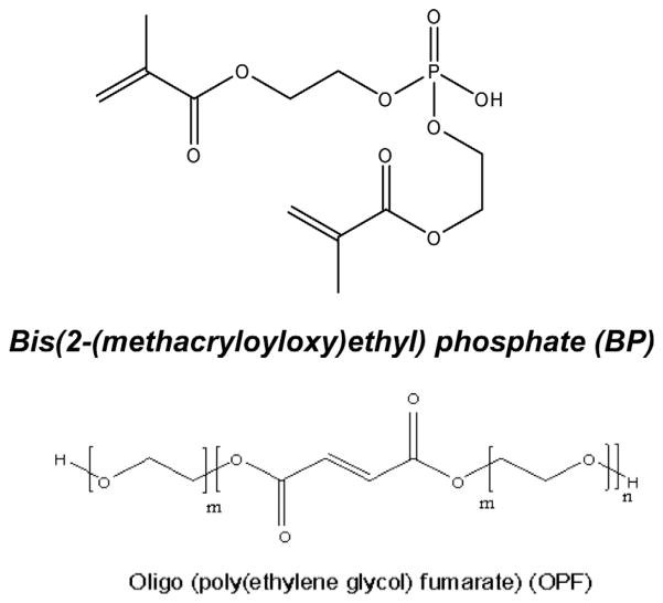
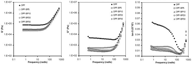
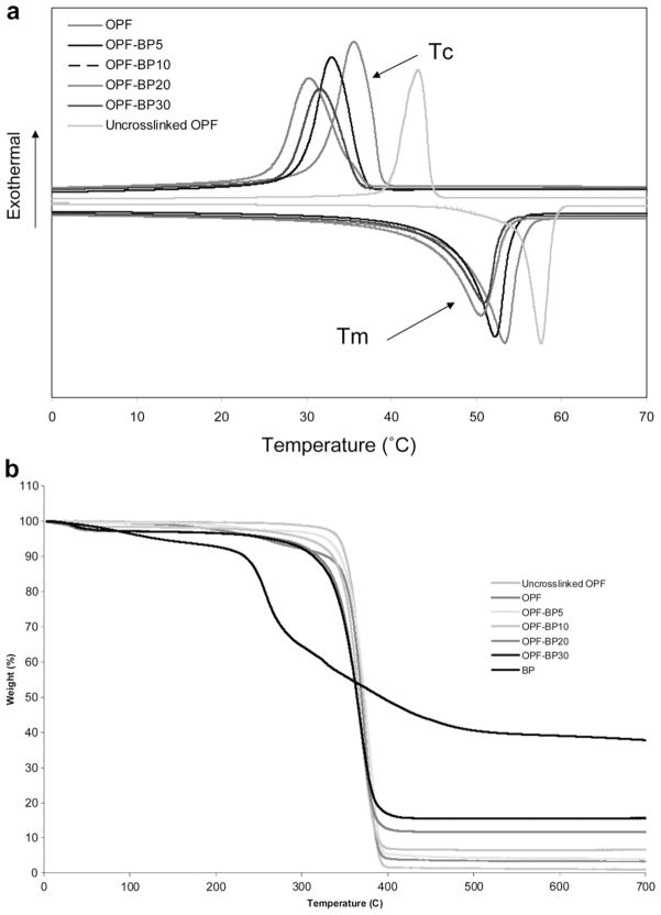
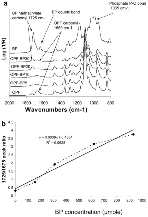
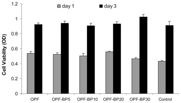
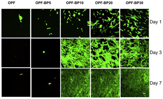
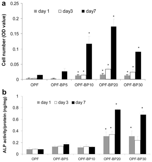
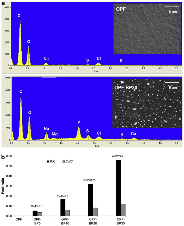
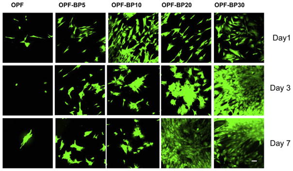
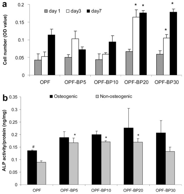
In this work, we have investigated the development of a synthetic hydrogel that contains a negatively charged phosphate group for use as a substrate for bone cell attachment and differentiation in culture. The photoreactive, phosphate-containing molecule, bis(2-(methacryloyloxy)ethyl)phosphate (BP), was incorporated into oligo(polyethylene glycol) fumarate hydrogel and the mechanical, rheological and thermal properties of the resulting hydrogels were characterized. Our results showed changes in hydrogel compression and storage moduli with incorporation of BP. The modification also resulted in decreased crystallinity as recorded by differential scanning calorimetry. Our data revealed that incorporation of BP improved attachment and differentiation of human fetal osteoblast (hFOB) cells in a dose-dependent manner. A change in surface chemistry and mineralization of the phosphate-containing surfaces verified by scanning electron microscopy and energy dispersive X-ray analysis was found to be important for hFOB cell attachment and differentiation. We also demonstrated that phosphate-containing hydrogels support attachment and differentiation of primary bone marrow stromal cells. These findings suggest that BP-modified hydrogels are capable of sustaining attachment and differentiation of both bone marrow stromal cells and osteoblasts that are critical for bone regeneration.


Conflict of interest statement

Conflicts of Interest

A non-provisional patent has been filed for photocrosslinkable oligo(polyethylene glycol) fumarate used in this research, and this technology has been licensed to BonWrx.

Figures

Fig. 1
Fig. 1
Chemical structures of oligo(polyethylene glycol) fumarate (OPF), and bis(2-methacryloyloxy)ethyl) phosphate (BP).
Fig. 2
Fig. 2
Storage modulus G′ (left panel), loss modulus G″ (middle panel) and viscosity η (right panel) vs. frequency of unmodified hydrogel (OPF) compared to hydrogels with varying concentrations of BP (OPF-BP).
Fig. 3
Fig. 3
(a) DSC curves for OPF and BP-modified hydrogels. Three consecutive heat/cool/heat cycles were used to determine melting points (Tm), crystallization temperature (Tc) and heats of fusion (ΔTm). (b) TGA curves used to determine onset of degradation (Td) for OPF and BP-modified hydrogels.
Fig. 4
Fig. 4
(a) Micro-ATR-FTIR of unmodified BP-modified hydrogels after crosslinking and lyophilization. With increasing BP concentration in hydrogel formulations, the peak intensity at 1725 cm−1 assigned to methacrylate groups from BP increased, and the peak intensity at 1650 cm−1 assigned to carbonyl from OPF decreased. (b) The linear relationship between the amounts of BP added to the OPF solution and the levels of covalently coupled BP in crosslinked hydrogels. The solid line is a linear fit of the data (R2 = 0.9628).
Fig. 5
Fig. 5
Toxicity of leaching materials from hydrogels after 1 and 3 d in culture. hFOB cells were plated on 24-well tissue culture plates and exposed to leaching materials from scaffolds via transwell inserts. The viability of the cells was tested using the MTS Cell Proliferation Assay by measuring the optical density (OD) at 490 nm. Values represent mean ± standard error (n = 3).
Fig. 6
Fig. 6
Osteoblast viability on hydrogels with different concentrations of BP in their formulation on days 1, 3 and 7. Viable cells are stained green and dead cells are stained red. Scale bar = 20 μm.
Fig. 7
Fig. 7
(a) Proliferation of hFOB cells on OPF hydrogels with varying amounts of BP at days 1, 3 and 7. Cell number was determined using the MTS Cell Proliferation Assay by measuring the optical density (OD) at 490 nm. (b) ALP activity of hFOB cells on OPF hydrogels with varying concentrations of BP in their formulation on days 1, 3 and 7. Values represent mean ± standard error (n = 3). *P < 0.005 significantly different as compared to unmodified OPF hydrogel.
Fig. 8
Fig. 8
(a) SEM images and EDAX spectra of OPF (unmodified) and OPF-BP30 (hydrogel with 930 μmol BP). (b) Comparison of calcium and phosphorus peaks on the hydrogel surfaces as a function of BP concentration. The peak heights of both calcium and phosphorous are normalized to the carbon peak height. The Ca/P ratio was obtained from area analysis.
Fig. 9
Fig. 9
MSC viability on hydrogels with different concentrations of BP in their formulation on days 1, 3 and 7. Viable cells are stained green and dead cells are stained red. Scale bar = 20 μm.
Fig. 10
Fig. 10
(a) Proliferation of MSCs on OPF hydrogels with varying concentrations of BP in their formulation on days 1, 3 and 7. Cell number was determined using the MTS Cell Proliferation Assay by measuring the optical density (OD) at 490 nm. *P < 0.05 significantly different as compared to unmodified OPF hydrogel at the same time point. (b) ALP activity of MSCs on OPF hydrogels with different concentrations of BP in osteogenic and non-ostegenic media after 7 days in culture. *P < 0.05 significantly different as compared to unmodified OPF hydrogel in the same media, #P < 0.05 significantly higher in osteogenic media. Values represent mean ± standard error (n = 3).

Similar articles

Cited by

References

    1. Deshpande AS, Fang PA, Zhang X, Jayaraman T, Sfeir C, Beniash E. Primary structure and phosphorylation of dentin matrix protein 1 (DMP1) and dentin phosphophoryn (DPP) uniquely determine their role in biomineralization. Biomacromolecules. 2011;12:2933–45. - PMC - PubMed
    1. Zouani OF, Chollet C, Guillotin B, Durrieu MC. Differentiation of pre-osteoblast cells on poly(ethylene terephthalate) grafted with RGD and/or BMPs mimetic peptides. Biomaterials. 2010;31:8245–53. - PubMed
    1. Zavgorodniy AV, Borrero-Lopez O, Hoffman M, Legeros RZ, Rohanizadeh R. Characterization of the chemically deposited hydroxyapatite coating on a titanium substrate. J Mater Sci Mater Med. 2011;22:1–9. - PubMed
    1. Zhang L, Ayukawa Y, Legeros RZ, Matsuya S, Koyano K, Ishikawa K. Tissue-response to calcium-bonded titanium surface. J Biomed Mater Res A. 2010;95:33–9. - PubMed
    1. Gandhi R, Davey JR, Mahomed NN. Hydroxyapatite coated femoral stems in primary total hip arthroplasty: a meta-analysis. J Arthroplasty. 2009;24:38–42. - PubMed

Publication types

MeSH terms

LinkOut - more resources