. 2012 Feb 8;4(120):120ra17.
doi: 10.1126/scitranslmed.3003316. Epub 2012 Jan 25.

Mechanisms of acquired crizotinib resistance in ALK-rearranged lung Cancers

Affiliations

Mechanisms of acquired crizotinib resistance in ALK-rearranged lung Cancers

Ryohei Katayama et al. Sci Transl Med. .

Abstract

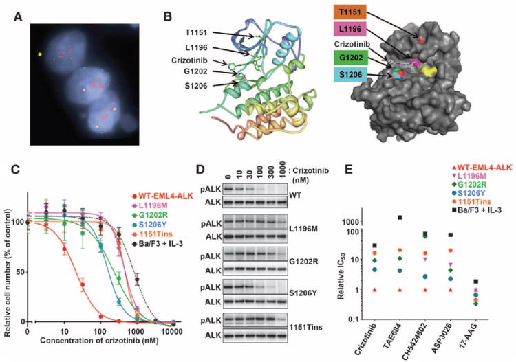
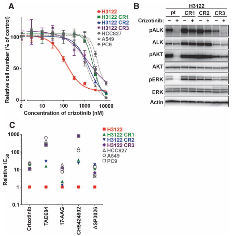
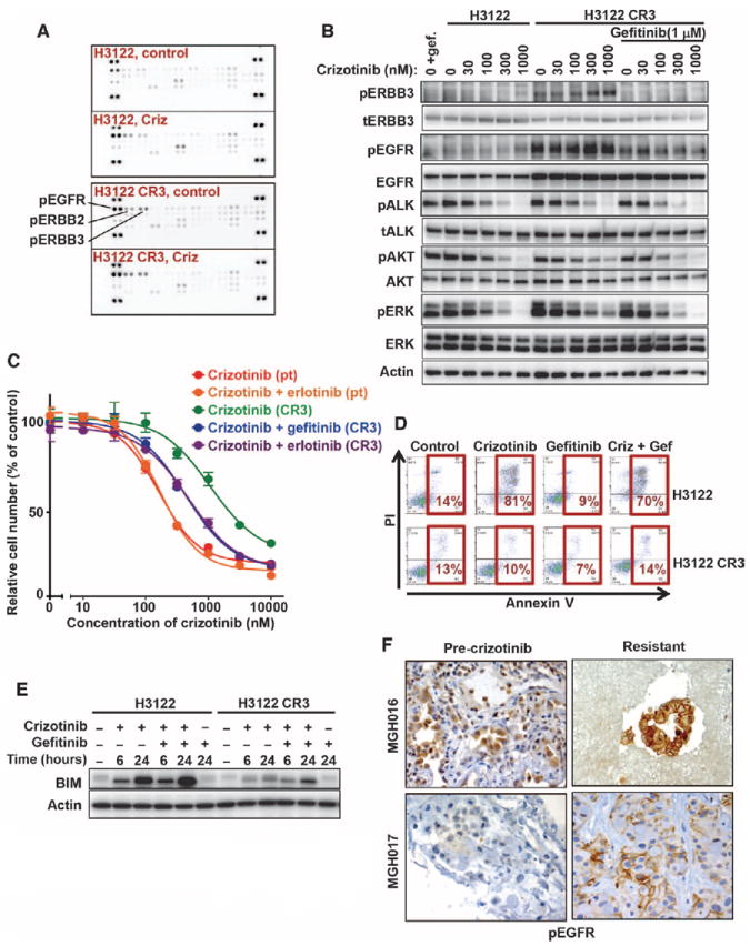
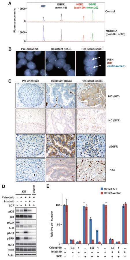
Most anaplastic lymphoma kinase (ALK)-positive non-small cell lung cancers (NSCLCs) are highly responsive to treatment with ALK tyrosine kinase inhibitors (TKIs). However, patients with these cancers invariably relapse, typically within 1 year, because of the development of drug resistance. Herein, we report findings from a series of lung cancer patients (n = 18) with acquired resistance to the ALK TKI crizotinib. In about one-fourth of patients, we identified a diverse array of secondary mutations distributed throughout the ALK TK domain, including new resistance mutations located in the solvent-exposed region of the adenosine triphosphate-binding pocket, as well as amplification of the ALK fusion gene. Next-generation ALK inhibitors, developed to overcome crizotinib resistance, had differing potencies against specific resistance mutations. In addition to secondary ALK mutations and ALK gene amplification, we also identified aberrant activation of other kinases including marked amplification of KIT and increased autophosphorylation of epidermal growth factor receptor in drug-resistant tumors from patients. In a subset of patients, we found evidence of multiple resistance mechanisms developing simultaneously. These results highlight the unique features of TKI resistance in ALK-positive NSCLCs and provide the rationale for pursuing combinatorial therapeutics that are tailored to the precise resistance mechanisms identified in patients who relapse on crizotinib treatment.


Conflict of interest statement

Competing interests: J.A.E. is a paid consultant for Ariad and Novartis; A.J.I. is a paid consultant for Pfizer and Abbott; A.T.S. is a paid consultant for Pfizer, Ariad, Chugai, and Novartis; B.J.S. has served on the advisory boards for Pfizer and Novartis. The other authors declare that they have no competing interests.

Figures

Fig. 1
Fig. 1. ALK gene amplification and multiple ALK resistance mutations in cancers with acquired crizotinib resistance
(A) FISH analysis of ALK demonstrates high-level ALK gene amplification in one resistant tumor. Amplified, rearranged ALK appears as a cluster of isolated red signals in this resistant specimen. (B) Three-dimensional mapping of each identified ALK mutation based on the crystal structure of ALK. Each of the four ALK mutations is mapped on a ribbon (left) or surface (right) diagram. In the surface structure model, each mutated residue is shown in a different color, and yellow depicts the DFG motif. Figures were drawn using the PyMOL software with the crystal structure information of Protein Data Bank ID 2XP2. (C) Ba/F3 cells were transformed by expression of either wild-type (WT) EML4-ALK or EML4-ALK harboring one of the four identified resistance mutations (L1196M, G1202R, S1206Y, or 1151Tins). Parental Ba/F3 cells (cultured with IL-3) or EML4-ALK–expressing Ba/F3 cells (cultured without IL-3) were treated with the indicated doses of crizotinib for 48 hours. Cell survival was measured using CellTiter-Glo. Each concentration was measured in sextuplicate, and the average and SD are shown. (D) Ba/F3 cells transformed by WT EML4-ALK or EML4-ALK harboring the indicated resistance mutation were treated with the indicated concentrations of crizotinib for 1 hour. Cell lysates were probed with phospho-ALK (pALK) and ALK-specific antibodies. (E) Differential sensitivity conferred by ALK TK mutations to next-generation ALK inhibitors and the hsp90 inhibitor 17-AAG. The relative IC50 of each drug across six different Ba/F3 cell lines, including parental, IL-3–dependent Ba/F3 cells as well as transformed Ba/F3 cells expressing the indicated EML4-ALK constructs, is shown. For each drug, the IC50 values for the various cell lines have been normalized to that of crizotinib-sensitive Ba/F3 cells expressing WT EML4-ALK. The values are the average from three independent experiments. The raw data from a representative experiment are shown in fig. S2 and tabulated in fig. S2F.
Fig. 2
Fig. 2. Heterogeneity of resistance mechanisms from cell lines with acquired resistance to crizotinib
(A) Three independently derived, crizotinib-resistant cell lines (H3122 CR1, CR2, and CR3) were treated with the indicated doses of crizotinib for 72 hours. As controls, parental H3122 cells and three ALK WT cell lines (HCC827, PC9, and A549) were also treated in parallel. Cell survival was measured using a CellTiter-Glo viability assay. Each concentration was measured in sextuplicate, and the average and SD are shown. (B) ALK signaling in crizotinib-resistant H3122 cell lines. Parental (pt) H3122 cells and the three different crizotinib-resistant H3122 cell lines (CR1, CR2, and CR3) were treated with 1 μM crizotinib for 6 hours. Cell extracts were immunoblotted with antibodies directed against the indicated proteins. (C) Differential sensitivity of H3122 CR1, CR2, and CR3 cells to ALK and hsp90 inhibitors. Shown is the relative IC50 for each drug across seven different cell lines, including the three crizotinib-resistant H3122 cell lines as well as three WT ALK controls (A549, HCC827, and PC9). For each drug, the IC50 values for the various cell lines have been normalized to that of parental H3122 cells. The values are the average from three independent experiments. The raw data from a representative experiment are shown in fig. S5.
Fig. 3
Fig. 3. Activation of EGFR in cell lines and patients with acquired resistance to crizotinib
(A) Parental H3122 and H3122 CR3 cells were incubated in the absence (control) or presence (Criz) of 1 μM crizotinib for 6 hours, and lysates were incubated with phospho-RTK arrays (R&D Systems). The positions of phospho-EGFR, phospho-ERBB2, and phospho-ERBB3 are indicated. (B) H3122 CR3 cells were treated for 6 hours with the indicated concentrations of crizotinib in the presence or absence of gefitinib. Cell lysates were probed with the indicated antibodies. (C) Sensitization of resistant H3122 CR3 cells by treatment with both crizotinib and an EGFR TKI (gefitinib or erlotinib). Parental H3122 and H3122 CR3 cells were treated with the indicated doses of crizotinib in the presence or absence of 2 μM gefitinib or 1 μM erlotinib for 72 hours. Cell survival was determined using the CellTiter-Glo viability assay. Each concentration was measured in sextuplicate, and the average and SD are shown. (D) Lack of apoptosis induction in H3122 CR3 cells treated with crizotinib (Criz) and gefitinib (Gef). Parental H3122 and H3122 CR3 cells were treated with 1 μM crizotinib, 2 μM gefitinib, or the combination. After 72 hours, cells were stained with Alexa Fluor 633–labeled annexin V and PI and analyzed by flow cytometry. The percentage of cells undergoing apoptosis is shown. (E) Defective up-regulation of BIM in H3122 CR3 cells treated with crizotinib and gefitinib. H3122 and H3122 CR3 cells were treated with 1 μM crizotinib, 2 μM gefitinib, or both drugs for 6 or 24 hours. Lysates were probed with BIM and actin-specific antibodies. (F) Increased EGFR activation in crizotinib-resistant tumors. Pre-crizotinib and crizotinib-resistant tumors were stained using a phospho-EGFR–specific antibody. Shown are two cases, MGH016 and MGH017, both of which demonstrate stronger plasma membrane staining of phospho-EGFR in the resistant cancer than in the corresponding pre-crizotinib sample.
Fig. 4
Fig. 4. Aberrant activation of KIT/SCF mediates acquired resistance to crizotinib
(A) Snapshot panel showing relatively increased amplitude of the WT KIT peak (arrow) in patient MGH0NZ compared to normal control, suggesting KIT gene amplification. This snapshot panel tests for mutations within KIT (blue), EGFR exon 19 (black), HER2 (red), and EGFR exon 20 (green). (B) Confirmation of KIT amplification by FISH analysis. KIT/centromere 7 FISH was performed on both pretreatment and resistant specimens from MGH0NZ. Amplified KIT appears as a cluster of red signals (arrows) and was detected in the solid but not the BAC component of the resistant specimen. Aqua signals indicate centromere 7. (C) IHC staining for KIT, SCF, phospho-EGFR (pEGFR), and Ki67 expression. Within the resistant specimen, the solid, but not the bronchioloalveolar, component showed strong KIT expression in the tumor cells and SCF expression in the stromal cells. Conversely, the bronchioloalveolar, but not the solid, component showed strong phospho-EGFR expression. Note that the KIT-positive cells in the BAC represent CD117-positive mast cells. (D). H3122 cells were infected with retrovirus expressing KIT or empty vector control. After 2 weeks of selection in puromycin, cells were treated for 6 hours with crizotinib (1 μM), human SCF (100 ng/ml), imatinib (1 μM), or a combination, as indicated. Cell extracts were immunoblotted to detect the indicated proteins. (E) Control and KIT-overexpressing H3122 cells were seeded in 12-well plates and treated with crizotinib (1 μM), human SCF (100 ng/ml), imatinib (1 μM), or their combination as indicated for 7 days. Cell viability was measured using a crystal violet assay. Experiments were performed in triplicate, and the average and SD are shown.

Comment in

Similar articles

Cited by

References

    1. Chiarle R, Voena C, Ambrogio C, Piva R, Inghirami G. The anaplastic lymphoma kinase in the pathogenesis of cancer. Nat Rev Cancer. 2008;8:11–23. - PubMed
    1. Janoueix-Lerosey I, Lequin D, Brugières L, Ribeiro A, de Pontual L, Combaret V, Raynal V, Puisieux A, Schleiermacher G, Pierron G, Valteau-Couanet D, Frebourg T, Michon J, Lyonnet S, Amiel J, Delattre O. Somatic and germline activating mutations of the ALK kinase receptor in neuroblastoma. Nature. 2008;455:967–970. - PubMed
    1. George RE, Sanda T, Hanna M, Fröhling S, Luther W, II, Zhang J, Ahn Y, Zhou W, London WB, McGrady P, Xue L, Zozulya S, Gregor VE, Webb TR, Gray NS, Gilliland DG, Diller L, Greulich H, Morris SW, Meyerson M, Look AT. Activating mutations in ALK provide a therapeutic target in neuroblastoma. Nature. 2008;455:975–978. - PMC - PubMed
    1. Chen Y, Takita J, Choi YL, Kato M, Ohira M, Sanada M, Wang L, Soda M, Kikuchi A, Igarashi T, Nakagawara A, Hayashi Y, Mano H, Ogawa S. Oncogenic mutations of ALK kinase in neuroblastoma. Nature. 2008;455:971–974. - PubMed
    1. Soda M, Choi YL, Enomoto M, Takada S, Yamashita Y, Ishikawa S, Fujiwara S, Watanabe H, Kurashina K, Hatanaka H, Bando M, Ohno S, Ishikawa Y, Aburatani H, Niki T, Sohara Y, Sugiyama Y, Mano H. Identification of the transforming EML4–ALK fusion gene in non-small-cell lung cancer. Nature. 2007;448:561–566. - PubMed

Publication types

MeSH terms