. 2012 Apr 15;302(8):R965-75.
doi: 10.1152/ajpregu.00680.2011. Epub 2012 Jan 25.

Integrative responses of neurons in parabrachial nuclei to a nauseogenic gastrointestinal stimulus and vestibular stimulation in vertical planes

Affiliations

Integrative responses of neurons in parabrachial nuclei to a nauseogenic gastrointestinal stimulus and vestibular stimulation in vertical planes

Takeshi Suzuki et al. Am J Physiol Regul Integr Comp Physiol. .

Abstract

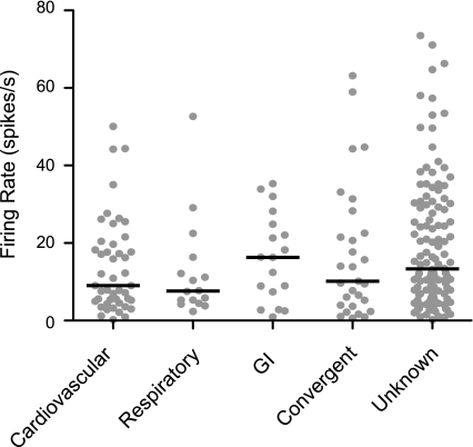
The parabrachial and adjacent Kölliker-Fuse (PBN/KF) nuclei play a key role in relaying visceral afferent inputs to the hypothalamus and limbic system and are, thus, believed to participate in generating nausea and affective responses elicited by gastrointestinal (GI) signals. In addition, the PBN/KF region receives inputs from the vestibular system and likely mediates the malaise associated with motion sickness. However, previous studies have not considered whether GI and vestibular inputs converge on the same PBN/KF neurons, and if so, whether the GI signals alter the responses of the cells to body motion. The present study, conducted in decerebrate cats, tested the hypothesis that intragastric injection of copper sulfate, which elicits emesis by irritating the stomach lining, modifies the sensitivity of PBN/KF neurons to vertical plane rotations that activate vestibular receptors. Intragastric copper sulfate produced a 70% median change in the gain of responses to vertical plane rotations of PBN/KF units, whose firing rate was modified by the administration of the compound; the response gains for 16 units increased and those for 17 units decreased. The effects were often dramatic: out of 51 neurons tested, 13 responded to the rotations only after copper sulfate was injected, whereas 10 others responded only before drug delivery. These data show that a subset of PBN/KF neurons, whose activity is altered by a nauseogenic stimulus also respond to body motion and that irritation of the stomach lining can either cause an amplification or reduction in the sensitivity of the units to vestibular inputs. The findings imply that nausea and affective responses to vestibular stimuli may be modified by the presence of emetic signals from the GI system.


Figures

Fig. 1.
Fig. 1.
Firing patterns of different unit types observed in the parabrachial and adjacent Kölliker-Fuse (PBN/KF), including cardiovascular (A–C), respiratory (D, E), and GI (F) units. A: record of arterial blood pressure (sampled at 100 Hz, top) and activity (sampled at 25,000 Hz, bottom) of a unit in lateral PBN with a cardiac-related firing pattern. B: averaged changes in blood pressure (top) and a poststimulus histogram showing associated changes in firing rate (bottom, bin width of 10 ms) of the same unit whose data are illustrated in A; 126 sweeps were pooled to generate traces. C: traces showing effects of mechanical stretch of carotid sinus (indicated by horizontal line) on arterial blood pressure (top) and activity of a unit in the medial PBN. A decrease in blood pressure occurred subsequent to the increase in the unit's firing rate, indicating that the excitation was evoked by afferent inputs from the carotid sinus and not by the change in blood pressure. D: activity of a KF neuron (bottom) that was synchronized to phrenic nerve discharges (top). E: activity of a neuron in the lateral PBN (bottom) that fired between phrenic nerve discharges (top). F: effect of copper sulfate infusion into the stomach (indicated by arrow) on arterial blood pressure (top) and activity of a unit in the medial PBN (bottom). A sustained increase in the neuron's firing rate occurred ∼2 min following the administration of copper sulfate, although there was no change in blood pressure.
Fig. 2.
Fig. 2.
Spontaneous firing rates of different types of units tested for responses to whole body rotations. Gray symbols indicate data for each neuron, whereas horizontal lines designate the median firing rate of each unit type.
Fig. 3.
Fig. 3.
Polar plots showing response vector orientations of different type of units: cardiovascular (A), respiratory (B), GI (C), convergent (D), and unknown (E). Response vector orientations were determined using wobble stimuli, usually delivered at 0.2 Hz. The response vector orientations were plotted using a head-centered coordinate system, with 0° corresponding to ipsilateral ear-down (IED) roll tilt, 90° to nose-down (ND) pitch, 180° to contralateral ear-down (CED) roll, and −90° to nose-up (NU) pitch.
Fig. 4.
Fig. 4.
Averaged responses of two units to 5° sinusoidal rotations in the pitch plane at 0.05–0.5 Hz. Each histogram contains 500 bins, such that bin width varies from 40 ms at 0.05 Hz to 4 ms at 0.5 Hz. A gray sine wave superimposed on each trace designates table movement. A: responses of a cardiovascular unit that occurred in phase with table movement. The number of sweeps averaged for each trace were: 32 in a, 12 in b, 8 in c, and 9 in d. B: responses of an unknown unit that lagged stimulus position; sine waves fit to the responses are indicated in red, so that response phases can be compared with table position (designated by a gray sine wave). The number of sweeps averaged for each trace were 19 in a, 9 in b, 4 in c, and 5 in d. ND, nose down; NU, nose up.
Fig. 5.
Fig. 5.
Bode plots illustrating the dynamic properties of responses of PBN/KF neurons to rotations in a fixed plane at multiple frequencies collected across all of the experiments. Response gain and phase are plotted with respect to stimulus position. A: bode plots for individual neurons; different unit types are indicated by lines with distinct colors: red, cardiovascular units; yellow, respiratory units; green, GI units; blue, convergent units; and gray, unknown units. Response gain at the lowest frequency for each unit (usually 0.05 Hz, but 0.02 Hz for 2 units and 0.1 Hz for 2 units) was standardized at 1 spike·s−1·degree−1, so that the change in gain with increasing stimulus frequency was evident. B: averaged data for each of the unit types (solid lines) and all neurons (dashed lines). Since a Bode plot was generated for only one respiratory neuron, averaged data are not provided. Error bars designate one SE.
Fig. 6.
Fig. 6.
Effects of copper sulfate administration on the averaged responses of two units to 7.5° wobble stimuli delivered at 0.2 Hz. Each histogram contains 500 bins; a gray waveform superimposed on each trace indicates the tilt table position, whereas a red waveform shows a sine wave fit to the response. In each panel, a indicates the response prior to intragastric copper sulfate, whereas b indicates the response after the drug was delivered. The shapes of five overlapped action potentials recorded from the units whose activity was binned in these histograms are illustrated in panels c and d; c shows action potential shape before copper sulfate administration, while d shows the action potential shape following administration. The spike shape was similar throughout the recording period, indicating that the same unit was sampled both before and after intragastric copper sulfate. A: responses of a neuron whose spontaneous firing rate decreased after copper sulfate administration. No modulation of unit activity was evident during rotations prior to intragastric copper sulfate (a, average of 11 sweeps, signal-to-noise ratio of 0.07), although a strong response was present afterwards (b, average of 8 sweeps, signal-to-noise ratio of 0.92). B: responses of a neuron whose spontaneous firing rate increased after copper sulfate administration. The robust modulation of unit activity evident before intragastric copper sulfate (a, average of 4 sweeps, signal-to-noise ratio of 2.14) was eliminated after the drug was injected (b, average of 15 sweeps, signal-to-noise ratio of 0.12).
Fig. 7.
Fig. 7.
A: effects of intragastric copper sulfate administration of the gain of responses to wobble stimulation. The absolute value of the percent change in gain following copper sulfate injection is indicated. Open symbols designate units whose response gain increased, whereas solid symbols indicate units whose response gain decreased. Data are segregated into two groups: those for units whose firing rate changed >30% following copper sulfate administration (GI input) and those whose firing rate remained stable (no GI input). Red circles indicate units with GI input whose spontaneous firing rate increased following copper sulfate administration, while black circles designate units whose firing rate decreased. Horizontal lines show median percentage changes in gain produced by intragastric copper sulfate. B: changes in response vector orientation produced by intragastric copper sulfate administration; data are provided only for the subset of neurons that responded to rotations before and after the drug was delivered. Horizontal lines show median values. C: bode plots showing average response dynamics for neurons before and after copper sulfate administration. Solid lines show responses prior to copper sulfate injection, whereas dashed lines indicate responses after drug delivery. Error bars indicate one SE. The average Bode plots include data from five neurons that responded to rotations only prior to intragastric copper sulfate and six neurons that only responded to rotations when the drug was present.
Fig. 8.
Fig. 8.
A: locations of PBN/KF neurons tested for responses to whole-body rotations. Locations of neurons are plotted on two transverse sections through the rostral (a) and caudal (b) half of PBN/KF, in accordance with Berman's atlas (7). Numbers at the top of each panel indicate distance in mm posterior (P) to stereotaxic zero. Different symbols are used to designate each unit type; red symbols indicate the neurons that responded to whole body rotations, whereas black symbols show units that failed to respond to tilt stimuli. B: coordinates of each recording site, based on histological reconstructions, with respect to stereotaxic zero (AP), the midline (ML), and the dorsal surface of the brain stem (Depth). Symbols are the same as in A. C: locations of the subset of PBN/KF neurons tested for the effects of copper sulfate administration on the responses to whole body rotations. Red symbols designate units whose response gain to wobble stimuli increased > 30% after intragastric copper sulfate, whereas blue symbols indicate units whose response gain decreased >30%; gray symbols show units whose responses to rotations were relatively unaffected. 5M, motor trigeminal nucleus; 5MT, motor roots of the trigeminal nerve; KF, Kölliker-Fuse nucleus; PBI, lateral parabrachial nucleus; PBm, medial parabrachial nucleus; scp, superior cerebellar peduncle; TAD, accessory dorsal tegmental nucleus; TDP, dorsal tegmental nucleus, pericentral division.

Similar articles

Cited by

References

    1. Ariumi H, Saito R, Nago S, Hyakusoku M, Takano Y, Kamiya H. The role of tachykinin NK-1 receptors in the area postrema of ferrets in emesis. Neurosci Lett 286: 123–126, 2000 - PubMed
    1. Baird JP, Travers SP, Travers JB. Integration of gastric distension and gustatory responses in the parabrachial nucleus. Am J Physiol Regul Integr Comp Physiol 281: R1581–R1593, 2001 - PubMed
    1. Balaban CD. Vestibular nucleus projections to the parabrachial nucleus in rabbits: Implications for vestibular influences on the autonomic nervous system. Exp Brain Res 108: 367–381, 1996 - PubMed
    1. Balaban CD, Jacob RG, Furman JM. Neurologic bases for comorbidity of balance disorders, anxiety disorders and migraine: Neurotherapeutic implications. Expert Rev Neurother 11: 379–394, 2011 - PMC - PubMed
    1. Balaban CD, McGee DM, Zhou J, Scudder CA. Responses of primate caudal parabrachial nucleus and Kolliker-Fuse nucleus neurons to whole body rotation. J Neurophysiol 88: 3175–3193, 2002 - PubMed

Publication types