Review
. 2012 Mar;132(3 Pt 2):763-75.
doi: 10.1038/jid.2011.450. Epub 2012 Jan 26.

Defining keratin protein function in skin epithelia: epidermolysis bullosa simplex and its aftermath

Affiliations
Review

Defining keratin protein function in skin epithelia: epidermolysis bullosa simplex and its aftermath

Pierre A Coulombe et al. J Invest Dermatol. 2012 Mar.

Abstract

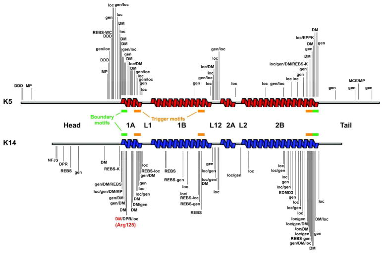
Epidermolysis bullosa simplex (EBS) is a rare genetic condition typified by superficial bullous lesions following incident frictional trauma to the skin. Most cases of EBS are due to dominantly acting mutations in keratin 14 (K14) or K5, the type I and II intermediate filament (IF) proteins that copolymerize to form a pancytoplasmic network of 10 nm filaments in basal keratinocytes of epidermis and related epithelia. Defects in K5-K14 filament network architecture cause basal keratinocytes to become fragile, and account for their rupture upon exposure to mechanical trauma. The discovery of the etiology and pathophysiology of EBS was intimately linked to the quest for an understanding of the properties and function of keratin filaments in skin epithelia. Since then, continued cross-fertilization between basic science efforts and clinical endeavors has highlighted several additional functional roles for keratin proteins in the skin, suggested new avenues for effective therapies for keratin-based diseases, and expanded our understanding of the remarkable properties of the skin as an organ system.


Conflict of interest statement

Conflict of interest statement: The authors declare no conflict of interest.

Figures

Figure 1
Figure 1. Keratin filaments: Structure, organization, and expression in the interfollicular epidermis
A) Schematic representation of the common domain structure shared by IF proteins including keratins. A central domain, comprised of α-helical coils 1A, 1B, 2A, and 2B separated by non-helical linkers L1, L12 and L2, is flanked by head and tail domains of unknown structure at its N- and C-termini. The boundaries of the rod domain (see red boxes) are highly conserved in primary structure. B) Visualization of filaments, reconstituted in vitro from purified human K5 and K14, by negative staining and electron microscopy. Bar: 150 nm. C) Double-labeling for keratin (red chromophore) and desmoplakin, a desmosome component (green chromophore), epitopes in human epidermal keratinocytes in culture. Keratin IFs are organized in a network that spans the entire cytoplasm and are attached at desmosomal cell-cell contacts between cells (arrowheads). n, nucleus. Bar: 50 μm. Micrograph courtesy of Dr. K. Green (Northwestern Univ., Chicago, IL). D) Histological cross-section of resin-embedded human trunk epidermis, revealing the basal (B), spinous (S), granular (G), and cornified (C) cell layers. Bar: 50 μm. n, nucleus. E, F) Differential distribution of keratin epitopes in the human epidermis. K14 occurs in the basal layer (E), where the epidermal progenitor cells reside. K10 occurs in the suprabasal, differentiating keratinocytes of epidermis (F). Basal lamina is depicted by a dashed line. Bar: 50μm. G) Ultrastructure of the boundary between the basal and suprabasal layer in mouse trunk epidermis, as seen in cross-section by routine transmission electron microscopy. Organization of keratin filaments as loose bundles correlates with the expression of K5–K14 in basal cells (see brackets), whereas the formation of denser, electron-dense filament bundles reflects the onset of K1–K10 expression in early differentiating cells (see arrowheads). Arrows point to desmosomes. Bar: 2 μm. n, nucleus. Adapted from (Miller et al., 2008).
Figure 2
Figure 2. Introduction to Epidermolysis Bullosa Simplex
A) Schematic representation of skin tissue (left) and detailed view of the bottom portion of the epidermis (right), highlighting the cytoplasmic network of keratin IFs (light green) attached to hemidesmosome cell-ECM (green) and desmosome cell-cell (magenta) contacts in basal keratinocytes. Arrows depict the plane of tissue rupture seen in the “simplex”, “junctional”, and “dystrophic” forms of EB. B) Example of trauma-induced bullous skin lesions (arrows) in the feet of a 2-month old child diagnosed with EBS. Picture kindly provided by Dr. Bernard Cohen (Johns Hopkins School of Medicine; see http://dermatlas.med.jhmi.edu). Adapted from (Coulombe et al., 2009).
Figure 3
Figure 3. Loss of keratin 14 elicits EBS-like features in newborn mouse skin
A, B) Picture of newborn mouse littermates, comparing K14 null and wildtype. A) The K14 null neonate exhibits massive skin blistering. Front paws and facial area are severely affected (see arrows). B) A wildtype littermate exhibits intact skin. C, D) Micrographs from hematoxylin/eosin-stained histological sections prepared from front paws of 2-day old K14 null (C) and wildtype (D) mice. Epidermal cleavage is obvious in the K14 null sample. Loss of epidermal integrity occurs “near” the basal layer of keratinocytes (see “blister”), the defining characteristic of EBS. Three basal keratinocytes are boxed in frame D. Abbreviations: epi, epidermis; hf, hair follicles. Bar: 100 μm. Adapted from (Coulombe et al., 2009).
Figure 4
Figure 4. Distribution of mutations in K5 and K14 according to EBS variants
The EBS-causing mutations that are available (September 2011) from the intermediate filament database (http://www.interfil.org; see (Szeverenyi et al., 2008)) are mapped to the secondary structure of K5 and K14 protein. Helix boundary motifs (green bars), as defined by Steinert et al. (Steinert et al., 1993), correspond to 15–20 residues segments that are very highly conserved across keratins and all other IF proteins. Trigger motifs (orange bars) are sequences capable of nucleating the formation of coiled-coil dimers (Wu et al., 2000). The Arg125 mutation hot spot in K14 (Coulombe et al., 1991a) is indicated in red. This schematic does not convey the frequency of mutations at any of the shown sites. See Table 1 for abbreviations and text for further detail.

Similar articles

Cited by

References

    1. Albers K, Fuchs E. The expression of mutant epidermal keratin cDNAs transfected in simple epithelial and squamous cell carcinoma lines. J Cell Biol. 1987;105:791–806. - PMC - PubMed
    1. Albers K, Fuchs E. Expression of mutant keratin cDNAs in epithelial cells reveals possible mechanisms for initiation and assembly of intermediate filaments. J Cell Biol. 1989;108:1477–93. - PMC - PubMed
    1. Anton-Lamprecht I. Genetically induced abnormalities of epidermal differentiation and ultrastructure in ichthyoses and epidermolyses: pathogenesis, heterogeneity, fetal manifestation, and prenatal diagnosis. J Invest Dermatol. 1983;81:149s–53s. - PubMed
    1. Atkinson SD, McGilligan VE, Liao H, Szeverenyi I, Smith FJ, Moore CB, et al. Development of Allele-Specific Therapeutic siRNA for Keratin 5 Mutations in Epidermolysis Bullosa Simplex. J Invest Dermatol. 2011;131:2079–86. - PubMed
    1. Beil M, Micoulet A, von Wichert G, Paschke S, Walther P, Omary MB, Van Veldhoven PP, Gern U, Wolff-Hieber E, Eggermann J, Waltenberger J, Adler G, Spatz J, Seufferlein T. Sphingosylphosphorylcholine regulates keratin network architecture and visco-elastic properties of human cancer cells. Nat Cell Biol. 2003;5:803–11. - PubMed

Publication types

MeSH terms