. 2012 May;81(10):993-1001.
doi: 10.1038/ki.2011.463. Epub 2012 Jan 25.

Identification of the vitamin D receptor in various cells of the mouse kidney

Affiliations
Free PMC article

Identification of the vitamin D receptor in various cells of the mouse kidney

Yongji Wang et al. Kidney Int. 2012 May.
Free PMC article

Abstract

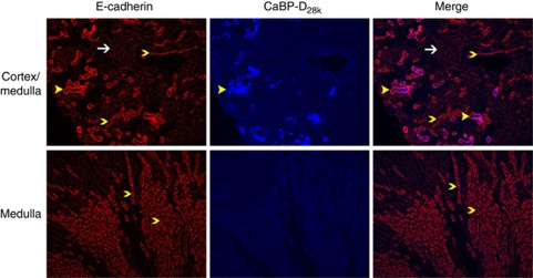
The kidney is the major, if not sole, site for the production of 1α,25-dihydroxyvitamin D(3) (1,25(OH)(2)D(3)), the biologically active form of vitamin D that can stimulate calcium reabsorption in the kidney and may provide renoprotective benefits. The biological effects of 1,25(OH)(2)D(3) are mediated through a nuclear hormone receptor, known as the vitamin D receptor (VDR). It is well accepted that the VDR is present in the distal renal convoluted tubule cells; however, whether VDR is present in other kidney cell types is uncertain. Using a highly specific and sensitive anti-VDR antibody, we determined its distribution in the mouse kidney by immunohistochemistry. Our results show that the VDR is not only present in the distal but is also found in the proximal tubules, but at 24-fold lower levels. The VDR was also found in the macula densa of the juxtaglomerular apparatus, glomerular parietal epithelial cells, and podocytes. In contrast, the VDR is either very low or absent in interstitial fibroblasts, glomerular mesangial cells, and juxtaglomerular cells. Thus, identification of VDR in the proximal tubule, macula densa, and podocytes suggests that 1,25(OH)(2)D(3) plays a direct role in these cells under normal conditions.


Figures

Figure 1
Figure 1
Colocalization of E-cadherin (red) and CaBP-D28k (blue) in mouse kidney (original magnification × 100). Distal renal tubule (yellow broad arrowhead) expressed the highest levels of CaBP-D28k and E-cadherin (upper panel). Cortical collecting duct (yellow arrowhead) expressed E-cadherin with a level comparable to that in the collecting ducts (yellow arrowhead denoted) in the medulla, suggesting those were the cortical collecting ducts (upper and lower panels). Please note that the structures that exhibited the low levels of E-cadherin and CaBP-D28k were the proximal renal tubules (white arrow denoted, upper panel).
Figure 2
Figure 2
Colocalization of vitamin D receptor (VDR) (red) and CaBP-D28k (blue) in the cortex (upper panel) and the medulla (lower panel) of mouse kidney (original magnification × 100). The distal renal tubules (yellow arrowhead) contained the highest level of VDR and CaBP-D28k. Collecting ducts (lower panel) in the medulla show a low level of CaBP-D28k. Potential proximal renal tubules were denoted by green broad arrowheads. Lack of VDR signals in renal tubules such as the proximal tubules and collecting ducts might be attributable to the loss of antibody incurred with sequential washings required for double staining protocol (Materials and Methods section).
Figure 3
Figure 3
Colocalization of vitamin D receptor (VDR) (red) and E-cadherin (green) in mouse kidney (original magnification × 100 or × 600). There was weak staining when mouse IgG isotypes were applied to the samples (upper panel). However, the staining was not found in the cell nucleus. The cell nuclei were stained by 4′,6-diamidino-2-phenylindole (DAPI) (blue). Distal renal tubule (yellow broad arrowhead) expressed the highest levels of VDR and E-cadherin (middle panel). Cortical collecting duct (yellow arrowhead) expressed E-cadherin and VDR with a relatively low level compared with the distal renal tubules (middle panel). Renal tubule lacking E-cadherin staining was the proximal renal tubule (white arrow). The lower panel (original magnification × 600) is confocal images of mouse renal tubules. Distal renal tubule was denoted by the yellow broad arrow, cortical collecting duct by the yellow arrowhead, and the proximal renal tubule by the white arrow.
Figure 4
Figure 4
The distribution of vitamin D receptor (VDR) in the mouse kidney (original magnification × 200 or × 600). (a) Distal renal tubules (DT, yellow broad arrowhead) of normal adult kidneys highly expressed VDR (red) but proximal renal tubules (PT, green arrowhead) and cortical collecting ducts (CCD, white broad arrowhead) expressed VDR with a low level. Glomerulus is denoted as gl. (b) VDR (red) was colocalized with 4′,6-diamidino-2-phenylindole (DAPI) (blue). This colocalization resulted in purple color in (b). Interstitial fibroblasts (ifb, white broad arrowhead) were located between the renal tubules, and their nuclei were stained by DAPI but not stained with the VDR antibody in (a). (c) Collecting ducts (CD, white broad arrowhead) in the medulla were positive for VDR. (d) Renal tubules with weak VDR staining in the cortex. The representative proximal tubules were denoted by the green broad arrowhead. (e) VDR was located in the distal tubule (DT, yellow broad arrowhead). The image was taken with the reduced exposure time compared with the image in (a). The images with less exposure showed that the VDR in the distal tubule was located in the nuclei of the tubule epithelial cells (e). (f, g) Confocal images of mouse kidney cortex sections stained for VDR and DAPI. The distal tubule was denoted with the yellow arrowhead and proximal tubule with the green arrowhead. (h) VDR antibody staining in the kidney sections of Demay VDR knockout mice served as negative controls. (i) Mouse isotype IgG did not generate significant immunostaining in the wild-type mouse kidney sections.
Figure 5
Figure 5
Colocalization of vitamin D receptor (VDR) (red) and E-cadherin (green) in normal human kidney cortex (original magnification × 200). (a, b) Human kidney cortex sections were stained for VDR and E-cadherin. Distal renal tubule (yellow broad arrowhead) expressed the highest levels of VDR, E-cadherin, and 4′,6-diamidino-2-phenylindole (DAPI). Cortical collecting duct (yellow arrowhead) expressed E-cadherin and VDR with a relatively low level compared with the distal renal tubules. Renal tubule lacking E-cadherin staining was the proximal renal tubule (white arrow). (c, d) Digitally zoomed in images. Proximal tubule (PT, white arrows) had no E-cadherin but contained VDR. Parietal epithelial cells (PEC, white broad arrows) expressed VDR as well. Please note that the VDR immunosignals were localized at the cytoplasm of the renal cells. (e) Human kidney cortex sections were stained for hnRNP, a nuclear protein. Please note that hnRNP was localized at the nuclei of renal cells. (f) There was no staining when mouse IgG isotypes were applied to the samples, indicating the secondary antibody did not react with human IgG. The cell nuclei were stained by DAPI (blue).
Figure 6
Figure 6
Vitamin D receptor (VDR) in mouse glomerulus and juxtaglomerular apparatus (JGA) (original magnification × 200 or × 600; a–e and h–i were digitally enlarged). (a) VDR was present in the glomerular parietal (white broad arrow) and visceral (white broad arrowhead) podocytes, macula densa (md), and distal renal tubule (DT). The VDR staining was depicted by red fluorescent color. (b) Colocalization of VDR (red) and nucleus (4′,6-diamidino-2-phenylindole (DAPI) staining, blue) in mouse glomerulus. The VDR/DAPI merge resulted in purple color. (c) VDR staining in the glomerulus (gl) of Demay VDR knockout mouse kidney. (d) JGA of the wild-type mouse kidney. VDR was present in the cells of macula densa (md) and distal renal tubule (DT). (e) Colocalization of VDR (red) and DAPI (blue) staining in the JGA of the wild-type mouse kidney. VDR was not found in the juxtaglomerular cells (jga) but present in the macula densa (md) and DT. (f) A confocal image showed the VDR staining (red) in the glomerular parietal epithelial cells (yellow arrow) and parietal (white broad arrow) and visceral (white broad arrowhead) podocytes. (g) A confocal image showed the colocalization of Wilms' tumor-1 (green) and VDR (red) in the glomerulus. VDR/DAPI merge resulted in yellow color. (h) Staining of mouse kidney sections with antibody to α smooth muscle actin (αSMA, red). The juxtaglomerular cells (jga) strongly expressed αSMA whereas the intraglomerular mesangial cells (blue arrowhead) or interstitial fibroblasts (white broad arrow) expressed αSMA with low abundance. (i) The αSMA staining was not colocalized with the WT-1 staining (green) (white broad arrowhead).
Figure 7
Figure 7
Vitamin D receptor (VDR) in mouse glomerulus and juxtaglomerular apparatus (original magnification × 600; a–d were digitally enlarged to original magnification × 1200). The left panels (white color) are the VDR antibody staining while the right panels are the colocalization for VDR, renin (red), and 4′,6-diamidino-2-phenylindole (blue). (a, b) The VDR and renin staining in the glomerulus of Demay VDR knockout mouse kidney. The VDR staining (white color) is not seen in the nuclei of renal cells. The renin staining (red color) is selectively seen in the juxtaglomerular cells. Note that some white staining could be seen in the cytoplasm of the proximal renal tubular cells or the outside of renal cells. The VDR knockout kidney samples stained with only the secondary antibody showed a similar pattern (not shown), suggesting that it is most likely the staining for mouse endogenous IgG. The IgG could be detected in the proximal tubular cells because of protein and peptide reabsorption at this particular site. (c, d) Omission of the primary VDR and renin antibodies resulted in the absence of the primary antibody-specific staining in the nuclei of kidney samples from wild-type mice. (e, f) VDR and renin staining in the glomerulus of wild-type mouse kidney. The VDR staining (white color) is seen in the nuclei of renal tubular cells and glomerular podocytes. The cells with a low level of VDR are indicated by using white broad arrows; the renin staining (red color) is selectively seen in the juxtaglomerular cells. The VDR staining is not seen in the nuclei of juxtaglomerular cells (yellow arrow). (g, h) The VDR and renin staining in the glomerulus of wild-type mouse kidney. The VDR staining (white color) is seen in the nuclei of renal tubular cells and glomerular podocytes. The VDR staining is not seen in the nuclei of juxtaglomerular cells (yellow arrow). Note that the interstitial fibroblasts are not stained by either VDR or renin antibodies.

Comment in

Similar articles

Cited by

References

    1. Reilly RF, Ellison DH. Mammalian distal tubule: physiology, pathophysiology, and molecular anatomy. Physiol Rev. 2000;80:277–313. - PubMed
    1. Yamamoto M, Kawanobe Y, Takahashi H, et al. Vitamin D deficiency and renal calcium transport in the rat. J Clin Invest. 1984;74:507–513. - PMC - PubMed
    1. DeLuca HF. Overview of general physiologic features and functions of vitamin D. Am J Clin Nutr. 2004;80:1689S–1696S. - PubMed
    1. Bouillon R, Carmeliet G, Verlinden L, et al. Vitamin D and human health: lessons from vitamin D receptor null mice. Endocr Rev. 2008;29:726–776. - PMC - PubMed
    1. Brunette MG, Chan M, Ferriere C, et al. Site of 1,25(OH)2 vitamin D3 synthesis in the kidney. Nature. 1978;276:287–289. - PubMed

Publication types

MeSH terms

Substances

LinkOut - more resources