

Postnatal infection is associated with widespread abnormalities of brain development in premature newborns
- PMID: 22278180
- PMCID: PMC3940469
- DOI: 10.1038/pr.2011.40
Postnatal infection is associated with widespread abnormalities of brain development in premature newborns
Abstract
Introduction: Infection is a risk factor for adverse neurodevelopmental outcome in preterm newborns. Our objective was to characterize the association of postnatal infection with adverse microstructural and metabolic brain development in premature newborns.
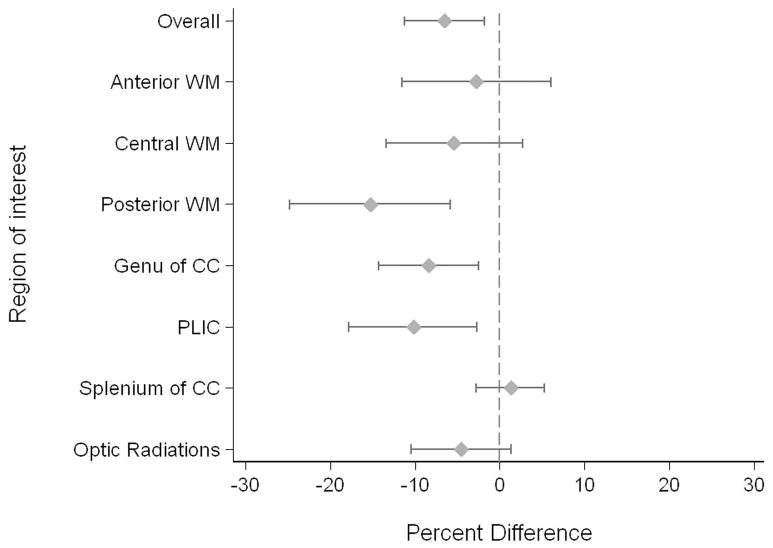
Results: In 34/117 newborns studied, clinical signs were accompanied by positive cultures whereas 17 had clinical signs of sepsis alone. White matter injury (WMI) was identified in 34 newborns. In multivariate regression models, infected newborns had brain imaging measures indicative of delayed brain development: lower N-acetylaspartate/choline, elevated average diffusivity (D(AV)), and decreased white matter fractional anisotropy. These widespread brain abnormalities were found in both newborns with positive-culture infection and in those with clinical infection.
Discussion: These findings suggest that postnatal infection, even without a positive culture, is an important risk factor for widespread abnormalities in brain development. These abnormalities extend beyond brain injuries apparent with conventional magnetic resonance injury (MRI).
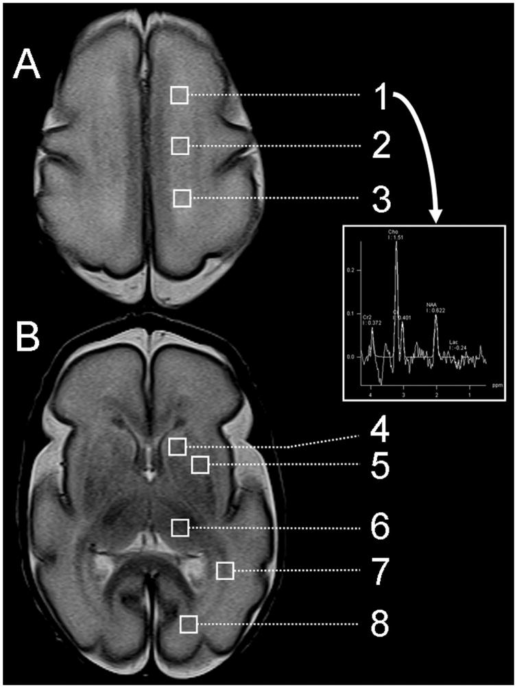
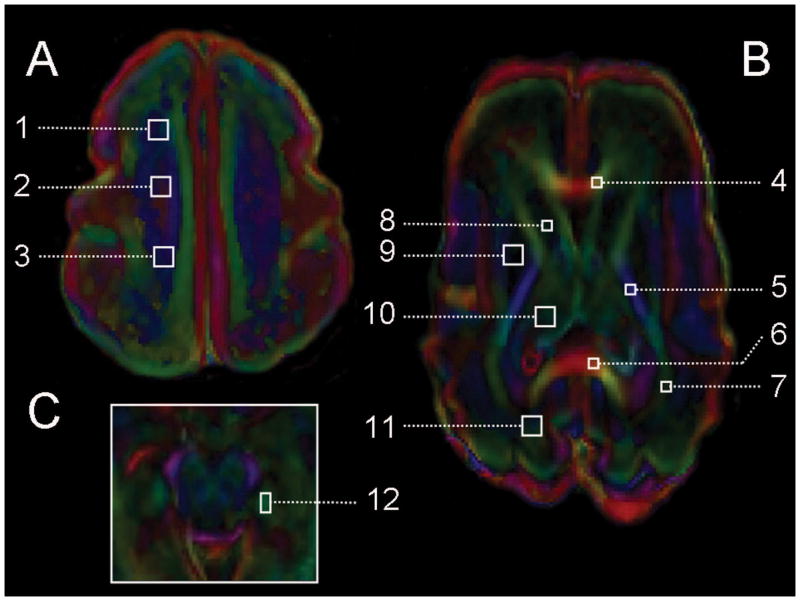
Methods: 117 preterm newborns (24-32 wk gestation) were studied prospectively at a median of 32.0 and 40.3 wk ostmenstrual age with MRI (WMI, hemorrhage), magnetic resonance (MR) spectroscopy (metabolism), and diffusion tensor imaging (microstructure). Newborns were categorized as having "no infection," "clinical infection," or "positive-culture infection." We compared brain injuries as well as metabolic and microstructural development across these infection groups.
Conflict of interest statement
The authors have no conflict of interest or potential financial interests to disclose.
Figures
References
-
- Miller SP, Ferriero DM, Leonard C, Piecuch R, Glidden DV, Partridge JC, Perez M, Mukherjee P, Vigneron DB, Barkovich AJ. Early brain injury in premature newborns detected with magnetic resonance imaging is associated with adverse early neurodevelopmental outcome. J Pediatr. 2005;147:609–616. - PubMed
-
- Shah DK, Doyle LW, Anderson PJ, Bear M, Daley AJ, Hunt RW, Inder TE. Adverse neurodevelopment in preterm infants with postnatal sepsis or necrotizing enterocolitis is mediated by white matter abnormalities on magnetic resonance imaging at term. J Pediatr. 2008;153:170–175. - PubMed
-
- Glass HC, Bonifacio SL, Chau V, Glidden D, Poskitt K, Barkovich AJ, Ferriero DM, Miller SP. Recurrent postnatal infections are associated with progressive white matter injury in premature infants. Pediatrics. 2008;122:299–305. - PubMed
-
- Stoll BJ, Hansen NI, Adams-Chapman I, Fanaroff AA, Hintz SR, Vohr B, Higgins RD. Neurodevelopmental and growth impairment among extremely low-birth-weight infants with neonatal infection. JAMA. 2004;292:2357–2365. - PubMed
Publication types
MeSH terms
Grants and funding
LinkOut - more resources
Full Text Sources
Other Literature Sources
Medical
