. 2012 Apr;86(7):3975-84.
doi: 10.1128/JVI.06828-11. Epub 2012 Jan 25.

The multibasic cleavage site in H5N1 virus is critical for systemic spread along the olfactory and hematogenous routes in ferrets

Affiliations

The multibasic cleavage site in H5N1 virus is critical for systemic spread along the olfactory and hematogenous routes in ferrets

Eefje J A Schrauwen et al. J Virol. 2012 Apr.

Abstract

The route by which highly pathogenic avian influenza (HPAI) H5N1 virus spreads systemically, including the central nervous system (CNS), is largely unknown in mammals. Especially, the olfactory route, which could be a route of entry into the CNS, has not been studied in detail. Although the multibasic cleavage site (MBCS) in the hemagglutinin (HA) of HPAI H5N1 viruses is a major determinant of systemic spread in poultry, the association between the MBCS and systemic spread in mammals is less clear. Here we determined the virus distribution of HPAI H5N1 virus in ferrets in time and space-including along the olfactory route-and the role of the MBCS in systemic replication. Intranasal inoculation with wild-type H5N1 virus revealed extensive replication in the olfactory mucosa, from which it spread to the olfactory bulb and the rest of the CNS, including the cerebrospinal fluid (CSF). Virus spread to the heart, liver, pancreas, and colon was also detected, indicating hematogenous spread. Ferrets inoculated intranasally with H5N1 virus lacking an MBCS demonstrated respiratory tract infection only. In conclusion, HPAI H5N1 virus can spread systemically via two different routes, olfactory and hematogenous, in ferrets. This systemic spread was dependent on the presence of the MBCS in HA.


Figures

Fig 1
Fig 1
In vitro phenotype of H5N1WT and H5N1ΔMBCS viruses. (A) Western blots of lysates of 293T cells untransfected or transfected with HAs of H5N1ΔMBCS virus and H5N1WT virus upon treatment with (lanes T) or without (lanes −) trypsin. (B) Replication of H5N1WT virus and H5N1ΔMBCS virus in MDCK cells in the presence or absence of trypsin (tryp). Geometric mean titers were calculated from two independent experiments; error bars indicate standard deviations. The lower limit of detection is indicated by the dotted line.
Fig 2
Fig 2
Weight loss and virus shedding in ferrets inoculated with H5N1WT or H5N1ΔMBCS viruses. (A) Mean body weights and standard deviations are depicted as percentage of body weight at time of inoculation for each group inoculated with H5N1WT virus and H5N1ΔMBCS virus. (B) Virus shedding from the upper respiratory tract of ferrets inoculated with H5N1WT virus or H5N1ΔMBCS virus from the nose and throat is indicated. Geometric mean titers are shown, with error bars provided to indicate the standard deviations. The lower limit of detection is 1.5 log10 TCID50.
Fig 3
Fig 3
Influenza virus antigen expression in respiratory epithelium and olfactory epithelium of ferrets inoculated with either H5N1WT virus or H5N1ΔMBCS virus. Influenza virus antigen in the respiratory epithelium and olfactory epithelium in ferrets inoculated with H5N1WT or H5N1ΔMBCS virus at 1, 3, 5, and 7 dpi. Tissue sections were stained with a monoclonal antibody against influenza A virus nucleoprotein, visible as red staining. In the respiratory epithelium, individual virus antigen-positive epithelial cells are observed at 1, 3, and 5 dpi in H5N1WT virus-inoculated ferrets and at 1 and 3 dpi in H5N1ΔMBCS virus-inoculated ferrets. In the olfactory epithelium, virus antigen in H5N1WT virus-inoculated ferrets was present from 1 to 7 dpi, with a peak at 3 dpi. At days 5 and 7 dpi, there was focal severe necrosis of olfactory epithelial cells with infiltration of neutrophils (arrows). In the H5N1ΔMBCS virus-inoculated ferrets, virus antigen was present from day 1 to 7. Magnification, ×400.
Fig 4
Fig 4
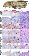
Detection of histological lesions or influenza A virus antigen expression in tissues from the nervous system of ferrets inoculated with H5N1WT virus. A cross section of a ferret head to illustrate the different anatomical sites is shown at the top. The nasal cavity contains the nasal turbinates, which are lined by respiratory mucosa, except at the back and top of the cavity, where they are lined by olfactory mucosa (position 1). The olfactory bulb (position 2) is separated from the nasal cavity by the cribriform plate and connected via the olfactory tract (position 3) to the cerebrum (position 4). The cerebellum (position 5), cerebrum, olfactory bulb, and olfactory tract are all surrounded by the meninges (position 6). (Left and middle columns) Immunohistochemistry for influenza A virus antigen in different tissues of the olfactory route. Magnifications, ×200 and ×400, left and middle columns, respectively. (Right column) Histological lesions in different tissues of the olfactory route. Magnification, ×400. Olfactory epithelium (position 1), which contained virus antigen from 1 dpi in olfactory receptor neurons, with focal necrosis of olfactory epithelial cells associated with infiltrating neutrophils (arrow); Bowman's glands (position 1), which contained virus antigen from 3 dpi, with necrosis of Bowman's gland epithelial cells associated with infiltrating neutrophils (arrow) along the nerve twigs (*); olfactory bulb (position 2), which contained virus antigen from 3 dpi, with infiltrating neutrophils (arrow) between the glomeruli (*) in the glomerular layer; olfactory tract (position 3), which contained virus antigen from 7 dpi, with neuronal necrosis and infiltrating neutrophils (arrows); cerebrum (position 4), which contained virus antigen from 5 dpi, with neuronal necrosis and infiltrating neutrophils (arrows); cerebellum (position 5), which contained virus antigen from 7 dpi, with neuronal necrosis and infiltrating neutrophils (arrow); meninges (position 6), which contained virus antigen from 7 dpi, with infiltration of many mononuclear cells and occasional neutrophils (arrow).
Fig 5
Fig 5
Detection of histological lesions (HE) and influenza A virus antigen (IHC) expression in extrarespiratory tract tissues in ferrets inoculated with H5N1WT virus. In the liver, virus antigen was observed in hepatocytes, with multifocal necrosis of hepatocytes and replacement by variable numbers of neutrophils and mononuclear cells. In the pancreas, virus antigen was observed in acinar cells, with necrosis of acinar cells, widespread edema, and infiltration of neutrophils and mononuclear cells. IHC, immunohistochemistry.
Fig 6
Fig 6
Attachment of H5N1WT virus to ferret respiratory epithelium and olfactory epithelium in the nose. No attachment of H5N1WT virus to the apical side of respiratory epithelium and abundant attachment of H5N1WT virus to the apical side of the olfactory epithelium. Tissue sections were incubated with H5N1WT virus; attachment is visible as red staining. Magnification, ×1,000.

References

    1. Aronsson F, Robertson B, Ljunggren HG, Kristensson K. 2003. Invasion and persistence of the neuroadapted influenza virus A/WSN/33 in the mouse olfactory system. Viral Immunol. 16:415–423 - PubMed
    1. Bertram S, Glowacka I, Steffen I, Kuhl A, Pohlmann S. 2010. Novel insights into proteolytic cleavage of influenza virus hemagglutinin. Rev. Med. Virol. 20:298–310 - PMC - PubMed
    1. Bodewes R, et al. 2011. Pathogenesis of influenza A/H5N1 virus infection in ferrets differs between intranasal and intratracheal routes of inoculation. Am. J. Pathol. 179:30–36 - PMC - PubMed
    1. Chen J, et al. 1998. Structure of the hemagglutinin precursor cleavage site, a determinant of influenza pathogenicity and the origin of the labile conformation. Cell 95:409–417 - PubMed
    1. Chutinimitkul S, et al. 2010. In vitro assessment of attachment pattern and replication efficiency of H5N1 influenza A viruses with altered receptor specificity. J. Virol. 84:6825–6833 - PMC - PubMed

Publication types

MeSH terms

Substances