. 2012 Apr;86(7):3809-18.
doi: 10.1128/JVI.06081-11. Epub 2012 Jan 25.

Vaccinia mature virus fusion regulator A26 protein binds to A16 and G9 proteins of the viral entry fusion complex and dissociates from mature virions at low pH

Affiliations

Vaccinia mature virus fusion regulator A26 protein binds to A16 and G9 proteins of the viral entry fusion complex and dissociates from mature virions at low pH

Shu-Jung Chang et al. J Virol. 2012 Apr.

Abstract

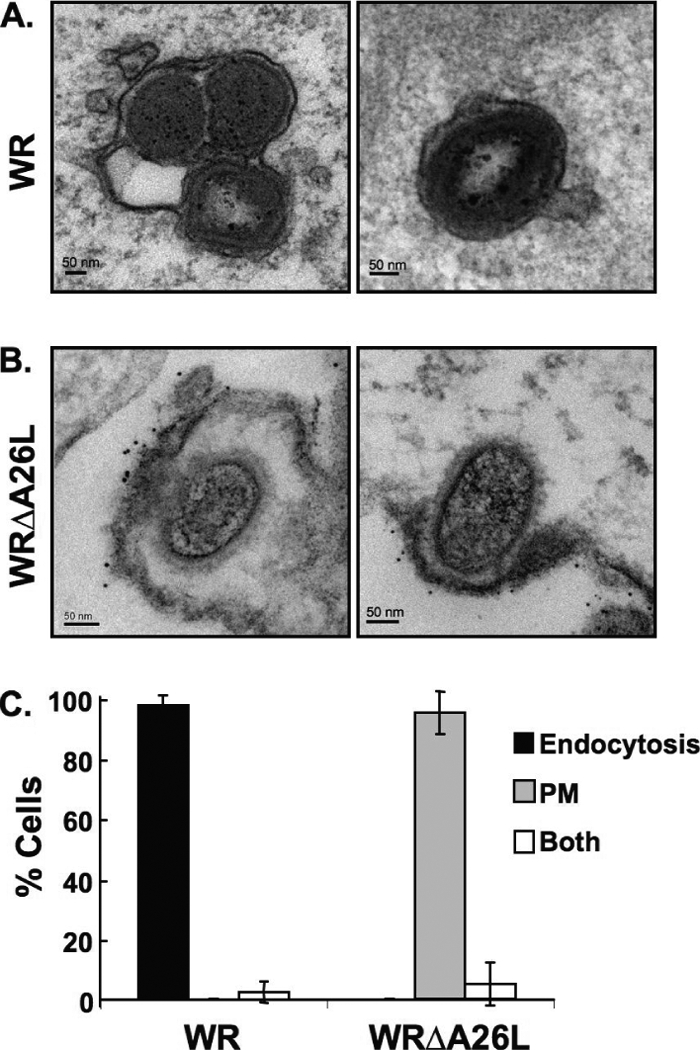
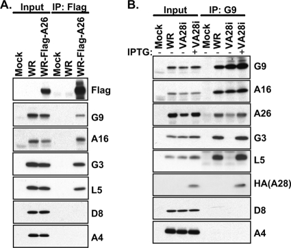
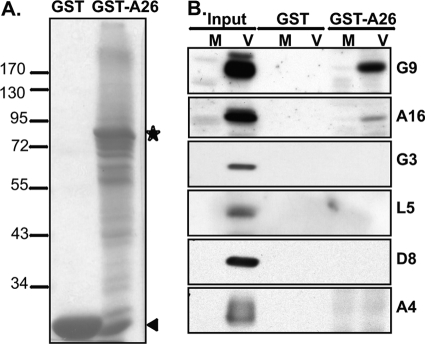
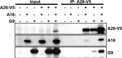
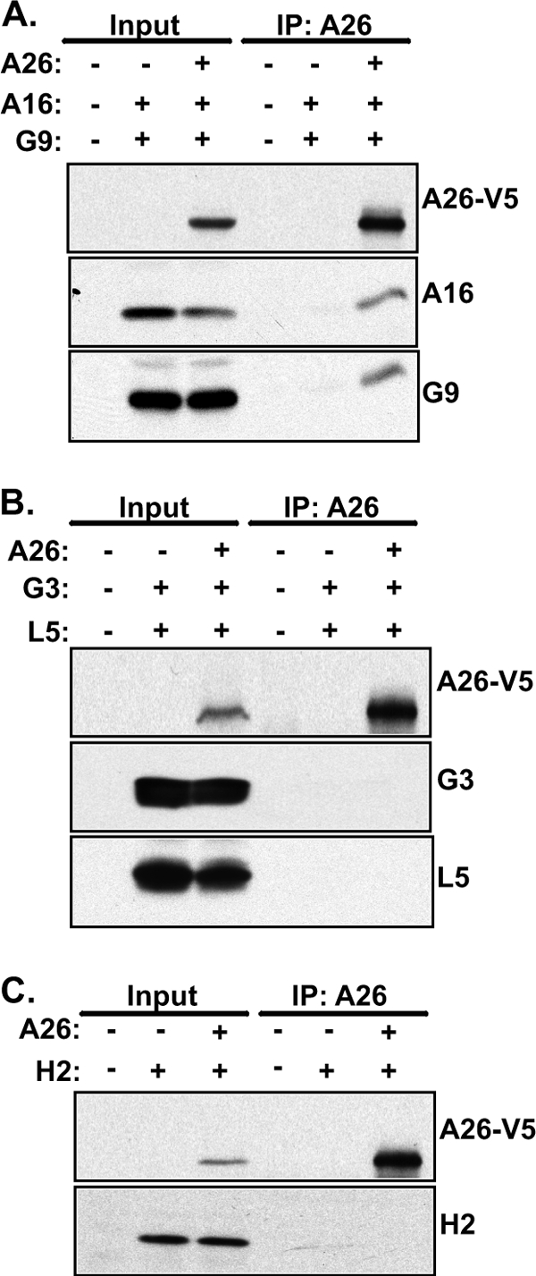
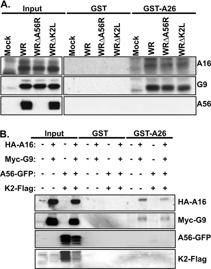
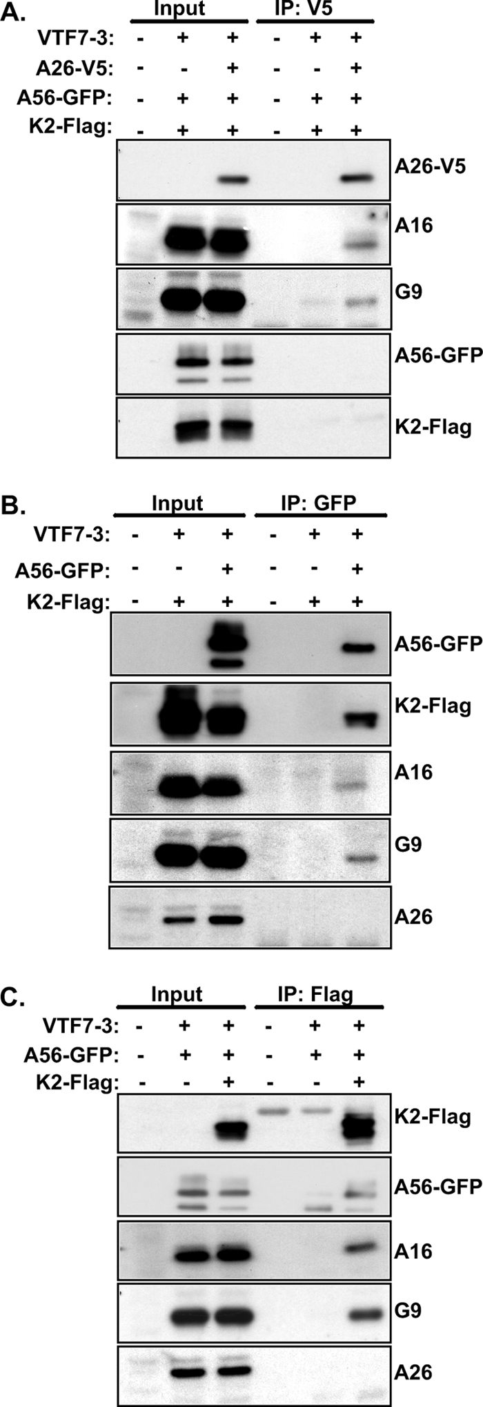
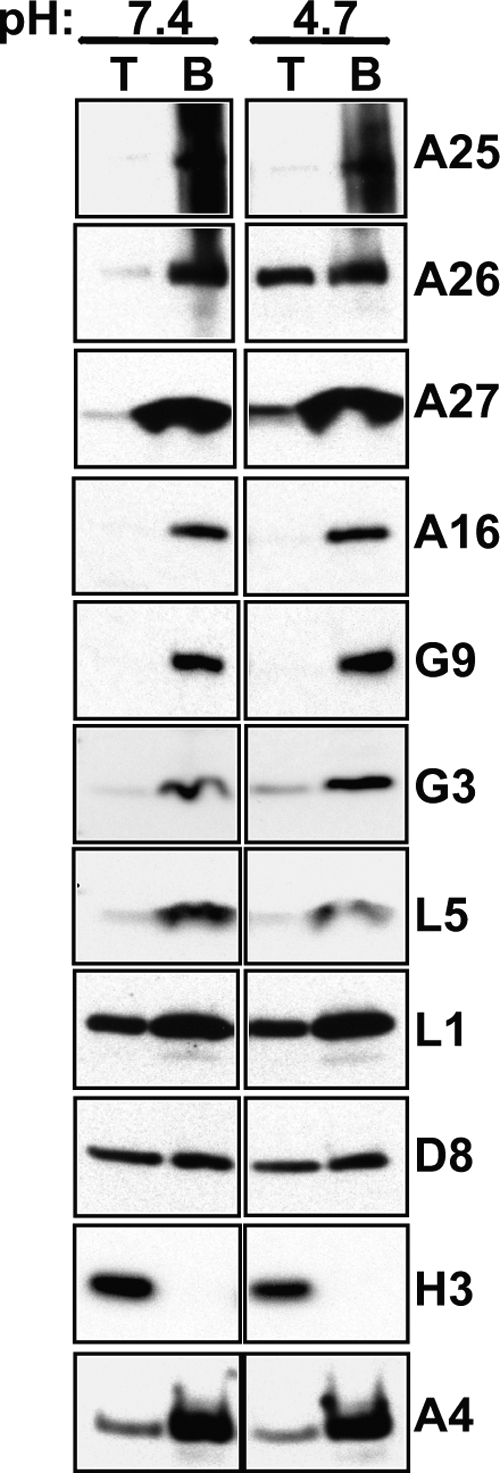
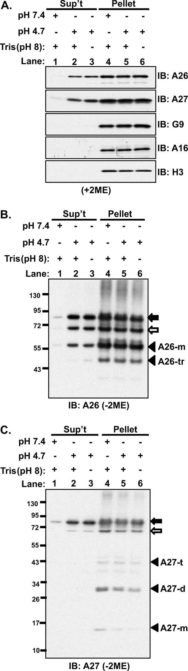
Vaccinia mature virus enters cells through either endocytosis or plasma membrane fusion, depending on virus strain and cell type. Our previous results showed that vaccinia virus mature virions containing viral A26 protein enter HeLa cells preferentially through endocytosis, whereas mature virions lacking A26 protein enter through plasma membrane fusion, leading us to propose that A26 acts as an acid-sensitive fusion suppressor for mature virus (S. J. Chang, Y. X. Chang, R. Izmailyan R, Y. L. Tang, and W. Chang, J. Virol. 84:8422-8432, 2010). In the present study, we investigated the fusion suppression mechanism of A26 protein. We found that A26 protein was coimmunoprecipitated with multiple components of the viral entry-fusion complex (EFC) in infected HeLa cells. Transient expression of viral EFC components in HeLa cells revealed that vaccinia virus A26 protein interacted directly with A16 and G9 but not with G3, L5 and H2 proteins of the EFC components. Consistently, a glutathione S-transferase (GST)-A26 fusion protein, but not GST, pulled down A16 and G9 proteins individually in vitro. Together, our results supported the idea that A26 protein binds to A16 and G9 protein at neutral pH contributing to suppression of vaccinia virus-triggered membrane fusion from without. Since vaccinia virus extracellular envelope proteins A56/K2 were recently shown to bind to the A16/G9 subcomplex to suppress virus-induced fusion from within, our results also highlight an evolutionary convergence in which vaccinia viral fusion suppressor proteins regulate membrane fusion by targeting the A16 and G9 components of the viral EFC complex. Finally, we provide evidence that acid (pH 4.7) treatment induced A26 protein and A26-A27 protein complexes of 70 kDa and 90 kDa to dissociate from mature virions, suggesting that the structure of A26 protein is acid sensitive.


Figures

Fig 1
Fig 1
Immunogold electron microscopy analyses of HeLa cells infected with vaccinia virus MVs. (A) HeLa cells were infected with vaccinia virus wild-type WR and WRΔA26L MVs as described in Materials and Methods. After infection, cells were fixed and stained with anti-vaccinia virus MV primary antibody and goat anti-rabbit antibody conjugated to 6-nm gold particles and analyzed by EM. MV particles of wild-type strain WR are enclosed within intracellular vesicles. (B) HeLa cells were infected with WRΔA26L MVs and analyzed by EM as described in panel A. MV particles of WRΔA26L fused with the cell plasma membrane that were decorated with the immunogold-labeled antibody. (C) Quantification of cells (>30) infected with vaccinia virus MVs through different entry routes. Cells containing MV entry through endocytosis or plasma membrane fusion pathways were quantified as described in Materials and Methods. Data represent percentages of cells that contain MVs within intracellular vesicles (endocytosis), MVs fused with the plasma membrane (PM), or both.
Fig 2
Fig 2
Coimmunoprecipitation of A26 protein with multiple components of the viral EFC in virus-infected cells. (A) Coimmunoprecipitation of A26 with EFC components in virus-infected cells. HeLa cells were either mock infected or infected with WR or WR-Flag-A26 virus at an MOI of 5 PFU/cell, harvested at 24 h p.i. for immunoprecipitation (IP) with anti-Flag-agarose, and analyzed by immunoblotting with various antibodies, shown at the right side of the gel. (B) Coimmunoprecipitation of G9 protein with A26 protein in virus-infected cells. HeLa cells were either mock infected or infected with wild-type WR or vA28i virus at an MOI of 5 PFU/cell, incubated in medium with or without 100 μM IPTG, harvested at 24 h p.i. for immunoprecipitation with anti-G9 (1:50) antibody, and analyzed as described for panel A. Input represents 1% of total cell lysates.
Fig 3
Fig 3
In vitro interaction of A26 protein with G9 and A16 proteins expressed from virus-infected cells. (A) Coomassie blue staining of recombinant GST and GST-A26 proteins purified from E. coli. The asterisk represents the full-length GST-A26 protein, and the arrowhead represents the GST protein. (B) GST-A26 pulled down A16 and G9 proteins from virus-infected cells. Recombinant GST and GST-A26 protein (30 μg) were incubated with cell lysates prepared from either mock-infected (M) or vaccinia virus WR-infected (V) HeLa cells, as described in Materials and Methods, harvested at 24 h p.i., and analyzed by immunoblotting with various antibodies, as shown in Fig. 2. Input represents 1% of total cell lysates.
Fig 4
Fig 4
A26 interacts individually with A16 and G9 but not with G3 and L5 proteins in vitro. (A) GST-A26 pulled down A16 and G9 individually. HEK293T cells were transfected with plasmids expressing HA-tagged A16, Myc-tagged G9, or both and harvested 24 h later. The lysates were subjected to GST pulldown analyses using 30 μg of recombinant GST and GST-A26 proteins as described in Materials and Methods and analyzed by immunoblotting with anti-HA (A16) (1:1,000) and anti-Myc (G9) (1:1,000) antibodies. (B) GST-A26 did not pull down G3 and L5. HEK293T cells were transfected with plasmids expressing vaccinia G3 and L5 proteins and harvested 24 h later, and the lysates were subjected to GST pulldown analyses as described above using anti-G3 (1:1,000) and anti-L5 (1:1,000) antibodies. Input represents 1% of total cell lysates.
Fig 5
Fig 5
A26 protein interacts with A16 and G9 proteins individually in transfected HEK293T cells. HEK293T cells were transiently transfected with plasmids expressing A26-V5, A16, or G9 protein individually or in combination. Coimmunoprecipitations were performed with anti-V5 antibody-conjugated agarose beads and analyzed by immunoblotting with anti-V5 (1:4,000), anti-A16 (1:1,000), and anti-G9 (1:5,000) antibodies. Input represents 1% of total cell lysates.
Fig 6
Fig 6
A26 does not interact with EFC component proteins H2, G3, and L5 in transfected HEK293T cells. HEK293T cells were transiently transfected with individual plasmids expressing A26-V5 along with A16 and G9 (A) or G3 and L5 (B) or H2 (C) protein, harvested for coimmunoprecipitations with anti-V5 agarose beads, and analyzed by immunoblotting with anti-V5 (1:4,000), anti-A16 (1:1,000), anti-G9 (1:5,000), anti-G3 (1:1,000), anti-L5 (1:1,000), and anti-H2 (1:1,000) antibodies. Input represents 1% of total cell lysates.
Fig 7
Fig 7
A26 protein in vitro binds to A16 and G9 proteins that are not in complex with A56/K2. (A) GST-A26 pulled down A16 and G9 proteins but not A56 protein. HeLa cells were infected with WR, WRΔA56R, and WRΔK2L at an MOI of 5 PFU/cell; cell lysates were harvested at 24 h p.i. for GST pulldown analyses with 30 μg of recombinant GST and GST-A26 proteins and analyzed by immunoblotting with anti-A16 (1:1,000), anti-G9 (1:5,000), and anti-A56 MAb (B2D10) (1:500). (B) GST-A26 did not pull down K2 protein. HEK293T cells were transiently transfected with plasmids expressing HA-A16, Myc-G9, A56-GFP, and K2-Flag proteins individually or in combination. The lysates were harvested for GST pulldown and analyzed by immunoblotting as described for panel A with anti-HA (1:1,000), anti-Myc (1:1,000), anti-GFP (1:4,000), and anti-Flag (1:1,000) antibodies. Input represents 1% of total cell lysates.
Fig 8
Fig 8
A26 protein binds to A16 and G9 proteins in vivo that are not in complex with A56/K2. (A) HEK293T cells were infected with VTF7-3 at an MOI of 5 PFU/cell and transfected with plasmids expressing A26-V5, A56-GFP, and K2-Flag proteins; cells were harvested at 24 h p.i. for coimmunoprecipitations with anti-V5 antibody conjugated to agarose beads and analyzed by anti-V5 (1:4,000), anti-A16(1:1,000), anti-G9 (1:5,000), anti-GFP (1:4,000), and anti-Flag (1:1,000) antibodies. (B) HEK293T cells were infected with VTF7-3 at an MOI of 5 PFU/cell, transfected with plasmids expressing A56-GFP and K2-Flag proteins, harvested at 24 h p.i. for coimmunoprecipitations with anti-GFP antibody, and analyzed by immunoblotting as described for panel A. (C) HEK293T cells were infected with VTF7-3 at an MOI of 5 PFU/cell, transfected with plasmids expressing A56-GFP and K2-Flag proteins as described for panel B for coimmunoprecipitations with anti-Flag antibody conjugated to agarose beads, and analyzed by immunoblotting as described for panel A. Input represents 1% of total cell lysates.
Fig 9
Fig 9
Effect of low-pH treatment on the hydrophilicity/hydrophobicity of A26 and A27 proteins. Vaccinia MV particles (8 μg) were solubilized in 2% Triton X-114 in buffer of pH 7.4 or pH 4.7, incubated at 37°C for 10 min, and then centrifuged at 300 × g for 3 min, as described in Materials and Methods. The aqueous (top fraction, T) and detergent (bottom fraction, B) phases were collected and analyzed by immunoblot analysis with various antibodies as described in Materials and Methods.
Fig 10
Fig 10
Low pH dissociates vaccinia A26 and A26-A27 protein complexes from MVs. (A). Purified vaccinia virus MV particles (24 μg) were treated with PBS at pH 7.4 or PBS at pH 4.7 at 37°C for 3 min with or without neutralization with 15 mM Tris-HCl (pH 8), followed by centrifugation. The supernatant (Sup't) and pellet fractions were collected, separated on SDS-PAGE gels under reducing (with 2-mercaptoethanol [+2ME]) conditions, and analyzed by immunoblotting (IB) using anti-A26 (1:1,000), anti-A27 (1:5,000), anti-G9 (1:5,000), anti-A16 (1:1,000), and anti-H3 (1:1,000) antibodies. (B and C) The samples described in panel A were separated under nonreducing (−2ME) conditions on 4 to 12% SDS-PAGE gels and analyzed by immunoblotting with anti-A26 (1:1,000) (B) and anti-A27 (1:5,000) (C) antibodies. The m, d, t, and tr suffixes with the A26 and A27 proteins represent monomer, dimer, trimer, and truncated, respectively.

Similar articles

Cited by

References

    1. Armstrong JA, Metz DH, Young MR. 1973. The mode of entry of vaccinia virus into L cells. J. Gen. Virol. 21:533–537 - PubMed
    1. Bengali Z, Townsley AC, Moss B. 2009. Vaccinia virus strain differences in cell attachment and entry. Virology 389:132–140 - PMC - PubMed
    1. Bisht H, Weisberg AS, Moss B. 2008. Vaccinia virus l1 protein is required for cell entry and membrane fusion. J. Virol. 82:8687–8694 - PMC - PubMed
    1. Bordier C. 1981. Phase separation of integral membrane proteins in Triton X-114 solution. J. Biol. Chem. 256:1604–1607 - PubMed
    1. Brown E, Senkevich TG, Moss B. 2006. Vaccinia virus F9 virion membrane protein is required for entry but not virus assembly, in contrast to the related L1 protein. J. Virol. 80:9455–9464 - PMC - PubMed

Publication types

Substances