. 2012 Mar;23(6):1129-39.
doi: 10.1091/mbc.E11-12-1018. Epub 2012 Jan 25.

GARP regulates the bioavailability and activation of TGFβ

Affiliations

GARP regulates the bioavailability and activation of TGFβ

Rui Wang et al. Mol Biol Cell. 2012 Mar.

Abstract

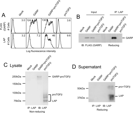
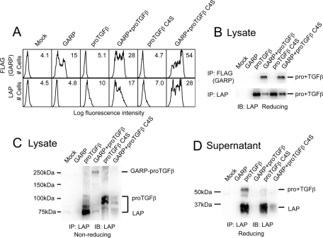
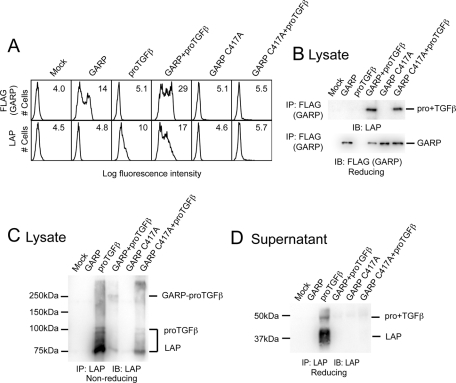
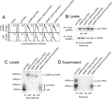
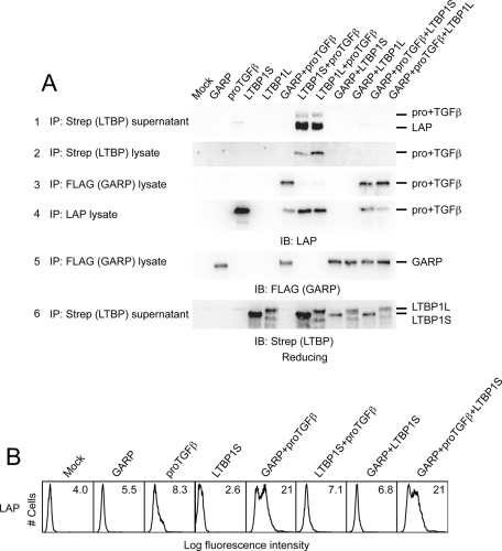
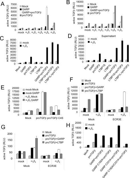
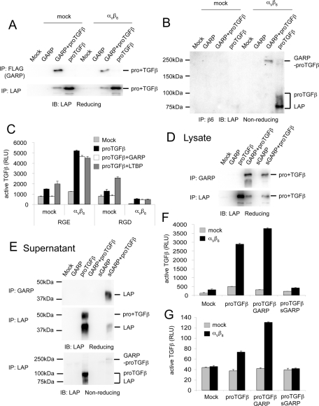
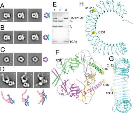
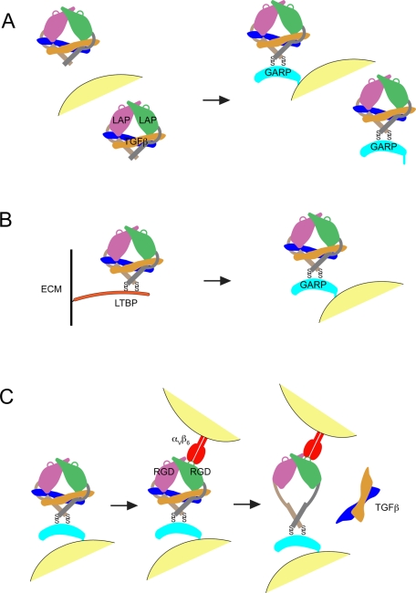
Glycoprotein-A repetitions predominant protein (GARP) associates with latent transforming growth factor-β (proTGFβ) on the surface of T regulatory cells and platelets; however, whether GARP functions in latent TGFβ activation and the structural basis of coassociation remain unknown. We find that Cys-192 and Cys-331 of GARP disulfide link to the TGFβ1 prodomain and that GARP with C192A and C331A mutations can also noncovalently associate with proTGFβ1. Noncovalent association is sufficiently strong for GARP to outcompete latent TGFβ-binding protein for binding to proTGFβ1. Association between GARP and proTGFβ1 prevents the secretion of TGFβ1. Integrin α(V)β(6) and to a lesser extent α(V)β(8) are able to activate TGFβ from the GARP-proTGFβ1 complex. Activation requires the RGD motif of latent TGFβ, disulfide linkage between GARP and latent TGFβ, and membrane association of GARP. Our results show that GARP is a latent TGFβ-binding protein that functions in regulating the bioavailability and activation of TGFβ.


Figures

FIGURE 1:
FIGURE 1:
GARP regulates the secretion of TGFβ1 by forming a complex with proTGFβ1 on the cell surface. (A) LAP is coexpressed with GARP on the cell surface. 293T cells were transfected with mock, FLAG-tagged GARP, FLAG-tagged GARP + proTGFβ1, or proTGFβ1. Surface expression of FLAG-GARP and LAP1 was measured using FACS. Numbers in each histogram show the mean fluorescence intensity. GARP markedly increased LAP1 expression on the cell surface. (B) LAP is associated with GARP. 293T cells were transfected with the indicated plasmids. The clarified lysates were immunoprecipitated with anti-LAP1 antibody. The resulting samples were subjected to Western blot analysis using an anti-FLAG antibody. One-fifteenth of the cell lysates used in IP were loaded as input. (C) GARP disulfide links to proTGFβ1. 293T cells were transfected with the indicated plasmids. The cell lysates were immunoprecipitated with the anti-LAP1 antibody, analyzed by 7.5% nonreduced SDS–PAGE, and Western blotted with a different LAP1 antibody. A 250-kDa band representing the GARP–proTGFβ1 complex was detected in the lysate prepared from GARP- and proTGFβ1- cotransfected cells. (D) Association of GARP and proTGFβ1 prevents the direct secretion of proTGFβ1 into the supernatant. 293T cells were transfected with the indicated plasmids. The supernatants were immunoprecipitated with the anti-LAP1 antibody and analyzed by 10% reducing SDS–PAGE.
FIGURE 2:
FIGURE 2:
Cys-4 of TGFβ1 disulfide links to GARP. (A) ProTGFβ1 C4S mutant is coexpressed with GARP on the cell surface. 293T cells were transfected with the indicated plasmids, and the surface FLAG-GARP and LAP1 expressions were measured by FACS. (B) ProTGFβ1 C4S mutant associates with GARP. 293T cells were transfected with the indicated plasmids. The cell lysates were immunoprecipitated with anti-FLAG or anti-LAP1 antibody, subjected to reducing SDS–10% PAGE, and blotted with a different anti-LAP1 antibody. (C) GARP disulfide links to Cys-4 of proTGFβ1. 293T cells were transfected with the indicated plasmids. The clarified lysates were immunoprecipitated with anti-LAP1 antibody, subjected to reducing SDS–7.5% PAGE, and blotted with a different anti-LAP1 antibody. (D) C4S mutation reduces the stability of the GARP–proTGFβ1 complex. 293T cells were transfected with the indicated plasmids. The supernatants were immunoprecipitated with anti-LAP1 antibody, subjected to reducing SDS–10% PAGE, and blotted with a different anti-LAP1 antibody.
FIGURE 3:
FIGURE 3:
C417A mutation abolishes the surface expression of GARP. (A) 293T cells were transfected with the indicated plasmids. Surface FLAG-GARP and LAP1 expression were measured by FACS. (B, C) The GARP C417A mutant associates with proTGFβ1. 293T cells were transiently transfected with the indicated plasmids. The clarified lysates were immunoprecipitated with the indicated antibodies, subjected to reducing SDS–10% PAGE (B) or nonreducing SDS–7.5% PAGE (C), and blotted with an anti-LAP1 antibody. (D) The GARP C417A mutant prevents secretion of proTGFβ1. 293T cells were transfected with the indicated plasmids. The supernatants were immunoprecipitated with an anti-LAP1 antibody, subjected to reducing SDS–10% PAGE, and blotted with a different anti-LAP1 antibody.
FIGURE 4:
FIGURE 4:
Cys-192 and Cys-331 of GARP disulfide link to proTGFβ1. (A) 293T cells were transfected with the indicated plasmids, and the surface FLAG-GARP and LAP1 expressions were measured by FACS. (B) The mutated GARPs associate with proTGFβ1. The cell lysates were immunoprecipitated with anti-FLAG antibody, subjected to reducing SDS–10% PAGE, and blotted with the indicated antibodies. (C) Cys-192 and Cys-331 of GARP disulfide link to proTGFβ1. 293T cells were transfected with the indicated plasmids. The clarified lysates were immunoprecipitated with anti-LAP1 antibody, subjected to reducing SDS–7.5% PAGE, and blotted with a different anti-LAP1 antibody. (D) C192A/C331A double mutation in GARP reduces the stability of the GARP–proTGFβ1 complex. 293T cells were transfected with the indicated plasmids. The supernatants were immunoprecipitated with an anti-LAP1 antibody, subjected to reducing SDS–10% PAGE, and blotted with a different anti-LAP1 antibody.
FIGURE 5:
FIGURE 5:
GARP outcompetes LTBP for proTGFβ binding. (A) 293T cells were transfected with the indicated plasmids. The clarified lysates and supernatants were immunoprecipitated with the indicated antibodies and blotted with the indicated antibodies. (B) 293T cells were transfected with the indicated plasmids. Surface LAP1 expression was measured by FACS.
FIGURE 6:
FIGURE 6:
Integrins αVβ6 and αVβ8 can activate TGFβ from the GARP–pro-TGFβ1 complex. (A) Mock or different αV integrin-expressing cells were transfected with the indicated plasmids and cocultured with TMLC to measure active TGFβ production. Data represent mean + SEM of triplicate samples. (B) 293T cells were transfected with indicated plasmids and cocultured with mock or αV integrin-expressing 293 cells, as well as the TMLC reporter cell line. (C, D) GARP and LTBP1 support αVβ6-mediated TGFβ activation at comparable levels. Mock or αVβ6-expressing cells were transfected with indicated plasmids. Cells (C) or the supernatants 24 h posttransfection (D) were cocultured with TMLC to assess active TGFβ production. (E, H) αVβ6 is unable to activate TGFβ from either the GARP–pro-TGFβ1 C4S complex (E) or the GARP C192A/C331A–pro-TGFβ1 complex (H). Mock or αVβ6-expressing cells were transfected with indicated plasmids and were cocultured with TMLC to assess active TGFβ production. (F, G). The ECR3E fragment does not interfere with αVβ6- or αVβ8-mediated TGFβ activation from the GARP–pro-TGFβ1 complex. Mock or αVβ6- or αVβ8-expressing cells were transfected with the indicated plasmids. The transfected cells were cocultured with TMLC to measure active TGFβ production.
FIGURE 7:
FIGURE 7:
The αVβ6-mediated TGFβ activation from the GARP–proTGFβ complex requires the RGD motif in LAP and membrane association of GARP. (A, B) αVβ6 does not interfere with the interaction between GARP and proTGFβ1. Mock or αVβ6-expressing cells were transfected with the indicated plasmids. The clarified lysates were immunoprecipitated with the indicated antibodies, subjected to reducing SDS–10% PAGE (A) or nonreducing SDS–7.5% PAGE (B), and blotted with an anti-LAP1 antibody. (C) An RGD peptide interferes with αVβ6-mediated TGFβ activation. Mock or αVβ6-expressing cells were transfected with the indicated plasmids and cocultured with TMLC in the presence of 0.5 mM RGE (GRGESP; control peptide) or RGD peptide (GRGDSP). (D, E) sGARP interacts with proTGFβ1. The extracellular domain of GARP was fused to a His-SBP tag to generate the soluble GARP construct. 293T cells were transfected with the indicated plasmids. The clarified lysates (D) and supernatants (E) were immunoprecipitated with our in-house GARP antibody (GARP2) or anti-LAP1 antibody and blotted with a different anti-LAP1 antibody. (F, G) Membrane association is required for GARP to support αVβ6- or αVβ8-mediated TGFβ activation. Cells were transfected with the indicated plasmids and cocultured with TMLC to assess active TGFβ production.
FIGURE 8:
FIGURE 8:
GARP–proTGFβ and GARP–proTGFβ–αVβ6 protein complexes. (A–D) Negative-stain EM of sGARP–proTGFβ1 C4S (A), sGARP–proTGFβ1 (B), proTGFβ1 for comparison (Shi et al., 2011; C), and αVβ6 integrin complex with sGARP–proTGFβ1 (D). Scale bars, 10 nm. Schematic representations are shown to the right (A–C) or below (D). (E) Nonreducing SDS–4–15% gradient PAGE of the αVβ6 complex with sGARP–proTGFβ1 from gel filtration (lane 1), αVβ6 alone (lane 2), and sGARP–proTGFβ1 alone (lane 3) stained with Coomassie blue. (F) Architecture of proTGFβ1 (Shi et al., 2011). (G, H) Architecture of the GARP homology model, shown at the same scale as proTGFβ and in an appropriate orientation for disulfide linkage to proTGFβ (G), and so the horseshoe is in the plane of the page (H). Cartoon representations, with relevant Cys side chains shown in orange spheres in (F–H), were made with PyMOL.
FIGURE 9:
FIGURE 9:
Models of how GARP helps regulate TGFβ activation. (A) GARP prevents secretion of proTGFβ and displays it on the cell surface. (B) GARP outcompetes LTBP for assembly into complexes with proTGFβ during biosynthesis. (C) αVβ6 integrin on the surface of one cell binds to the RGD motif in LAP and collaborates with GARP on another cell to generate tensile force across the complex and thereby induce the conformational changes in LAP, which lead to the release and hence activation of TGFβ.

Similar articles

Cited by

References

    1. Abe M, Harpel JG, Metz CN, Nunes I, Loskutoff DJ, Rifkin DB. An assay for transforming growth factor-β using cells transfected with a plasminogen activator inhibitor-1 promoter-luciferase construct. Anal Biochem. 1994;216:276–284. - PubMed
    1. Aluwihare P, Mu Z, Zhao Z, Yu D, Weinreb PH, Horan GS, Violette SM, Munger JS. Mice that lack activity of αVβ6- and αVβ8-integrins reproduce the abnormalities of TGFβ1- and TGFβ3-null mice. J Cell Sci. 2009;122:227–232. - PMC - PubMed
    1. Andersson J, Tran DQ, Pesu M, Davidson TS, Ramsey H, O'shea JJ, Shevach EM. CD4+ FoxP3 +regulatory T cells confer infectious tolerance in a TGF-β-dependent manner. J Exp Med. 2008;205:1975–1981. - PMC - PubMed
    1. Annes JP, Chen Y, Munger JS, Rifkin DB. Integrin αVβ6-mediated activation of latent TGF-β requires the latent TGF-β binding protein-1. J Cell Biol. 2004;165:723–734. - PMC - PubMed
    1. Annes JP, Munger JS, Rifkin DB. Making sense of latent TGFβ activation. J Cell Sci. 2003;116:217–224. - PubMed

Publication types