. 2012 Mar;139(5):948-57.
doi: 10.1242/dev.067579. Epub 2012 Jan 25.

SHP-2 acts via ROCK to regulate the cardiac actin cytoskeleton

Affiliations

SHP-2 acts via ROCK to regulate the cardiac actin cytoskeleton

Yvette Langdon et al. Development. 2012 Mar.

Abstract

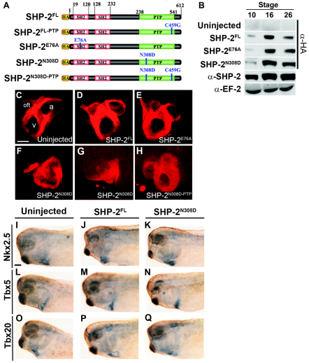
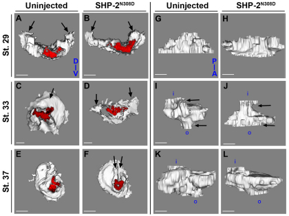
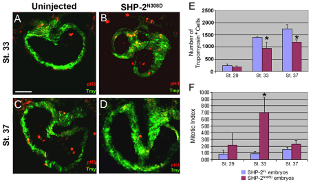
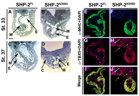
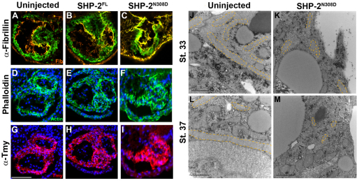
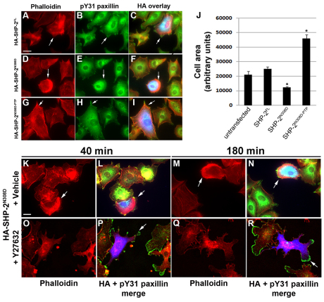
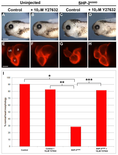
Noonan syndrome is one of the most common causes of human congenital heart disease and is frequently associated with missense mutations in the protein phosphatase SHP-2. Interestingly, patients with acute myelogenous leukemia (AML), acute lymphoblastic leukemia (ALL), juvenile myelomonocytic leukemia (JMML) and LEOPARD syndrome frequently carry a second, somatically introduced subset of missense mutations in SHP-2. To determine the cellular and molecular mechanisms by which SHP-2 regulates heart development and, thus, understand how Noonan-associated mutations affect cardiogenesis, we introduced SHP-2 encoding the most prevalent Noonan syndrome and JMML mutations into Xenopus embryos. Resulting embryos show a direct relationship between a Noonan SHP-2 mutation and its ability to cause cardiac defects in Xenopus; embryos expressing Noonan SHP-2 mutations exhibit morphologically abnormal hearts, whereas those expressing an SHP-2 JMML-associated mutation do not. Our studies indicate that the cardiac defects associated with the introduction of the Noonan-associated SHP-2 mutations are coupled with a delay or arrest of the cardiac cell cycle in M-phase and a failure of cardiomyocyte progenitors to incorporate into the developing heart. We show that these defects are a result of an underlying malformation in the formation and polarity of cardiac actin fibers and F-actin deposition. We show that these defects can be rescued in culture and in embryos through the inhibition of the Rho-associated, coiled-coil-containing protein kinase 1 (ROCK), thus demonstrating a direct relationship between SHP-2(N308D) and ROCK activation in the developing heart.


Figures

Fig. 1.
Fig. 1.
Noonan-associated mutation of SHP-2 leads to heart defects in Xenopus. (A) Schematic of SHP-2 constructs. Note that constructs used in Xenopus in vivo experiments were not epitope tagged with HA. (B) Western blot analysis of embryo lysates from embryos injected with the respective HA epitope-tagged SHP-2 constructs at stages 10 (gastrula), 16 (early neurula) and 26 (late neurula) probed with an anti-HA antibody, an anti-SHP-2 antibody that recognizes both endogenous and introduced versions of SHP-2, and an anti-EF-2 antibody as a loading control. (C-H) Whole-mount antibody staining of cleared stage 36 embryos with anti-tropomyosin antibody (Tmy). Anterior is to the left, posterior to the right. Scale bar: 100 μm. (I-Q) Whole-mount in situ hybridization of cleared stage 33 from uninjected (I,L,O), SHP-2FL (2 ng; J,M,P) and SHP-2N308D (2 ng; K,N,Q) embryos probed for Nkx2.5 (I-K), Tbx5 (L-N) or Tbx20 (O-Q). Scale bar: 1 mm. a, atrium; oft, outflow tract; v, ventricle.
Fig. 2.
Fig. 2.
3D modeling of uninjected and SHP-2N308D cardiac tissue. (A-L) 3D modeling of Tmy expression in stage 29 (A,B,G,H), stage 33 (C,D,I,J) and stage 37 (E,F,K,L) Xenopus embryos. In A-F, hearts are viewed from anterior to posterior and red denotes the most anterior section of the heart. In G-L, hearts are viewed from the dorsal side. Arrows in A-F denote ends of cardiac trough. Note that in C the trough has closed across the dorsal midline in uninjected control but remains open in SHP-2N308D heart (2 ng; D). Edges of the trough come together by stage 37 (F) in the SHP-2N308D heart but have not closed across the ventral midline (arrows). Also, at stage 33 (J), the SHP-2N308D heart is much wider in anterior portions but is much thinner in posterior portions (arrows) versus corresponding control (I). Note the relative orientation of the outflow tract (o) to the inflow tract (i) in I versus J, indicative of a delay in looping. a, anterior; d, dorsal; p, posterior; v, ventral. Scale bars: 50 μm.
Fig. 3.
Fig. 3.
SHP-2N308D leads to a delay of the cardiac cell cycle. (A-D) Transverse heart sections through stage 33 (A,B) and stage 37 (C,D) Xenopus embryos stained with Tmy (green) and anti-phospho histone H3 (red), from uninjected (A,C) and SHP-2N308D-derived (B,D) embryos (2 ng). Scale bar: 50 μm. (E,F) Graphs representing total number of Tmy-positive cells (E) and the mitotic index (F) in SHP-2FL- (blue bars) and SHP-2N308D- (red bars) derived embryos at stages 29, 33 and 37. Bars represent the average of at least three embryos. Error bars represent s.d. *P≤0.05 (comparison of SHP-2FL and SHP-2N308D embryos).
Fig. 4.
Fig. 4.
A subpopulation of cardiac cells does not differentiate or incorporate into hearts of SHP-2N308D Xenopus embryos. (A-D) Transverse Toluidine Blue-stained histological sections of cardiac regions from stage 33 (A,B) and stage 37 (C,D) SHP-2FL (A,C) and SHP-2N308D (B,D) embryos (2 ng). c, unincorporated cardiac cells; E, endocardium; M, myocardium. Scale bar: 50 μm. (E-J) Transverse sections through stage 37 hearts of SHP-2FL (E,G,I) and SHP-2N308D (F,H,J) embryos (2 ng) stained with anti-MHC (green), anti-TBX5 (red) and DAPI (blue). Arrows mark TBX5 positive cells that have not incorporated into the heart. Scale bar: 50 μm.
Fig. 5.
Fig. 5.
SHP-2N308D leads to alterations in cardiac myofibril formation. (A-I) Transverse sections of stage 37 hearts from uninjected (A,D,G), SHP-2FL (B,E,H) or SHP-2N308D (C,F,I) Xenopus embryos (2 ng) immunostained for fibrillin (orange) and Tmy (green) (A-C), cardiac actin as detected by conjugated phalloidin (green; D-F) and tropomyosin (red) and DAPI (blue) (G-I). Scale bar: 100 μm. (J-M) Representative transmission electron micrographs of transverse images from stage 33 (J,K) or stage 37 (L,M) heart tissue derived from uninjected (J,L) or SHP-2N308D (L,M) embryos. Cardiac muscle fibrils are highlighted. Scale bars: 1 μm.
Fig. 6.
Fig. 6.
SHP-2N308D regulates actin deposition. (A-I) Cells were transfected with HA-SHP-2FL (A-C), HA-SHP-2N308D (D-F) or HA-SHP-2N308D-PTP (G-I), plated on FN and stained for phalloidin (red) (A,D,G), pY31 paxillin (green) (B,E,H), and HA (blue) to identify transfected cells (C,F,I). Arrows denote lamellipodia and focal adhesion complexes. (J) 2D cell area as a measure of cell spreading was calculated from 150-200 cells transfected with the designated construct, using ImageJ software tools. Error bars represent s.e.m.*P≤0.05. (K-R) Inhibition of ROCK activity rescues SHP-2N308D cell rounding phenotype. SHP-2N308D-transfected cells (blue; L,N,P,R) treated with Y27632 (O-R) for either 40 (O,P) or180 (Q,R) minutes displayed reduced actin arc formation and re-established focal adhesion complexes (arrows) compared with vehicle-treated cells (K-N). Scale bars: 10 μm.
Fig. 7.
Fig. 7.
ROCK inhibition rescues the cardiac morphogenetic defects seen in SHP-2N308D embryos. (A-D) Brightfield images of stage 40 Xenopus embryos from appropriate conditions. Note that pericardial edema in SHP-2N308D embryo (3.6 ng; C) is absent in sibling incubated with Y27632 (D) and uninjected controls (A,B). Scale bar: 2 mm. (E-H) Immunofluorescent images of cleared hearts stained with anti-MHC, indicating the rescue of cardiac malformation and looping defects in SHP-2N308D embryo (G) compared with sibling incubated in Y27632 (H) and uninjected controls (E,F). Scale bar: 100 μm. (I) Graph of percentage of normal heart morphology at stage 40 in control and SHP-2N308D sibling embryos treated with 10 μM ROCK inhibitor Y27632. Statistical significance is denoted as *P≤0.0001, **P=0.0002, ***P=0.0001 using Fisher’s exact two-tailed test. Anterior is to the left, posterior to the right in all images. a, atrium; oft, outflow tract; v, ventricle.

Similar articles

Cited by

References

    1. Ahuja P., Perriard E., Pedrazzini T., Satoh S., Perriard J. C., Ehler E. (2007). Re-expression of proteins involved in cytokinesis during cardiac hypertrophy. Exp. Cell Res. 313, 1270–1283 - PubMed
    1. Arber S., Barbayannis F. A., Hanser H., Schneider C., Stanyon C. A., Bernard O., Caroni P. (1998). Regulation of actin dynamics through phosphorylation of cofilin by LIM-kinase. Nature 393, 805–809 - PubMed
    1. Bennett A. M., Hausdorff S. F., O’Reilly A. M., Freeman R. M., Neel B. G. (1996). Multiple requirements for SHPTP2 in epidermal growth factor-mediated cell cycle progression. Mol. Cell. Biol. 16, 1189–1202 - PMC - PubMed
    1. Bentires-Alj M., Paez J. G., David F. S., Keilhack H., Halmos B., Naoki K., Maris J. M., Richardson A., Bardelli A., Sugarbaker D. J., et al. (2004). Activating mutations of the noonan syndrome-associated SHP2/PTPN11 gene in human solid tumors and adult acute myelogenous leukemia. Cancer Res. 64, 8816–8820 - PubMed
    1. Brown D. D., Christine K. S., Showell C., Conlon F. L. (2007). Small heat shock protein Hsp27 is required for proper heart tube formation. Genesis 45, 667–678 - PMC - PubMed

Publication types

MeSH terms

Substances