. 2012 Mar;139(5):906-16.
doi: 10.1242/dev.074252. Epub 2012 Jan 25.

Dusky-like functions as a Rab11 effector for the deposition of cuticle during Drosophila bristle development

Affiliations

Dusky-like functions as a Rab11 effector for the deposition of cuticle during Drosophila bristle development

Ranganayaki Nagaraj et al. Development. 2012 Mar.

Abstract

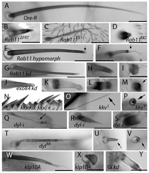
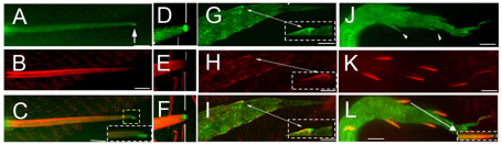
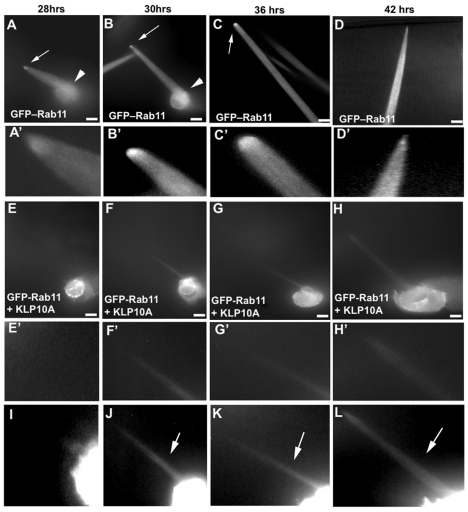
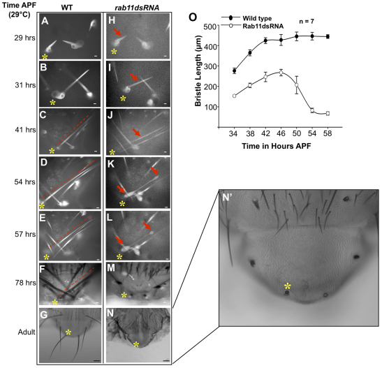
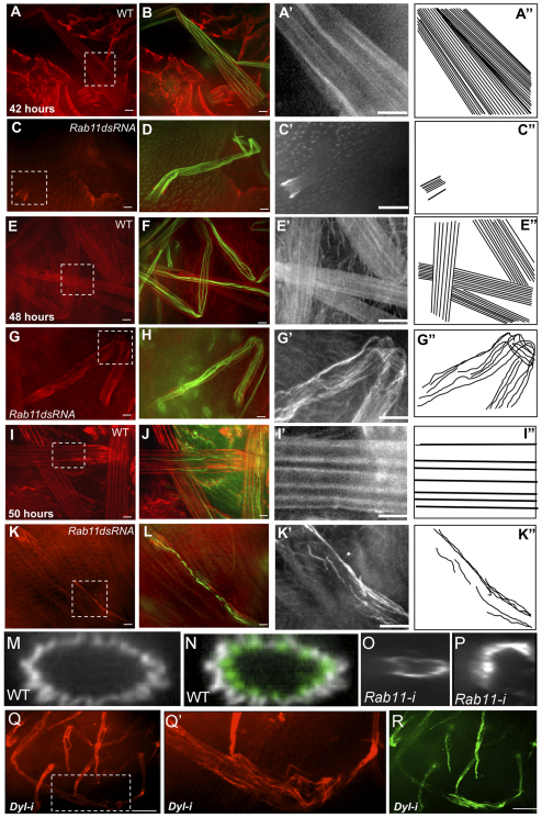
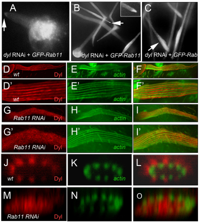
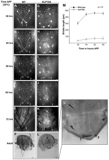
The morphogenesis of Drosophila sensory bristles is dependent on the function of their actin and microtubule cytoskeleton. Actin filaments are important for bristle shape and elongation, while microtubules are thought to mediate protein and membrane trafficking to promote growth. We have identified an essential role for the bristle cuticle in the maintenance of bristle structure and shape at late stages of bristle development. We show that the small GTPase Rab11 mediates the organized deposition of chitin, a major cuticle component in bristles, and disrupting Rab11 function leads to phenotypes that result from bristle collapse rather than a failure to elongate. We further establish that Rab11 is required for the plasma membrane localization of the ZP domain-containing Dusky-like (Dyl) protein and that Dyl is also required for cuticle formation in bristles. Our data argue that Dyl functions as a Rab11 effector for mediating the attachment of the bristle cell membrane to chitin to establish a stable cuticle. Our studies also implicate the exocyst as a Rab11 effector in this process and that Rab11 trafficking along the bristle shaft is mediated by microtubules.


Figures

Fig. 1.
Fig. 1.
Bristle phenotypes. (A) Scutellar bristle from wild-type (Ore-R) adult. (B) Thoracic bristle from putative Rab11ΔFRT null clone. (C) Thoracic bristle from putative Rab11ex1 null clone. (D) Abdominal bristle from putative Rab11ex1 null clone. (E,F) Thoracic bristles from Rab11 hypomorph (Rab1193Bi/Rab11ex1) adults. Arrows point to pigmentation defect at the tip. (G-I) Rab11 knockdown (kd) thoracic bristles. Knockdown starting 19 hours APF (G), 12 hours APF (H), or at white prepupal stage (I). (J-M) exo84 kd bristles (arrow points to bristle in M). (N) Wing bristles from flies with disrupted kkv function (kkv RNAi; kkv/+). (O,P) Scutellar bristles from putative kkv1 clones (arrows point to bristles). (Q-S) Thoracic bristles from dyl kd (w; dyl RNAi/+; neur-Gal4/+). Arrow points to affected bristle in Q. (T-V) Bristles from a dylML02088 escaper fly (arrows point to bristles). (W,X) Thoracic bristles from flies expressing Klp10A. Expression starting 15 hours APF (W) or at white prepupal stage (X). (Y) Thoracic bristle from fly with p150glued kd from white prepupal stage to adult. All images were taken at 10× magnification. Scale bars: 100 μm.
Fig. 2.
Fig. 2.
GFP-Rab11 localization in bristles and laterals. GFP-Rab11 (green) localization was visualized by immunostaining with anti-GFP antibody. F-actin (red) was visualized by phalloidin staining. (A) GFP-Rab11 is found in the bristle cell body and shaft. It is enriched at the tip (arrow). (B) F-actin for the bristle in A. (C) Merge of A and B. The tip is magnified in the inset. (D-F) Localization of GFP-Rab11 with respect to actin filaments. (D) GFP-Rab11 is enriched at the tip (arrow). (E) F-actin for bristle shown in D. (F) Merge of D and E. Note that Rab11 at the tip is distal to the F-actin. (G-I) Early stages of lateral outgrowth (28 hours APF, 25°C). (G) GFP-Rab11. (H) F-actin. (I) Merged image. Note the very early tip enrichment of GFP-Rab11. Insets are magnifications of the indicated regions (arrowed). (J-L) Middle stage of lateral outgrowth (32 hours APF, 25°C). (J) GFP-Rab11. Arrowheads point to Rab11 at tip. (K) F-actin. (L) Merged image. All images were taken at 60× magnification. Scale bars: 10 μm.
Fig. 3.
Fig. 3.
Rab11 intracellular transport is microtubule dependent. Time-lapse imaging of bristles expressing GFP-Rab11 and Klp10A under neur-Gal4 as compared with control bristles expressing only GFP-Rab11. (A-C) GFP-Rab11 localization in a wild-type bristle cell during its growth phase. Note the tip enrichment of Rab11 (arrows). Note the cell body GFP-Rab11 (arrowheads). (D) GFP-Rab11 localization in a wild-type bristle cell near the end of its growth phase. Note the decreased tip enrichment. (A′-D′) Magnified images of the bristle tip from A-D, respectively. (E-G) GFP-Rab11 localization in a bristle expressing Klp10A during its growth phase (starting ∼24 hours APF). (H) GFP-Rab11 localization in a bristle expressing Klp10A after its growth phase. (E′-H′) Magnified images of the bristle tip from E-H, respectively. (I-L) Highly enhanced versions of images E-H that allow the bristle shaft structure (arrows) to be seen. Images are of a single bristle tracked in each set. Images were taken at 60× magnification and represent a projection of optical sections. In control images C and D, the entire length of the bristle cell is not shown. Scale bars: 10 μm.
Fig. 4.
Fig. 4.
Rab11 is essential for bristle stability. (A-N′) Time-lapse imaging of wild-type and Rab11 dsRNA-expressing bristles. All images except G and N (2.5×) were taken at 20× (oil immersion) magnification. Images are shown of the same pupa tracked from 29 hours APF to adult. Asterisks and arrows mark the bristle being tracked; dashed line is parallel to the bristle. Images represent single optical sections. (A-G) Wild-type pupal thoracic bristles (control). (H-N′) Rab11 kd bristles (kd starting at white prepupal stage). Arrows in K and L indicate regions of bristle that showed deformities. (N′) Enlargement of N. Scale bars: 10 μm in A-F,H-M; 100 μm in G,N. (O) The growth of wild-type and Rab11 dsRNA-expressing bristles over time as determined by measuring the length of bristles (μm) at different times of development (hours APF at 29°C). Mean ± s.d.
Fig. 5.
Fig. 5.
Chitin organization in bristles. (A-L) Chitin (red) and actin (green) in wild-type bristles and Rab11 kd bristles at 42, 48 and 50 hours APF. Magnifications of chitin staining (boxed regions) are shown in grayscale in A′,C′,E′,G′,I′,K′, and interpretive drawings of these in A″,C″,E″,G″ I″,K″. (M,N) Projection of z-stacks from images of wild-type bristles stained for chitin (grayscale) and F-actin (green) localization. (O,P) Projection of z-stacks of Rab11 kd bristles showing uneven chitin deposition along the bristle circumference. (Q-R) Chitin (red) and F-actin (green) in dyl kd bristles. (Q′) Enlargement with enhanced contrast of boxed region from Q. Scale bars: 10 μm.
Fig. 6.
Fig. 6.
Dyl and Rab11 localization in bristles. (A-C) GFP-Rab11 localization in bristle expressing dyl RNAi at (A) 29 hours, (B) 40 hours and (C) 56 hours APF at 29°C. Arrows in A and B indicate tip accumulation of GFP-Rab11 (magnified in inset in B). Arrow in C shows the accumulation of GFP-Rab11 in a blebbing region of a bristle. (D-O) Dyl localization in wild-type and Rab11 mutant bristles visualized by staining with anti-Dyl antibody. Localization of (D) Dyl and (E) F-actin in wild-type bristles. (F) Merge of D and E. (D′-F′) Magnifications of D-F, respectively. (G,H) Localization of Dyl (G) and F-actin (H) in Rab11 kd bristles. (I) Merge of G and H. (G′-I′) Magnifications of G-I, respectively. (J-O) z projections showing Dyl (J,M) and F-actin (K,N) localization around the circumference of wild-type (J,K; merge in L) and Rab11 kd (M,N; merge in O) bristles.
Fig. 7.
Fig. 7.
Microtubules are essential for bristle growth. (A-L′) Time-lapse imaging of wild-type and Klp10A-expressing bristles. Images of the pupal bristles and adult bristles were taken at 20× and 2.5× magnification, respectively. (A-F) Wild-type bristles 1 and 2 tracked over 40 hours of development from pupa to adult stage. (G-L) Klp10A-expressing bristles 1 and 2 tracked over 40 hours of development. (L′) Magnification of L. Scale bars: 10 μm in A-E,G-K; 100 μm in F,L. (M) Growth of wild-type and Klp10A-expressing bristles over time. Mean ± s.d. Images are projections of single optical sections.

Similar articles

Cited by

References

    1. Abdu U., Bar D., Schupbach T. S. (2006). spn-F encodes a novel protein that affects oocyte patterning and bristle morphology in Drosophila. Development 133, 1477–1484 - PubMed
    1. Band A. M., Ali H., Vartiainen M. K., Welti S., Lappalainen P., Olkkonen V. M., Kuismanen E. (2002). Endogenous plasma membrane t-SNARE syntaxin 4 is present in rab11 positive endosomal membranes and associates with cortical actin cytoskeleton. FEBS Lett. 531, 513–519 - PubMed
    1. Bitan A., Guild G. M., Bar D. D., Abdu U. (2010). Asymmetric microtubule function is an essential requirement for polarized organization of the Drosophila bristle. Mol. Cell. Biol. 30, 496–507 - PMC - PubMed
    1. Bogard N., Lan L., Xu J., Cohen R. S. (2007). Rab11 maintains connections between germline stem cells and niche cells in the Drosophila ovary. Development 134, 3413–3418 - PubMed
    1. Cant K., Knowles B. A., Mooseker M. S., Cooley L. (1994). Drosophila singed, a fascin homolog, is required for actin bundle formation during oogenesis and bristle extension. J. Cell Biol. 125, 369–380 - PMC - PubMed

Publication types

MeSH terms

LinkOut - more resources