

Glycerophosphodiester phosphodiesterase domain containing 5 (GDPD5) expression correlates with malignant choline phospholipid metabolite profiles in human breast cancer
- PMID: 22279038
- PMCID: PMC4126590
- DOI: 10.1002/nbm.2766
Glycerophosphodiester phosphodiesterase domain containing 5 (GDPD5) expression correlates with malignant choline phospholipid metabolite profiles in human breast cancer
Abstract
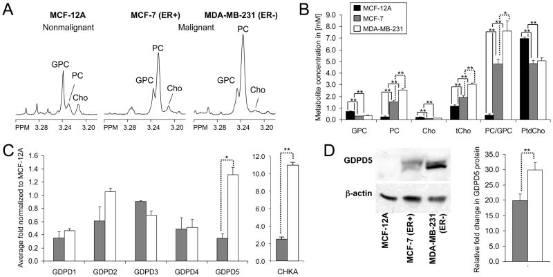
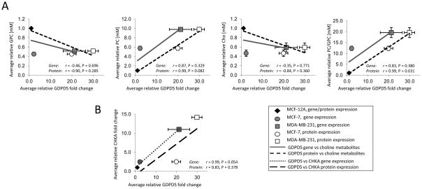
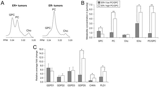
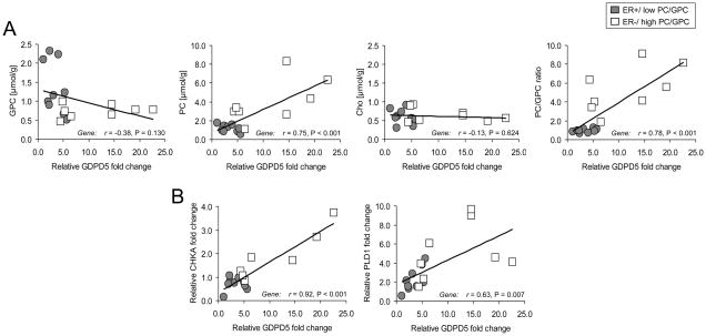
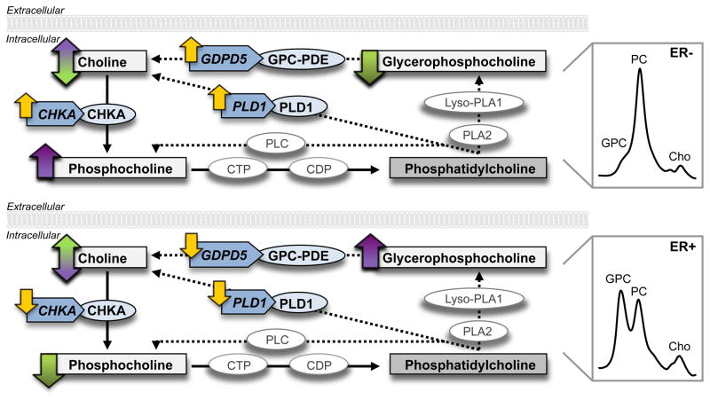
Altered choline phospholipid metabolism is a hallmark of cancer, leading to malignant choline metabolite profiles consisting of low glycerophosphocholine (GPC) and high phosphocholine (PC) in human breast cancers. Glycerophosphocholine phosphodiesterase (GPC-PDE) catalyzes the degradation of GPC to free choline and glycerol-3-phosphate. The gene(s) encoding for the GPC-PDE(s) responsible for GPC degradation in breast cancers have not yet been identified. Here, we demonstrate for the first time that the GPC-PDE encoded by glycerophosphodiester phosphodiesterase domain containing 5 (GDPD5) is associated with breast cancer malignancy. Two human breast cancer cell lines (n = 8 and n = 10) and primary human breast tumor samples (n = 19) were studied with combined MRS and quantitative reverse transcription-polymerase chain reaction to investigate several isoforms of GDPD expression with respect to choline phospholipid metabolite levels. Of the five GDPDs tested, GDPD5 was found to be significantly overexpressed in highly malignant estrogen receptor negative (ER(-)) compared with weakly malignant estrogen receptor positive (ER(+)) human breast cancer cells (p = 0.027) and breast tumors from patients (p = 0.015). GDPD5 showed significantly positive correlations with PC (p < 0.001), total choline (tCho) (p = 0.007) and PC/GPC (p < 0.001) levels in human breast tumors. GDPD5 showed a trend towards a negative correlation with GPC levels (p = 0.130). Human breast cancers with malignant choline metabolite profiles consisting of low GPC and high PC levels highly co-expressed GDPD5, choline kinase alpha (CHKA) and phosphatidylcholine-specific phospholipase D1 (PLD1), whereas cancers containing high GPC and relatively low PC levels displayed low co-expression of GDPD5, CHKA and PLD1. GDPD5, CHKA and PLD1 were significantly overexpressed in highly malignant ER(-) tumors in our patient cohort. Our study identified GDPD5 as a GPC-PDE that probably participates in the regulation of choline phospholipid metabolism in breast cancer, which possibly occurs in cooperation with CHKA and PLD1.
Copyright © 2012 John Wiley & Sons, Ltd.
Conflict of interest statement
The author(s) declare that they have no competing interests.
Figures
Similar articles
-
Interplay of choline metabolites and genes in patient-derived breast cancer xenografts.Breast Cancer Res. 2014 Jan 21;16(1):R5. doi: 10.1186/bcr3597. Breast Cancer Res. 2014. PMID: 24447408 Free PMC article.
-
Molecular Effects of Doxorubicin on Choline Metabolism in Breast Cancer.Neoplasia. 2017 Aug;19(8):617-627. doi: 10.1016/j.neo.2017.05.004. Epub 2017 Jun 24. Neoplasia. 2017. PMID: 28654865 Free PMC article.
-
Silencing of the glycerophosphocholine phosphodiesterase GDPD5 alters the phospholipid metabolite profile in a breast cancer model in vivo as monitored by (31) P MRS.NMR Biomed. 2014 Jun;27(6):692-9. doi: 10.1002/nbm.3106. Epub 2014 Apr 24. NMR Biomed. 2014. PMID: 24764256 Free PMC article.
-
Choline metabolism-based molecular diagnosis of cancer: an update.Expert Rev Mol Diagn. 2015 Jun;15(6):735-47. doi: 10.1586/14737159.2015.1039515. Epub 2015 Apr 28. Expert Rev Mol Diagn. 2015. PMID: 25921026 Free PMC article. Review.
-
Therapeutic targets and biomarkers identified in cancer choline phospholipid metabolism.Pharmacogenomics. 2006 Oct;7(7):1109-23. doi: 10.2217/14622416.7.7.1109. Pharmacogenomics. 2006. PMID: 17054420 Review.
Cited by
-
Interplay of choline metabolites and genes in patient-derived breast cancer xenografts.Breast Cancer Res. 2014 Jan 21;16(1):R5. doi: 10.1186/bcr3597. Breast Cancer Res. 2014. PMID: 24447408 Free PMC article.
-
The effect of antitumor glycosides on glioma cells and tissues as studied by proton HR-MAS NMR spectroscopy.PLoS One. 2013 Oct 23;8(10):e78391. doi: 10.1371/journal.pone.0078391. eCollection 2013. PLoS One. 2013. PMID: 24194925 Free PMC article.
-
Metabolic Portraits of Breast Cancer by HR MAS MR Spectroscopy of Intact Tissue Samples.Metabolites. 2017 May 16;7(2):18. doi: 10.3390/metabo7020018. Metabolites. 2017. PMID: 28509845 Free PMC article. Review.
-
Choline-releasing glycerophosphodiesterase EDI3 drives tumor cell migration and metastasis.Proc Natl Acad Sci U S A. 2012 May 22;109(21):8155-60. doi: 10.1073/pnas.1117654109. Epub 2012 May 8. Proc Natl Acad Sci U S A. 2012. PMID: 22570503 Free PMC article.
-
Proton MR spectroscopy in the breast: Technical innovations and clinical applications.J Magn Reson Imaging. 2019 Oct;50(4):1033-1046. doi: 10.1002/jmri.26700. Epub 2019 Mar 7. J Magn Reson Imaging. 2019. PMID: 30848037 Free PMC article. Review.
References
-
- Glunde K, Jie C, Bhujwalla ZM. Molecular causes of the aberrant choline phospholipid metabolism in breast cancer. Cancer Res. 2004;64:4270–4276. - PubMed
-
- Katz-Brull R, Lavin PT, Lenkinski RE. Clinical utility of proton magnetic resonance spectroscopy in characterizing breast lesions. J Natl Cancer Inst. 2002;94:1197–1203. - PubMed
-
- Meisamy S, Bolan PJ, Baker EH, Bliss RL, Gulbahce E, Everson LI, Nelson MT, Emory TH, Tuttle TM, Yee D, Garwood M. Neoadjuvant chemotherapy of locally advanced breast cancer: predicting response with in vivo (1)H MR spectroscopy--a pilot study at 4 T. Radiology. 2004;233:424–431. - PubMed
-
- Glunde K, Jacobs MA, Bhujwalla ZM. Choline metabolism in cancer: implications for diagnosis and therapy. Expert Rev Mol Diagn. 2006;6:821–829. - PubMed
Publication types
MeSH terms
Substances
Grants and funding
LinkOut - more resources
Full Text Sources
Medical
Research Materials
