

doi: 10.1101/gad.182980.111.
Anti-apoptotic Mcl-1 is essential for the development and sustained growth of acute myeloid leukemia
Affiliations
- PMID: 22279045
- PMCID: PMC3273836
- DOI: 10.1101/gad.182980.111
Item in Clipboard
Anti-apoptotic Mcl-1 is essential for the development and sustained growth of acute myeloid leukemia
Genes Dev.
.
Abstract
Acute myeloid leukemia (AML) frequently relapses after initial treatment. Drug resistance in AML has been attributed to high levels of the anti-apoptotic Bcl-2 family members Bcl-x(L) and Mcl-1. Here we report that removal of Mcl-1, but not loss or pharmacological blockade of Bcl-x(L), Bcl-2, or Bcl-w, caused the death of transformed AML and could cure disease in AML-afflicted mice. Enforced expression of selective inhibitors of prosurvival Bcl-2 family members revealed that Mcl-1 is critical for survival of human AML cells. Thus, targeting of Mcl-1 or regulators of its expression may be a useful strategy for the treatment of AML.
Figures
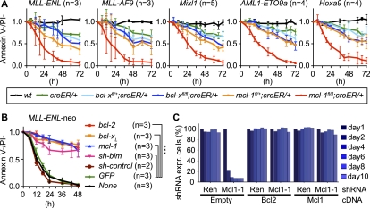
Impact of conditional deletion of bcl-x or mcl-1 in transformed myeloid and AML cells in vitro. (A) Cells of the indicated genotypes, transformed with the oncogenes indicated, were grown for 72 h in medium with or without tamoxifen. Viable (AnnexinV−/PI−) cells were enumerated by flow cytometry. Graphs represent the means ± SEM of the ratio of viable tamoxifen-treated cells versus viable untreated cells (n = 3–5 independently transduced and sorted cell lines for each transforming oncogene and genotype). (B) MLL-ENL/neo transformed mcl-1fl/fl;creER/+ cells were secondarily transduced with retroviruses encoding GFP alone or GFP plus Bcl-2, Bcl-xL, Mcl-1, or shRNA for knockdown of Bim and purified on the basis of GFP expression. Cells were then cultured in the presence or absence of tamoxifen and their survival was measured; data are presented as in A. (***) P < 0.001. (C) RNAi-mediated suppression of Mcl-1 in MLL-AF9 plus N-Ras AML cells. Graphs represent the percentages of cells coexpressing shRNA and the cDNAs indicated over time following doxycycline treatment.
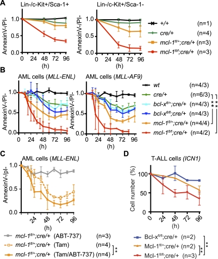
Impact of conditional in vitro deletion of bcl-x or mcl-1 in normal hematopoietic stem and progenitor cells and in vivo derived AML cells. (A) Normal LSK (left panel) and Lin−c-Kit+Sca-1− myeloid progenitors (right panel) were cultured in the presence or absence of tamoxifen and cell survival was determined; data are presented as in Figure 1A. (B) Leukemic cells from mice bearing MLL-ENL-induced (left panel) or MLL-AF9-induced (right panel) AML were cultured in the presence or absence of tamoxifen and cell survival was determined; data are presented as described in Figure 1A. (**) P < 0.01. (C) MLL-ENL transformed AML cells of the indicated genotypes were grown in medium containing tamoxifen, ABT-737 (1 μM), or tamoxifen plus ABT-737 (1 μM). Cell survival was determined, and data are presented as described in Figure 1A. (**) P < 0.01. (D) Leukemic cells from mice bearing ICN1-induced T-ALL of the indicated genotypes were cultured in the presence or absence of tamoxifen. Cell survival was determined, and data are presented as described in Figure 1A. (**) P < 0.01.
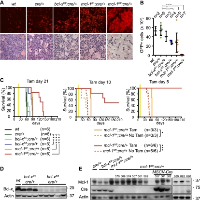
Conditional deletion of mcl-1 causes regression of AML in tumor-burdened mice. (A) Histological examination of bone marrow from mice burdened with MLL-ENL-induced AML of the indicated genotypes. The top panels show apoptosis of cells (detected by TUNEL staining) after 3 d of treatment with tamoxifen. The bottom panels show the presence of leukemic blasts after 5 d of treatment with tamoxifen. (B) Symptomatic (elevated leukocyte counts, thrombocytopenia, and anemia) mice bearing MLL-ENL-induced AML of the indicated genotypes were treated for 5 d with tamoxifen. Shown are the total numbers of GFP+ leukemic cells collected from two femora and two tibiae on the sixth day. (*) P < 0.05; (**) P < 0.01; (***) P < 0.001. (C) Survival of AML-burdened mice that were treated with tamoxifen (treatment window indicated by yellow bar) 21 d (left panel) 10 d (middle panel) or 5 d (right panel) after transplantation with MLL-ENL transformed AML cells of the indicated genotypes. Untreated mice (dotted lines) from the same cohort treated with tamoxifen at 10 d or 5 d all developed disease, thus confirming the presence of AML at that time pointl (*) P < 0.05; (**) P < 0.01; (***) P < 0.001. (D,E) Western blot analysis to detect Bcl-xL (D) or Mcl-1 and CreER proteins (E) in leukemic cells of the indicated genotypes from mice that relapsed with AML after tamoxifen treatment.
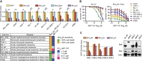
Impact of functional inactivation of Mcl-1 by inducible expression of Bim-derived BH3-like ligands that selectively neutralize different prosurvival Bcl-2 family members in human leukemia-derived cell lines and primary human AML cells. (A) Mouse AML cells and human leukemia-derived cell lines (U937, THP-1, ML-1, HL-60, MV4,11, OCI-AML3, HEL92.1.7, KU-812, and K562) were transduced with lentiviral constructs that allow inducible expression of BimS4E (negative control, no binding to Bcl-2-like proteins), BimS2A (binding only Mcl-1), BimSBad (binding Bcl-2, Bcl-xL, and Bcl-w but not Mcl-1 or A1), or BimSwt (binding all Bcl-2 prosurvival proteins) and were either left untreated or treated with doxycycline. Graphs represent the ratio of GFP+ cells (doxycycline-treated compared with untreated) detected by flow cytometry of at least three experiments for each cell line. In the table, cell lines derived from AML are boxed; the heat map summarizes the sensitivity of all cell lines to BimS2A expression or ABT-737 treatment. (B) The cell lines indicated were treated with the indicated doses of ABT-737 alone (left panel) or in combination with induced BimS2A (inhibits Mcl-1) expression (right panel). (C) Primary human AML cells were transduced with BimS4E-encoding (negative control), BimS2A-encoding (inhibits Mcl-1), or BimSwt-encoding (inhibits all prosurvival Bcl-2 family members) lentiviruses and were either left untreated or treated with doxycycline. Shown is a graph representing the ratio of doxycycline-treated compared with untreated GFP+ AML blast cells from one representative experiment for each patient sample. Western blot analysis to detect Bax and Bak protein levels in primary AML cells. Low levels of Bax and Bak were detected in sample #5; low levels of Bax and abnormal size of Bak were detected in sample #2.
Comment in
-
Selectively targeting Mcl-1 for the treatment of acute myelogenous leukemia and solid tumors.Genes Dev. 2012 Feb 15;26(4):305-11. doi: 10.1101/gad.186189.111. Genes Dev. 2012. PMID: 22345513 Free PMC article.
References
-
- Anastassiadis K, Glaser S, Kranz A, Berhardt K, Stewart AF 2010. A practical summary of site-specific recombination, conditional mutagenesis, and tamoxifen induction of CreERT2. Methods Enzymol 477: 109–123 - PubMed
-
- Bouillet P, Robati M, Bath ML, Strasser A 2005. Polycystic kidney disease prevented by transgenic RNA interference. Cell Death Differ 12: 831–833 - PubMed
Publication types
MeSH terms
Substances
Grants and funding
LinkOut - more resources
Full Text Sources
Other Literature Sources
Medical
Molecular Biology Databases
Research Materials