. 2012 Jan 15;26(2):190-202.
doi: 10.1101/gad.179663.111.

MASTR directs MyoD-dependent satellite cell differentiation during skeletal muscle regeneration

Affiliations

MASTR directs MyoD-dependent satellite cell differentiation during skeletal muscle regeneration

Mayssa H Mokalled et al. Genes Dev. .

Abstract

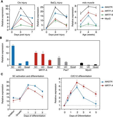
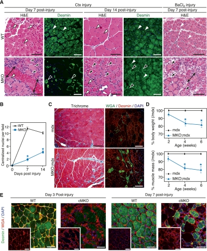
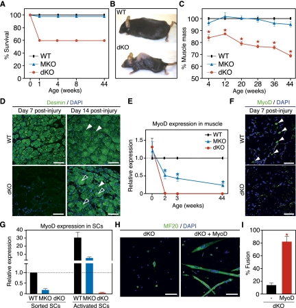
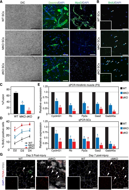
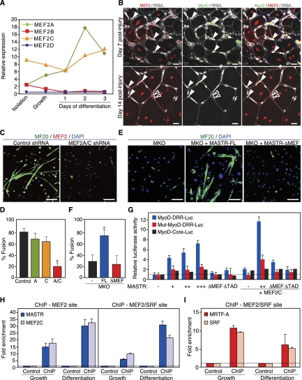
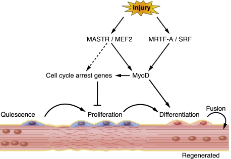
In response to skeletal muscle injury, satellite cells, which function as a myogenic stem cell population, become activated, expand through proliferation, and ultimately fuse with each other and with damaged myofibers to promote muscle regeneration. Here, we show that members of the Myocardin family of transcriptional coactivators, MASTR and MRTF-A, are up-regulated in satellite cells in response to skeletal muscle injury and muscular dystrophy. Global and satellite cell-specific deletion of MASTR in mice impairs skeletal muscle regeneration. This impairment is substantially greater when MRTF-A is also deleted and is due to aberrant differentiation and excessive proliferation of satellite cells. These abnormalities mimic those associated with genetic deletion of MyoD, a master regulator of myogenesis, which is down-regulated in the absence of MASTR and MRTF-A. Consistent with an essential role of MASTR in transcriptional regulation of MyoD expression, MASTR activates a muscle-specific postnatal MyoD enhancer through associations with MEF2 and members of the Myocardin family. Our results provide new insights into the genetic circuitry of muscle regeneration and identify MASTR as a central regulator of this process.


Figures

Figure 1.
Figure 1.
Up-regulation of MASTR and MRTF-A during muscle regeneration. (A) Shown is the expression of MASTR, MRTF-A, and MRTF-B in the TA muscle following Ctx and barium chloride (BaCl2) injuries and in mdx mice. qPCR shows MASTR and MRTF-A up-regulation 3 d after Ctx and BaCl2 injection. MASTR and MRTF-A are also up-regulated in the TA muscle of 2-, 4-, and 6-wk-old mdx mice compared with wild-type (WT) mice. For Ctx and BaCl2 injury, relative expression at days 3 and 7 post-injury is normalized to GAPDH levels and to baseline level at day 0. For mdx mice, relative expression is normalized to GAPDH levels and to wild-type expression for each time point. MyoD expression is used as control for muscle regeneration and parallels MASTR and MRTF-A expression patterns. (B) Expression of MASTR and MRTF-A in sorted SCs. Sorted SCs (SC), mononuclear unsorted cells (UCs), and quadriceps mRNA samples (Quad) were used for qPCR. Relative expression is normalized to the levels of ribosomal 18S RNA. The levels of Pax7 and MyoD are shown as controls. (C) Up-regulation of MASTR and MRTF-A expression during SC and C2C12 differentiation. Sorted SCs, activated SCs maintained under growth conditions, and differentiating SCs at days 1, 2, or 3 of differentiation were used for qPCR. MASTR and MRTF-A are expressed in sorted and activated SCs, and are up-regulated at days 1 and 2 of differentiation. Similarly, qPCR on differentiating C2C12 cultures shows approximately sixfold to sevenfold up-regulation of MASTR and MRTF-A by day 2 of differentiation.
Figure 2.
Figure 2.
Aberrant muscle regeneration of MASTR-null mice. (A) Regeneration of MKO muscle following chemical injury. TA muscle from wild-type (WT) and MKO mice was injected with Ctx and BaCl2 and assayed for regeneration by H&E and by Desmin immunohistochemistry at days 7 and 14 post-injury. Regenerating and degenerating fibers are indicated by white and black arrowheads, respectively. At day 7 post-Ctx or post-BaCl2 injury, wild-type muscle contains regenerating myofibers that are centrally nucleated and heterogeneous in size. MKO muscle shows fewer regenerating fibers and is mostly composed of necrotic fibers and regions of fibrotic tissue and inflammatory cells (black arrows). By day 14 post-Ctx injury, regenerating wild-type fibers are mature, centrally nucleated, and homogeneous in size, whereas regenerating MKO fibers are heterogeneous in size and decreased in number. Bar, 65 μm. (B) Quantitation of regeneration shows a significant decrease in the number of centralized nuclei in MKO muscle compared with wild-type muscle at days 7 and 14 post-injury. Analysis was performed on five animals for each genotype and time point and on five sections from each animal; (*) P < 0.01. (C) Enhanced mdx regeneration defect by MASTR deletion. Trichrome staining and immunohistochemistry for Desmin and WGA show enhanced muscle damage and fibrosis in 4-wk-old MKO;mdx mice compared with mdx mice. Bar, 125 μm. (D) MKO;mdx mice have decreased body mass and muscle mass compared with littermate mdx mice; (*) P < 0.01. (E) Impaired regeneration of cMKO mice following Ctx injury. Staining for Desmin and WGA shows defective TA muscle regeneration of cMKO mice at days 3 and 7 post-Ctx injury. Bar, 45 μm.
Figure 3.
Figure 3.
Cooperative regulation of MyoD expression, muscle mass, and muscle regeneration by MASTR and MRTF-A. (A) Partial lethality of dKO mice at birth. Survival curve shows ∼40% perinatal lethality of dKO mice compared with wild-type (WT) or MKO mice. Mice that survived the first week of postnatal life were viable and fertile. (B) Loss of muscle mass and kyphosis of dKO mice by 44 wk of age. (C) Decreased muscle mass in dKO mice. Percent lean mass was measured by NMR from wild-type, MKO, and dKO mice. dKO mice are significantly leaner than wild-type or MKO littermates. Lean mass content decreased with age and reached ∼70% in 44-wk-old dKO mice. MKO mice do not show a significant decrease in muscle mass at baseline; (*) P < 0.01. (D) Defective regeneration of dKO muscle following Ctx injury. Regeneration of the TA muscle was examined by Desmin immunohistochemistry at days 7 and 14 after Ctx administration. Regenerating and degenerating fibers are indicated by white and black arrowheads, respectively. Bar, 65 μm. (E) Down-regulation of MyoD mRNA levels in MKO and dKO muscle. Hindlimb neonatal muscle and TA muscle from 2-, 3-, and 44-wk-old mice were used for qPCR. MyoD levels were normalized to GAPDH and wild-type MyoD levels for each time point. MyoD expression was significantly diminished in MKO muscle and was undetectable in dKO muscle, starting from 2 wk of age; (*) P < 0.01. (F) Down-regulation of MyoD protein levels in dKO mice. MyoD immunohistochemistry on injured TA muscle from wild-type and dKO mice reveals a dramatic decrease in the number of MyoD-positive cells in dKO muscle at day 7 post-injury. (G) Down-regulation of MyoD mRNA levels in MKO and dKO SCs. Sorted and activated SCs from wild-type, MKO, and dKO mice were used for qPCR. Relative expression was normalized to MyoD levels in wild-type sorted SCs. MyoD levels were down-regulated in MKO SCs and were undetectable in dKO SCs. (H) Rescue of the dKO SC differentiation defect by MyoD expression. dKO SCs were infected with control or MyoD-expressing retrovirus, allowed to differentiate for 4 d, and stained with anti-myosin antibody to assay differentiation. MyoD overexpression rescues the dKO differentiation defect. Bar, 90 μm. (I) Quantification of the percent myotube fusion confirms that MyoD overexpression rescues the dKO SC differentiation defect; (*) P < 0.01.
Figure 4.
Figure 4.
Increased proliferation and defective differentiation of MKO and dKO SCs. (A) Defective differentiation of MKO and dKO SCs in vitro. Bright-field microscope images show impaired differentiation of MKO SCs and enhanced differentiation defect of dKO SCs, compared with wild-type (WT) cells. Desmin immunohistochemistry confirms aberrant differentiation of MKO and dKO SCs. Extensive multinucleated myotube networks are observed in wild-type but not mutant SC cultures. MyoD immunohistochemistry shows that the number of MyoD-positive cells is decreased in MKO SC cultures compared with wild-type cells. Bars: for DIC, 200 μm; for Desmin, 65 μm; for MyoD, 90 μm. (B) Increased proliferation of MKO and dKO cells. Wild-type, MKO, and dKO SCs were maintained under growth conditions and pulsed with BrdU for 1 h. Anti-BrdU immunocytochemistry shows that the number of BrdU-positive cells (white arrowheads) is increased in MKO and dKO SCs at days 2, 3, and 4 of culture. Bar, 70 μm. (C) Quantitation of MKO and dKO SC differentiation. Percent myotube fusion, calculated from A, confirms the differentiation defect of MKO and dKO SCs; (*) P < 0.01. (D) Quantitation of MKO and dKO SC proliferation. Percent BrdU-positive cells, calculated from B, confirms the increase in proliferation of MKO SCs compared with wild-type cells; (*) P < 0.01. (E,F) Down-regulation of genes involved in cell cycle arrest following MASTR deletion. Wild-type, MKO, and dKO TA muscle (E) and SC cultures (F) were used for qPCR. Expression of CyclinG1, Retinoblastoma (Rb), protein phosphatase 2 (Pp2a), growth arrest-specific 2 (Gas2), and growth arrest and DNA damage-inducible 45a (Gadd45a) mRNA is significantly diminished following MASTR/MRTF-A deletion; (*) P < 0.01. (G) Increased cell proliferation in the cMKO muscle following Ctx injury. PCNA immunohistochemistry shows an increase in the number of PCNA-positive cells (arrowheads) in the cMKO TA muscle at days 3 and 7 post-injury.
Figure 5.
Figure 5.
Activation of the MyoD-DRR enhancer by MASTR, MEF2, and MRTF-A. (A) Regulation of the MEF2 family during SC activation and proliferation. Shown is the expression of MEF2A, MEF2B, MEF2C, and MEF2D in SCs at isolation, under growth conditions, and at days 1, 2, and 3 of differentiation by qPCR. Relative expression was normalized to 18S RNA levels. (B) Expression of MEF2 in activated MyoD-positive SCs. Immunohistochemistry was performed on TA muscle at days 7 and 14 post-Ctx injury using anti-MEF2 antibody that detects MEF2A, MEF2C, and MEF2D isoforms. MyoD and WGA staining shows colocalization of MEF2-positive cells to activated SCs. White arrowheads indicate cells that are double positive for MEF2 and MyoD, whereas black arrowheads represent cells that are positive for MEF2 and negative for MyoD. Bar, 10 μm. (C) Defective SC differentiation following knockdown of MEF2A and MEF2C in vitro. SCs infected with a combination of MEF2A and MEF2C shRNA viruses show delayed differentiation and myotube formation compared with control cells infected with an empty shRNA virus. Bar, 65 μm. (D) Quantitation of the percent myotube fusion confirms that MEF2A/C knockdown inhibits SC differentiation. Knockdown of either MEF2A or MEF2C alone does not cause a significant differentiation defect; (*) P < 0.01. (E) Rescue of the MKO SC differentiation defect by MASTR-FL, but not by MASTR-ΔMEF. MKO SCs were transfected with control, MASTR-FL-expressing, or MASTR-ΔMEF-expressing vectors; allowed to differentiate for 4 d under low-serum culture conditions; and stained with anti-Myosin antibody to assay differentiation. Unlike MASTR-FL overexpression, which results in myotube formation in MKO SCs, MASTR-ΔMEF fails to rescue the MKO SC differentiation defect. Bar, 90 μm. (F) Quantitation of the percent myotube fusion confirms that overexpression of MASTR-FL, but not MASTR-ΔMEF, rescues the MKO SC differentiation defect; (*) P < 0.01. (G) Activation of the MyoD-DRR enhancer by MASTR and MEF2. C2C12 cells were transfected with the MyoD-DRR-Luciferase vector and expression vectors encoding various combinations of MASTR and MEF2C and assayed for luciferase activity. The MyoD-core-Luciferase vector was used as a negative control. The Mut-MyoD-DRR-Luciferase vector, in which the MEF2 and MEF2/SRF-binding sites were mutated, was also used as a control. MASTR alone or with MEF2C activates the MyoD-DRR but not the MyoD-core vectors. MASTR mutants that lack either the MEF2-binding (ΔMEF) or the transactivation (ΔTAD) domains do not induce MyoD-DRR activation. Mutation of the MEF2- and MEF2/SRF-binding sites on the DRR enhancer blocks reporter activation. (H) Binding of MASTR and MEF2 to the MyoD-DRR enhancer. Proliferating and differentiating C2C12 cells were transfected with MASTR and MEF2C and used for ChIP assay. Enrichment of MASTR and MEF2C at the MyoD DRR enhancer was quantified by qPCR and normalized to enrichment in C2C12 cells transfected with empty vectors. MASTR and MEF2C are enriched at the MEF2- and MEF2/SRF-binding sites of the DRR enhancer. (I) Binding of MRTF-A and SRF to the MyoD-DRR enhancer. Proliferating and differentiating C2C12 cells were transfected with MRTF-A and SRF and used for ChIP assay. Enrichment of MRTF-A and SRF at the MyoD-DRR enhancer was quantified by qPCR and normalized to enrichment in C2C12 cells transfected with empty vectors. Comparable enrichment was obtained for MRTF-A and SRF under both proliferation and differentiation conditions.
Figure 6.
Figure 6.
Schematic model for MASTR, MEF2, and MRTF-A function during SC differentiation. MASTR coactivates MEF2 and cooperates with MRTF-A to regulate MyoD expression, SC differentiation, and skeletal muscle regeneration in response to injury.

Similar articles

Cited by

References

    1. Asakura A, Lyons GE, Tapscott SJ 1995. The regulation of MyoD gene expression: Conserved elements mediate expression in embryonic axial muscle. Dev Biol 171: 386–398 - PubMed
    1. Asakura A, Hirai H, Kablar B, Morita S, Ishibashi J, Piras BA, Christ AJ, Verma M, Vineretsky KA, Rudnicki MA 2007. Increased survival of muscle stem cells lacking the MyoD gene after transplantation into regenerating skeletal muscle. Proc Natl Acad Sci 104: 16552–16557 - PMC - PubMed
    1. Bachinski LL, Sirito M, Bohme M, Baggerly KA, Udd B, Krahe R 2010. Altered MEF2 isoforms in myotonic dystrophy and other neuromuscular disorders. Muscle Nerve 42: 856–863 - PMC - PubMed
    1. Banks GB, Chamberlain JS 2008. The value of mammalian models for duchenne muscular dystrophy in developing therapeutic strategies. Curr Top Dev Biol 84: 431–453 - PubMed
    1. Beauchamp JR, Heslop L, Yu DS, Tajbakhsh S, Kelly RG, Wernig A, Buckingham ME, Partridge TA, Zammit PS 2000. Expression of CD34 and Myf5 defines the majority of quiescent adult skeletal muscle satellite cells. J Cell Biol 151: 1221–1234 - PMC - PubMed

Publication types

MeSH terms

LinkOut - more resources