. 2012 Apr;107(8):2212-9.
doi: 10.1152/jn.00768.2011. Epub 2012 Jan 25.

Serotonin modulates multiple calcium current subtypes in commissural interneurons of the neonatal mouse

Affiliations

Serotonin modulates multiple calcium current subtypes in commissural interneurons of the neonatal mouse

Matthew D Abbinanti et al. J Neurophysiol. 2012 Apr.

Abstract

Calcium currents are critical to the intrinsic properties of neurons and the networks that contain them. These currents make attractive targets for neuromodulation. Here, we examine the serotonergic modulation of specific calcium current subtypes in neonatal (P0-5) intersegmental commissural interneurons (CINs), members of the hindlimb locomotor central pattern generator in the mouse spinal cord. Previous work in our lab showed that serotonin (5-HT) excited CINs in part by reducing a calcium current and thus indirectly reducing the calcium-activated potassium current (Diaz-Rios et al. 2007). We have determined which calcium currents are targets of serotonin modulation. Utilizing whole cell voltage clamp and toxins to specific calcium current subtypes, we found that N- and P/Q-type currents comprise over 60% of the overall calcium current. Blockade of each of these subtypes alone with either ω-conotoxin GVIA or ω-agatoxin TK was unable to occlude 5-HT's reduction of the calcium current. However, coapplication of both blockers together fully occluded 5-HT's reduction of the calcium current. Thus, 5-HT decreases both N- and P/Q-type calcium current to excite neonatal CINs.


Figures
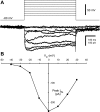
Fig. 1.
Fig. 1.
Barium current in a neonatal commissural interneuron (CIN). A: representative current traces in a P4 CIN in recording solution containing barium replacing calcium in response to steps from −60 mV. B: I-V plot of the peak barium current in this same cell. Note the current is activated around −40 mV and peaks at 0 mV.
Fig. 2.
Fig. 2.
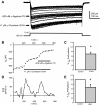
N- and P/Q-type calcium current are present in P0-5 CINs. A: current traces in response to voltage steps from −60 to 0 mV elicited every 30 s. Toxin labels illustrate the rapid drops in current in response to their application. B: plot of peak of the current in the traces above vs. time. Application of toxins is depicted by black bars running below the plot. Note the rapid reduction in current upon application of ω-conotoxin and ω-agatoxin. C: rundown-corrected percent reduction of IBa by application of 1 μM ω-conotoxin GVIA. D: instantaneous dI/dt vs. time for the same experiment determined by change in current amplitude between each trace (30 s). Peaks indicate points of rapid decrease in current amplitude, coinciding with the application of the blockers in B. E: rundown-corrected percent reduction of IBa by application of 200 nM ω-agatoxin TK. *P < 0.05.
Fig. 3.
Fig. 3.
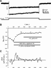
5-HT decreases IBa. A: current traces in response to voltage steps from −60 to 0 mV before (a), during (b), and after (c) 10 μM 5-HT application. B: plot of peak amplitude vs. time from current traces taken once every 30 s. Black bars indicate the application of 5-HT. Application of an additional 10 μM 5-HT did not produce any additional reduction. Points labeled a, b, and c are the traces labeled in A. C: 5-HT reduces the rundown-corrected IBa. *P < 0.05.
Fig. 4.
Fig. 4.
Blocking P/Q-type ICa does not occlude 5-HT's reduction of the current. A: IBa traces elicited by steps from −60 to 0 mV in control conditions (a), during application of 200 nM ω-agatoxin TK (b), and during coapplication of 200 nM ω-agatoxin TK and 10 μM 5-HT. B: plot of peak IBa vs. time with sweeps (as in A) taken every 30 s. Black bars indicate application of ω-agatoxin TK and 5-HT. Measurements from sweeps in A are indicated by lowercase letters (a, b, c). C: plot of dI/dt vs. time for the same experiment. Note the peaks just after agatoxin and 5-HT application.
Fig. 5.
Fig. 5.
Blocking N-type ICa does not occlude 5-HT's reduction in the current. A: IBa traces elicited by steps from −60 to 0 mV in control conditions (a), during application of 1 μM ω-conotoxin GVIA (b), and during coapplication of 1 μM ω-conotoxin GVIA and 10 μM 5-HT. B: plot of peak IBa vs. time with sweeps taken every 30 s. Black bars indicate application of ω-conotoxin GVIA and 5-HT. Measurements from sweeps in A are indicated by lowercase letters (a, b, c). C: plot of instantaneous dI/dt. Note peaks just after application of conotoxin and 5-HT.
Fig. 6.
Fig. 6.
Blocking N- and P/Q-type calcium channels together occludes 5-HT's effects. A: current traces in response to steps from −60 to 0 mV in control conditions (a), during coapplication of 1 μM ω-conotoxin GVIA and 200 nM ω-agatoxin TK (b), and application of 5-HT in addition to conotoxin and agatoxin (c). B: peak current amplitudes measured every 30 s. Measurements from traces in A are indicated by lowercase letters (a, b, c). Black bars indicate application of noted drug. C: plot of instantaneous dI/dt. Note peaks just after application of conotoxin and agatoxin but no peak following application of 10 μM 5-HT.

References

    1. Alviña K, Khodakhah K. Selective regulation of spontaneous activity of neurons of the deep cerebellar nuclei by N-type calcium channels in juvenile rats. J Physiol 586: 2523–2538, 2008 - PMC - PubMed
    1. Bayliss DA, Umemiya M, Berger AJ. Inhibition of N- and P-type calcium currents and the after-hyperpolarization in rat motoneurones by serotonin. J Physiol 485: 635–647, 1995 - PMC - PubMed
    1. Berkefeld H, Sailer CA, Bildl W, Rohde V, Thumfart JO, Eble S, Klugbauer N, Reisinger E, Bischofberger J, Oliver D, Knaus HG, Schulte U, Fakler B. BKCa-Cav channel complexes mediate rapid and localized Ca2+-activated K+ signaling. Science 314: 615–620, 2006 - PubMed
    1. Buschges A, Wikstrom MA, Grillner S, El Manira A. Roles of high-voltage-activated calcium channel subtypes in a vertebrate spinal locomotor network. J Neurophysiol 84: 2758–2766, 2000 - PubMed
    1. Butt SJ, Harris-Warrick RM, Kiehn O. Firing properties of identified interneuron populations in the mammalian hindlimb central pattern generator. J Neurosci 22: 9961–9971, 2002 - PMC - PubMed

Publication types

MeSH terms

LinkOut - more resources