Comparative Study
. 2012 Jan 25;32(4):1244-52.
doi: 10.1523/JNEUROSCI.2277-11.2012.

Distinct roles for specific leptin receptor signals in the development of hypothalamic feeding circuits

Affiliations
Comparative Study

Distinct roles for specific leptin receptor signals in the development of hypothalamic feeding circuits

Sebastien G Bouret et al. J Neurosci. .

Abstract

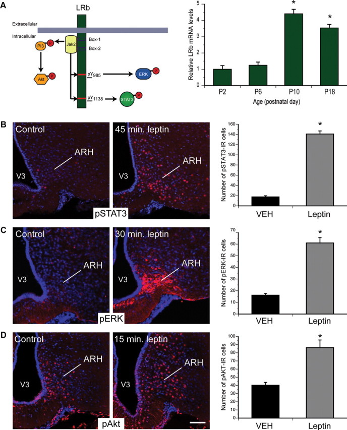
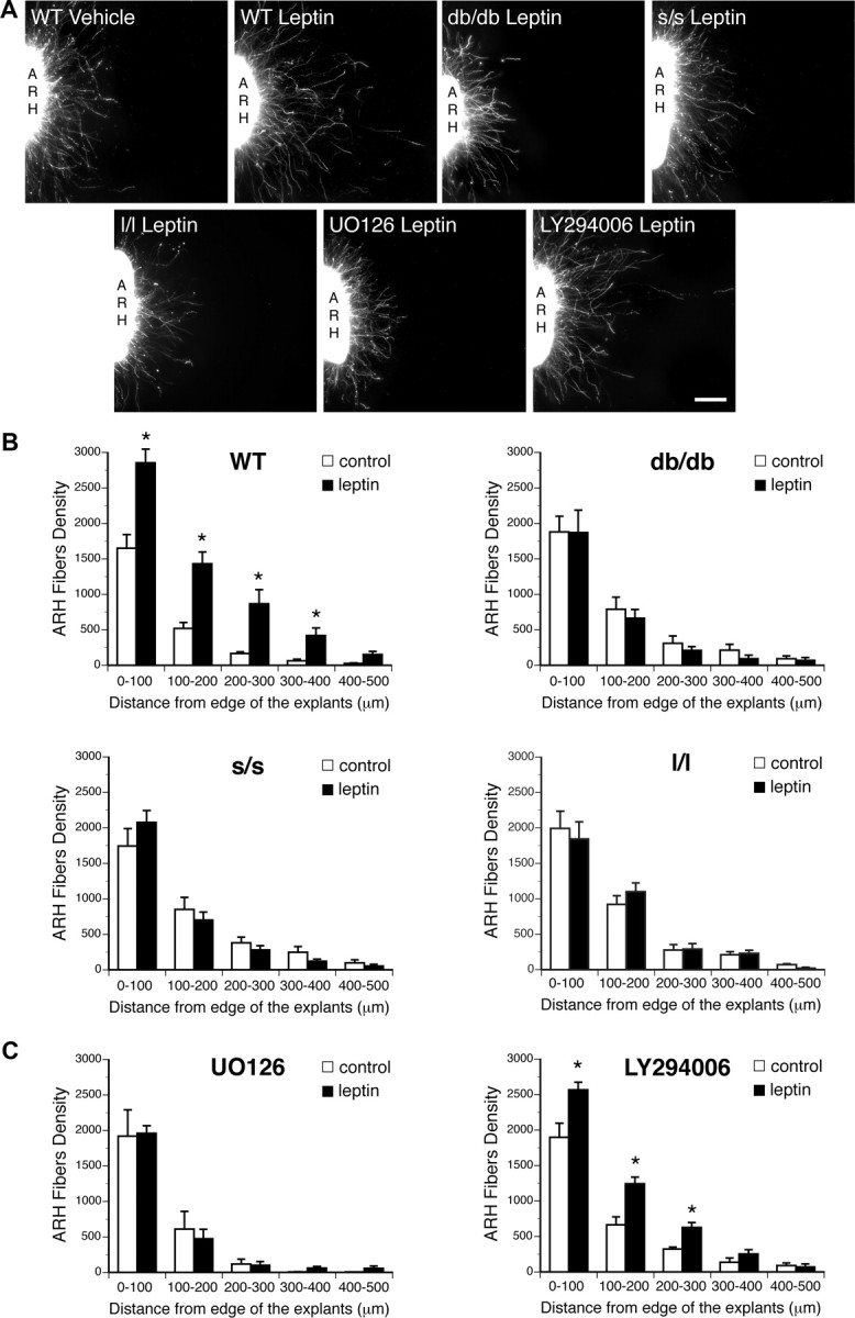
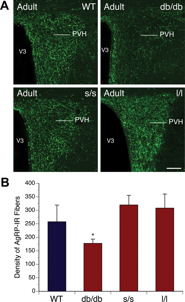
Circulating hormones influence multiple aspects of hypothalamic development and play a role in directing formation of neural circuits. Leptin is secreted by adipocytes and functions as a key developmental signal that promotes axon outgrowth from the arcuate nucleus (ARH) during a discrete developmental critical period. To determine the cellular mechanisms by which leptin impacts development of hypothalamic circuits, we examined roles for leptin receptor (LepRb) signals in neonatal mice. LepRb, ERK, and STAT3 signaling were required for leptin-stimulated neurite outgrowth from ARH explants in vitro. Neonatal mice with disrupted LepRb→ERK signaling displayed impaired ARH projections but were able to compensate by adulthood. LepRb→STAT3 signaling also plays a role in early circuit formation and controls the ultimate architecture of POMC, but not AgRP, projections. Thus, the developmental actions of leptin on feeding circuits are dependent on LepRb, and distinct signaling pathways are responsible for directing formation of NPY and POMC projections.


Figures

Figure 1.
Figure 1.
LepRb signaling in neonatal ARH neurons. A, Real-time PCR was used to compare levels of the long form of leptin receptor (LepRb) mRNA in the ARH of WT mice killed on P2, P6, P10, and P18 (n = 4 per group). B–D, Confocal images and quantitative comparisons of pSTAT3-IR, pERK-IR, and pAkt-IR cells (red fluorescence) 45 min (B), 30 min (C), and 15 min (D) after intraperitoneal administration of leptin (10 mg/kg) or vehicle alone (VEH) on P10 pups (n = 4 per group). V3, Third ventricle. The values shown are mean ± SEM. A, *p < 0.05 versus P2 and P6; B–D, *p < 0.05 versus vehicle. Scale bar, 25 μm.
Figure 2.
Figure 2.
Effect of leptin signaling blockade on leptin-induced ARH neurite outgrowth in vitro. A, B, Isolated organotypic cultures of ARH from P6 WT, db/db, s/s, and l/l mice (n = 5–7 per case) were incubated for 72 h with serum-free medium containing leptin (5 μg/ml) or lacking leptin and then immunostained with β III tubulin, a marker of neurites. A, C, Isolated organotypic cultures of ARH from P6 WT mice (n = 5–7 per case) were incubated for 72 h with serum-free medium containing leptin (5 μg/ml) or lacking leptin in the presence or absence of U0126 (10 μm) or LY294002 (5 μm) and then immunostained with β III tubulin. The values shown are mean ± SEM. *p < 0.05 between control and leptin-treated explants. Scale bar, 90 μm.
Figure 3.
Figure 3.
Development of ARH projections in mice mutant for LepRb signaling. Confocal images (A) and quantification (B) of ARH fibers, labeled with the anterograde tracer DiI, innervating the PVH of P12 WT, db/db, s/s, and l/l mice (n = 4–6 per group). The values shown are mean ± SEM. *p < 0.05 versus WT; #p < 0.05 versus db/db. Scale bar, 180 μm.
Figure 4.
Figure 4.
Leptin receptor signaling in ARH POMC neurons during postnatal development. Confocal images of pSTAT3 and pERK-IR cells (red fluorescence) 45 min (A) and 30 min (B) after intraperitoneal administration of leptin (10 mg/kg) or vehicle alone on P10 pups in which POMC-containing neurons have been labeled with the GFP (n = 4 per group). Arrows point to double-labeled neurons. Graphs represent the mean number of POMC neurons (gray bars) and mean number of double-labeled cells per hemisection after intraperitoneal administration of leptin (10 mg/kg, black bars) or vehicle alone (white bars). The values shown are mean ± SEM. *p < 0.05 versus vehicle. me, Median eminence; V3, third ventricle. Scale bar: A, 140 μm; B, 100 μm.
Figure 5.
Figure 5.
Leptin receptor signaling in ARH NPY neurons during postnatal development. Confocal images of pSTAT3-IR and pERK-IR cells (red fluorescence) 45 min (A) and 30 min (B) after intraperitoneal administration of leptin (10 mg/kg) or vehicle alone on P10 pups in which NPY-containing neurons have been labeled with the hrGFP (n = 4 per group). Arrows point to double-labeled neurons. Graphs represent the mean number of NPY neurons (gray bars) and mean number of double-labeled cells per hemisection after intraperitoneal administration of leptin (10 mg/kg, black bars) or vehicle alone (white bars). The values shown are mean ± SEM. *p < 0.05 versus vehicle. me, Median eminence; V3, third ventricle. Scale bar: A, 110 μm; B, 130 μm.
Figure 6.
Figure 6.
Arcuate AgRP neural projections in adult mice mutant for LepRb signaling. Confocal images (A) and quantification (B) of AgRP-IR fibers in the PVH of adult (P60) WT, db/db, s/s, and l/l mice (n = 4–6 per group). V3, Third ventricle. The values shown are mean ± SEM. *p < 0.05 versus db/db. Scale bar, 180 μm.
Figure 7.
Figure 7.
aMSH neural projections in adult mice mutant for LepRb signaling. Confocal images (A) and quantification (B) of aMSH-IR fibers in the PVH of adult (P60) WT, db/db, s/s, and l/l mice (n = 4–6 per group). V3, Third ventricle. The values shown are mean ± SEM. *p < 0.05 versus db/db and s/s. Scale bar, 180 μm.

References

    1. Ahima RS, Flier JS. Leptin. Annu Rev Physiol. 2000;62:413–437. - PubMed
    1. Ahima RS, Prabakaran D, Flier JS. Postnatal leptin surge and regulation of circadian rhythm of leptin by feeding. Implications for energy homeostasis and neuroendocrine function. J Clin Invest. 1998;101:1020–1027. - PMC - PubMed
    1. Bates SH, Stearns WH, Dundon TA, Schubert M, Tso AW, Wang Y, Banks AS, Lavery HJ, Haq AK, Maratos-Flier E, Neel BG, Schwartz MW, Myers MG., Jr STAT3 signalling is required for leptin regulation of energy balance but not reproduction. Nature. 2003;421:856–859. - PubMed
    1. Bjørbaek C, Buchholz RM, Davis SM, Bates SH, Pierroz DD, Gu H, Neel BG, Myers MG, Jr, Flier JS. Divergent roles of SHP-2 in ERK activation by leptin receptors. J Biol Chem. 2001;276:4747–4755. - PubMed
    1. Björnholm M, Münzberg H, Leshan RL, Villanueva EC, Bates SH, Louis GW, Jones JC, Ishida-Takahashi R, Bjørbaek C, Myers MG., Jr Mice lacking inhibitory leptin receptor signals are lean with normal endocrine function. J Clin Invest. 2007;117:1354–1360. - PMC - PubMed

Publication types