Comparative Study
. 2012 Jan 25;32(4):1253-60.
doi: 10.1523/JNEUROSCI.4652-11.2012.

Calcium binding by synaptotagmin's C2A domain is an essential element of the electrostatic switch that triggers synchronous synaptic transmission

Affiliations
Comparative Study

Calcium binding by synaptotagmin's C2A domain is an essential element of the electrostatic switch that triggers synchronous synaptic transmission

Amelia R Striegel et al. J Neurosci. .

Abstract

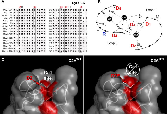
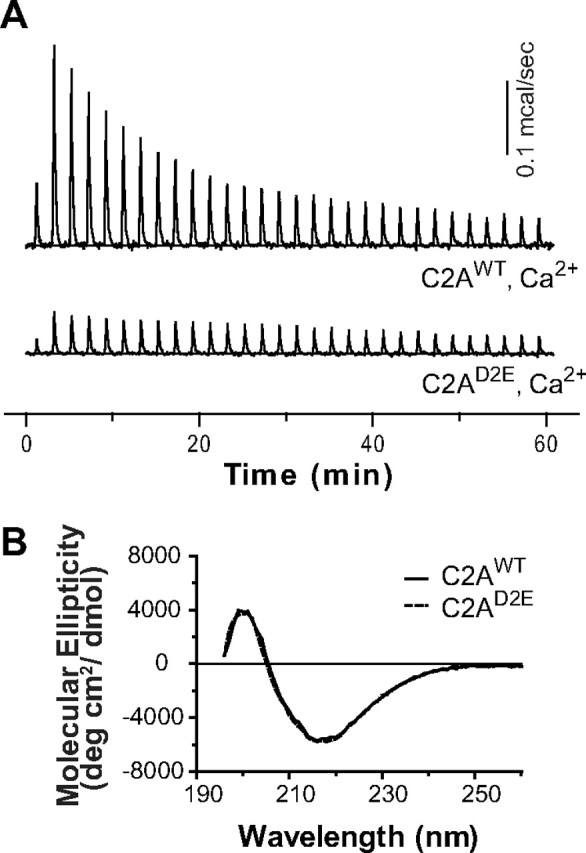
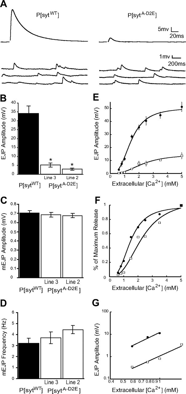
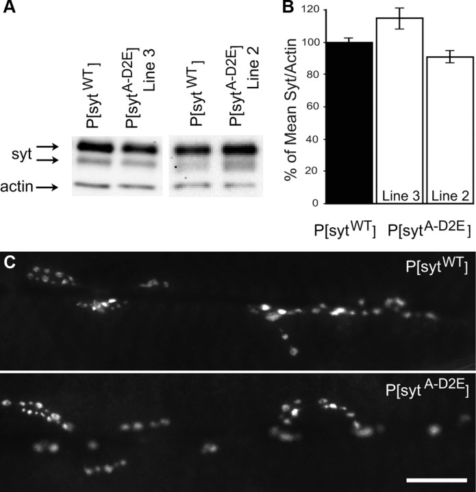
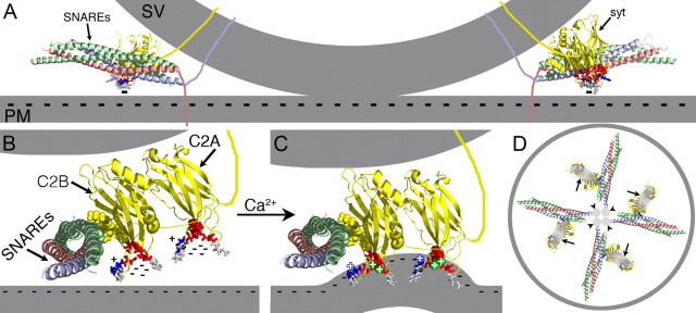
Synaptotagmin is the major calcium sensor for fast synaptic transmission that requires the synchronous fusion of synaptic vesicles. Synaptotagmin contains two calcium-binding domains: C2A and C2B. Mutation of a positively charged residue (R233Q in rat) showed that Ca2+-dependent interactions between the C2A domain and membranes play a role in the electrostatic switch that initiates fusion. Surprisingly, aspartate-to-asparagine mutations in C2A that inhibit Ca2+ binding support efficient synaptic transmission, suggesting that Ca2+ binding by C2A is not required for triggering synchronous fusion. Based on a structural analysis, we generated a novel mutation of a single Ca2+-binding residue in C2A (D229E in Drosophila) that inhibited Ca2+ binding but maintained the negative charge of the pocket. This C2A aspartate-to-glutamate mutation resulted in ∼80% decrease in synchronous transmitter release and a decrease in the apparent Ca2+ affinity of release. Previous aspartate-to-asparagine mutations in C2A partially mimicked Ca2+ binding by decreasing the negative charge of the pocket. We now show that the major function of Ca2+ binding to C2A is to neutralize the negative charge of the pocket, thereby unleashing the fusion-stimulating activity of synaptotagmin. Our results demonstrate that Ca2+ binding by C2A is a critical component of the electrostatic switch that triggers synchronous fusion. Thus, Ca2+ binding by C2B is necessary and sufficient to regulate the precise timing required for coupling vesicle fusion to Ca2+ influx, but Ca2+ binding by both C2 domains is required to flip the electrostatic switch that triggers efficient synchronous synaptic transmission.


Figures

Figure 1.
Figure 1.
The C2A domain of synaptotagmin 1 has five highly conserved aspartate residues that coordinate Ca2+. A, Alignment of C2A from Ca2+-binding synaptotagmin isoforms: synaptotagmin 1 from Drosophila (Dsyt1), bee (Asyt1), Manduca (Ma syt1), squid (Lsyt1), C. elegans (Csnt1), chicken (Gsyt1), mouse (Mu syt1), rat (Rsyt1), and human synaptotagmins (Hsyt) 1–3, 5–7, 9, and 10. Conserved residues are shown in gray and identical residues are in bold. The five conserved aspartate residues that coordinate the binding of Ca2+ ions are boxed and labeled as D1–D5. The conserved residues that mediate Ca2+-dependent interactions with negatively charged membranes are also indicated by M, R, and F. B, Schematic representation of loops 1 and 3 that form the Ca2+-binding pocket of the C2A domain. Adapted from Fernandez et al. (2001) to highlight the aspartates that coordinate Ca2+ (D1–D5) as well as the residues that interact with membranes (M, R, F). C, Molecular model of the C2A Ca2+-binding pocket illustrating the potential effect of the C2AD2E mutation. Coloring the oxygen atoms of the aspartate residues that coordinate Ca2+ by element revealed the negatively charged Ca2+-binding sites on the solvent accessible surface (red). In rat syt 1, asp178 (left, D2) participates in the coordination of the first Ca2+ (Ca1 site indicated by dotted line, see also B). Using the mutagenesis function in PyMol, we modeled the consequences of altering asp178 to a glutamate (right, D2E). We predicted that the bulging out of the solvent accessible surface (right, enlarged red bulge above white dotted line) could prevent Ca2+ binding to the Ca1 site.
Figure 2.
Figure 2.
The C2AD2E mutation inhibits Ca2+ binding by C2A without disrupting protein folding. A, ITC analysis of Ca2+ binding to the isolated C2A domain of WT and mutant Drosophila synaptotagmin 1; a representative Ca2+ titration is shown (n = 3). C2AWT bound three calcium ions (Table 1). The heat of binding of Ca2+ by C2AD2E was so small that the data could not be accurately fit. B, C2AD2E is correctly folded. The CD spectra of the mutant domain (C2AD2E) was identical to wild-type (C2AWT).
Figure 3.
Figure 3.
Synchronous evoked release is severely impaired in C2A Ca2+-binding mutants, but spontaneous release remains unchanged. A, Representative traces of EJPs and mEJPs recorded in saline containing 1.5 mm [Ca2+]. B, Mean EJP amplitude was markedly decreased in P[sytA-D2E] mutants compared with P[sytWT] controls (mean ± SEM, *p ≪ 0.001, one-way ANOVA). Neither mEJP amplitude (C, mean ± SEM, p > 0.7, one-way ANOVA) nor frequency (D, mean ± SEM, p > 0.15, one-way ANOVA) varied significantly between P[sytA-D2E] mutants compared with P[sytWT] controls. E, EJP amplitude versus [Ca2+] fit with the Hill equation. F, Ca2+ dose–response data normalized to the maximal response in each line to illustrate the decrease in apparent Ca2+ affinity in the P[sytA-D2E] mutant. G, EJP amplitudes within the nonsaturating range of Ca2+ on a double log plot demonstrate that the Ca2+ cooperativity of release is not changed in the P[sytA-D2E] mutants. A linear regression line was used to determine the slope. Error bars are SEM. Black circles indicate P[sytWT], white squares indicate P[sytA-D2E].
Figure 4.
Figure 4.
Synaptotagmin expression is similar in P[sytA-D2E] mutants compared with P[sytWT] controls. A, Representative Western blots of the CNS of third instar larvae probed with anti-synaptotagmin and anti-actin antibodies. B, Synaptotagmin:actin ratio normalized to the mean ratio of the transgenic control, P[sytWT]. There was no significant difference between genotypes (mean ± SEM, p > 0.3, one-way ANOVA; P[sytWT], n = 15; P[sytA-D2E]: line 3, n = 6; line 2, n = 7). C, Synaptotagmin is properly localized to the larval neuromuscular junction in both mutant and control transgenic synaptotagmin lines. Scale bar, 20 μm.
Figure 5.
Figure 5.
Ca2+ binding by C2A is an essential component of the electrostatic switch. The crystal structure of the core complex [PDB file 1SFC, containing syntaxin (red), SNAP-25 (green), and VAMP/synaptobrevin (blue)], the NMR structures of the C2A (PDB file 1BYN) and C2B (PDB file 1K5W) domains of synaptotagmin (yellow), and Ca2+ (green circles with plus signs) are shown to scale using PyMOL. The membranes, the transmembrane domains, and the link between C2A and C2B were added in Adobe Photoshop. A, Cross-section of a docked vesicle showing two SNARE complexes and their associated synaptotagmin molecules (syt). SC, Synaptic vesicle; PM, plasma membrane. B, One synaptotagmin/SNARE complex viewed from the site of vesicle/presynaptic membrane apposition. A Ca2+-independent docking/priming interaction between the C2B polylysine motif (yellow, space-filled residues) and SNAP-25 (green, space-filled residues) (Rickman et al., 2004; Loewen et al., 2006b) holds the C2B Ca2+-binding site immediately adjacent to the presynaptic membrane with the C2A Ca2+-binding site further removed. In the absence of Ca2+, the conserved aspartate residues (red residues: sytC2A-D2, space-filled; the rest as sticks) within the pockets create a high concentration of negative charge (cluster of minus signs), resulting in electrostatic repulsion of the presynaptic membrane that prevents any membrane interactions by the tips of the C2 domains. C, Upon Ca2+ binding, the electrostatic repulsion of the pockets is neutralized, thereby initiating the electrostatic switch: a strong attraction of the negatively charged membrane by the bound Ca2+ (green circles with plus signs) and the basic residues at the tips of Ca2+-binding pockets (blue stick residues, blue plus signs). Insertion of the hydrophobic residues (gray stick residues) at the tips of the C2 domains into the core of the presynaptic membrane then triggers fusion by promoting a local Ca2+-dependent positive curvature of the plasma membrane (Martens et al., 2007; Hui et al., 2009; Paddock et al., 2011). The C2 domain interactions with the membrane likely pull the synaptic vesicle (upper gray membrane) toward the presynaptic membrane (lower gray membrane). The Ca2+-induced increase in positive charge at the end of the C2B domain also likely increases the strength of the electrostatic interaction between the C2B polylysine motif and the SNARE complex, resulting in simultaneous binding of the SNARE complex and the presynaptic membrane (Davis et al., 1999; Bhalla et al., 2006; Loewen et al., 2006a; Dai et al., 2007). D, Membrane penetration by multiple synaptotagmins (large gray ovals, arrows) would pull the plasma membrane toward the vesicle in a ring around the SNARE transmembrane domains (small gray circles, arrowheads) facilitating fusion.

References

    1. Bai J, Wang P, Chapman ER. C2A activates a cryptic Ca2+-triggered membrane penetration activity within the C2B domain of synaptotagmin I. Proc Natl Acad Sci U S A. 2002;99:1665–1670. - PMC - PubMed
    1. Bhalla A, Chicka MC, Tucker WC, Chapman ER. Ca(2+)-synaptotagmin directly regulates t-SNARE function during reconstituted membrane fusion. Nat Struct Mol Biol. 2006;13:323–330. - PubMed
    1. Brand AH, Perrimon N. Targeted gene expression as a means of altering cell fates and generating dominant phenotypes. Development. 1993;118:401–415. - PubMed
    1. Broadie K, Bellen HJ, DiAntonio A, Littleton JT, Schwarz TL. Absence of synaptotagmin disrupts excitation-secretion coupling during synaptic transmission. Proc Natl Acad Sci U S A. 1994;91:10727–10731. - PMC - PubMed
    1. Brose N, Petrenko AG, Südhof TC, Jahn R. Synaptotagmin: a calcium sensor on the synaptic vesicle surface. Science. 1992;256:1021–1025. - PubMed

Publication types

MeSH terms

LinkOut - more resources