Comparative Study
. 2012 Jan 25;32(4):1261-72.
doi: 10.1523/JNEUROSCI.3838-11.2012.

Similar intracellular Ca2+ requirements for inactivation and facilitation of voltage-gated Ca2+ channels in a glutamatergic mammalian nerve terminal

Affiliations
Comparative Study

Similar intracellular Ca2+ requirements for inactivation and facilitation of voltage-gated Ca2+ channels in a glutamatergic mammalian nerve terminal

Kun-Han Lin et al. J Neurosci. .

Abstract

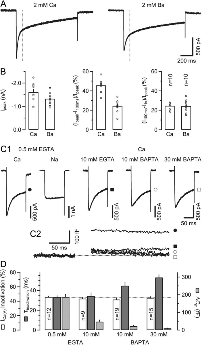
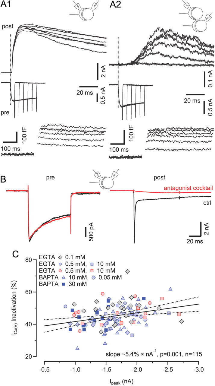
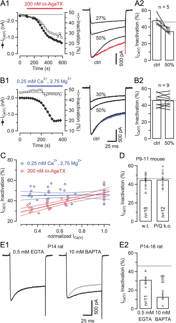
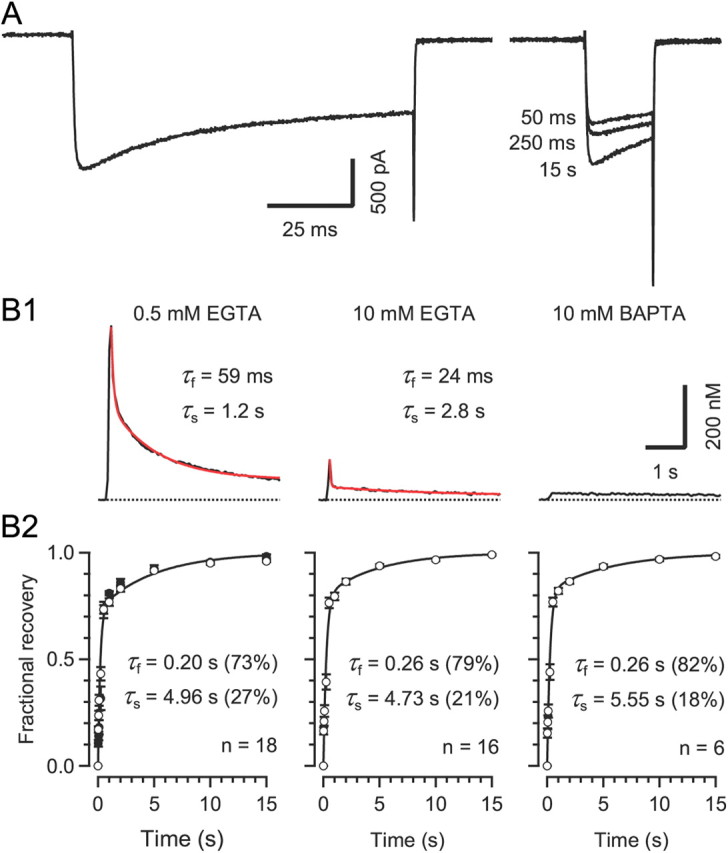
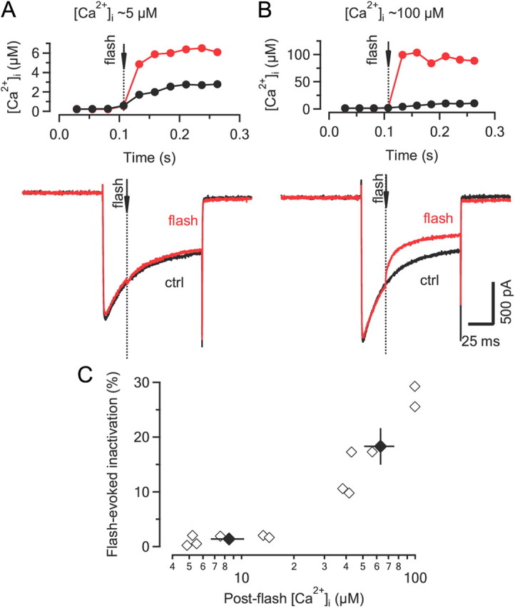
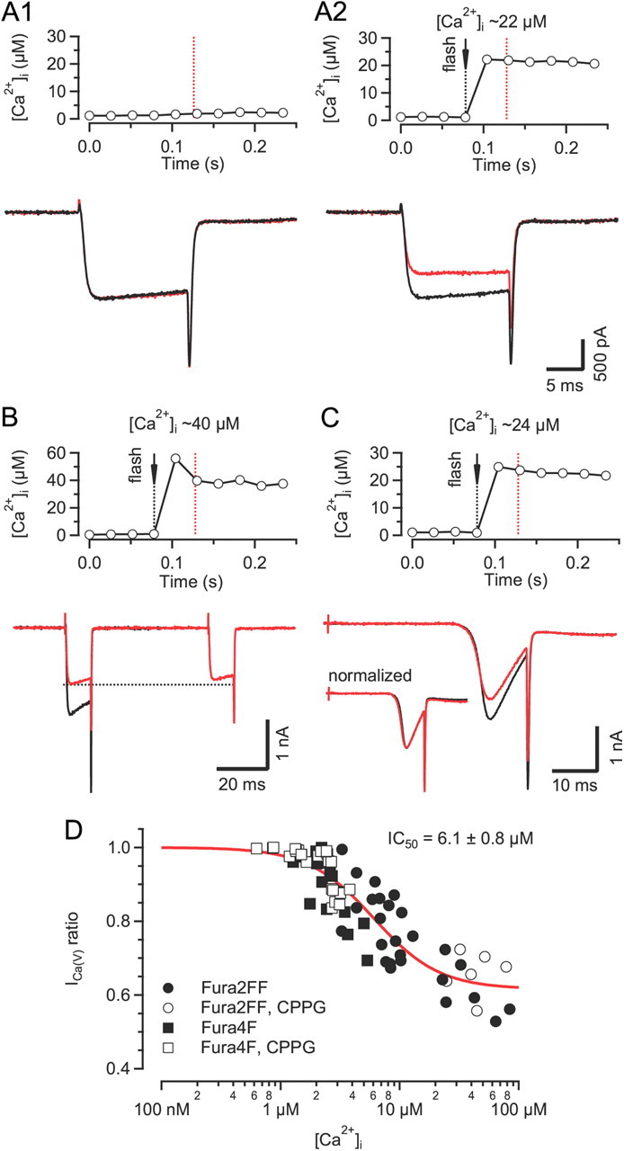
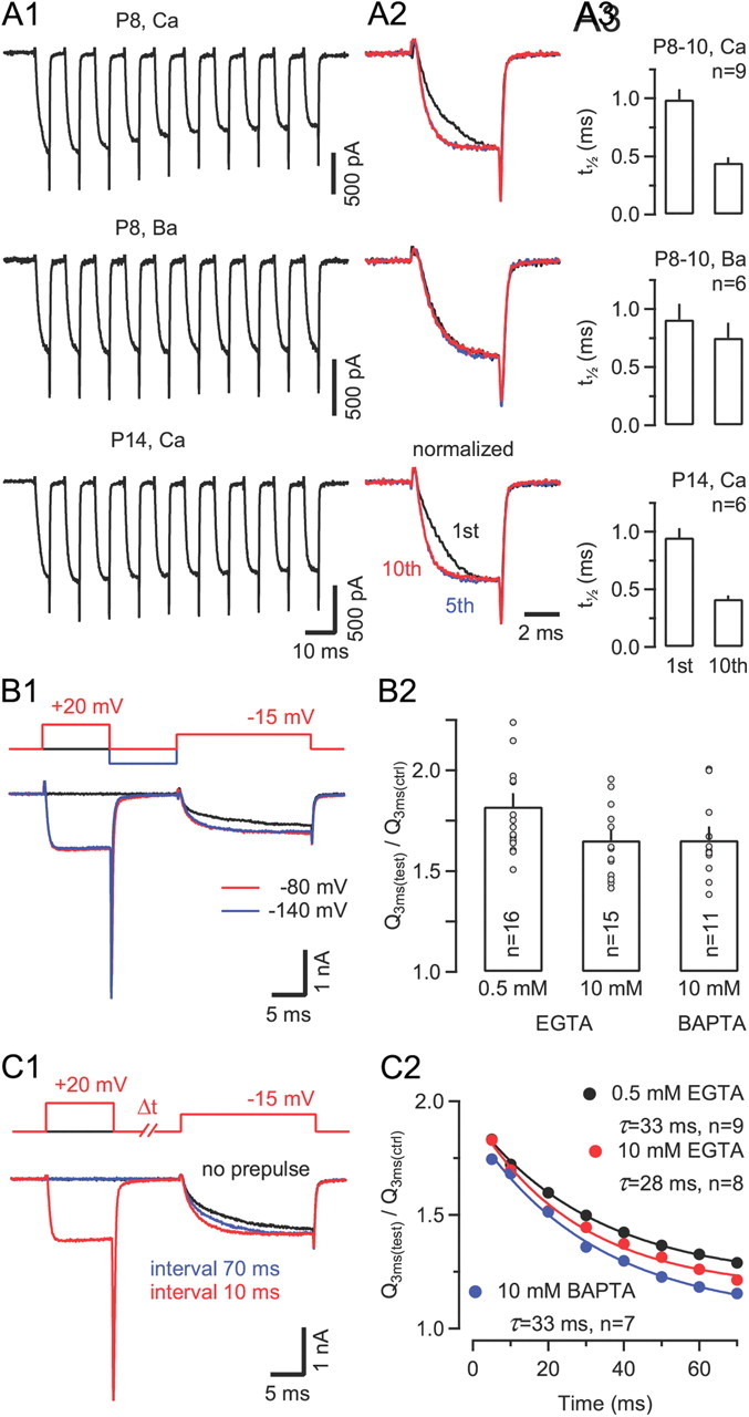
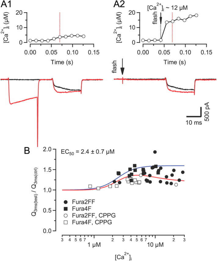
Voltage-gated Ca2+ channels (VGCCs) of the P/Q-type, which are expressed at a majority of mammalian nerve terminals, show two types of Ca2+-dependent feedback regulation-inactivation (CDI) and facilitation (CDF). Because of the nonlinear relationship between Ca2+ influx and transmitter release, CDI and CDF are powerful regulators of synaptic strength. To what extent VGCCs inactivate or facilitate during spike trains depends on the dynamics of free Ca2+ ([Ca2+]i) and the Ca2+ sensitivity of CDI and CDF, which has not been determined in nerve terminals. In this report, we took advantage of the large size of a rat auditory glutamatergic synapse--the calyx of Held--and combined voltage-clamp recordings of presynaptic Ca2+ currents (ICa(V)) with UV-light flash-induced Ca2+ uncaging and presynaptic Ca2+ imaging to study the Ca2+ requirements for CDI and CDF. We find that nearly half of the presynaptic VGCCs inactivate during 100 ms voltage steps and require several seconds to recover. This inactivation is caused neither by depletion of Ca2+ ions from the synaptic cleft nor by metabotropic feedback inhibition, because it is resistant to blockade of metabotropic and ionotropic glutamate receptors. Facilitation of ICa(V) induced by repetitive depolarizations or preconditioning voltage steps decays within tens of milliseconds. Since Ca2+ buffers only weakly affect CDI and CDF, we conclude that the Ca2+ sensors are closely associated with the channel. CDI and CDF can be induced by intracellular photo release of Ca2+ resulting in [Ca2+]i elevations in the low micromolar range, implying a surprisingly high affinity of the Ca2+ sensors.


Figures

Figure 1.
Figure 1.
Two kinetically distinct components of ICa(V) inactivation during sustained depolarizations: fast and Ca2+ dependent and slow and Ca2+ independent. A, Presynaptic ICa(V) elicited by 1 s depolarizations from Vh = −80 mV and recorded with either Ca2+ (voltage steps to 0 mV, left) or Ba2+ (voltage steps to –10 mV, right) as the charge carrier. Inactivation of ICa(V) was biphasic under both conditions. However, the rapidly inactivating component was strongly reduced in amplitude when recording in Ba2+. B, Pooled results showing average values of peak amplitudes (left) and normalized fractions of fast (middle) and slowly (right) inactivating components of ICa(V). Average amplitudes of ICa(V) decreased slightly when substituting extracellular Ca2+ with Ba2+ (p = 0.11). External Ba2+ profoundly reduced the amplitude of the fast inactivating component of ICa(V) (p < 0.001) while leaving the slowly inactivating component unaltered (p = 0.9). C, Supplementing the pipette solution with the fast Ca2+ chelator BAPTA at ≥10 mm slowed the inactivation time course but had little effect on the amount of ICa(V) inactivation during 100 ms steps to 0 mV. C1, Presynaptic ICa(V) recorded with either Ca2+ or Na+ as the charge carrier (left) and different concentrations of Ca2+ chelators in the patch pipette (right). For comparison, ICa(V) recorded in divalent free external solution is shown (second trace). C2, Corresponding ΔCm traces. Filled and empty symbols denote the same recordings as in C1. As expected, ΔCm responses were strongly attenuated in the presence of ≥10 mm intracellular Ca2+ chelator. D, Pooled results showing average values for the amount of inactivation, inactivation time constant and vesicle exocytosis. The amount of inactivation during 100 ms depolarizations was similar for all four recording conditions (p = 0.58, ANOVA), while the rate of inactivation of ICa(V) decreased with high concentrations of BAPTA in the pipette solution.
Figure 2.
Figure 2.
Neither depletion of Ca2+ from the synaptic cleft nor feedback inhibition of VGCCs via metabotropic receptors account for ICa(V) inactivation during sustained 100 ms depolarizations. A, Paired recordings from calyx terminals and synaptically coupled (A1) or neighboring (A2) MNTB principal cell somata. Vesicle exocytosis was elicited by applying step depolarizations from Vh = −80 to 0 mV of various durations (5–30 ms). Dotted lines mark the onset of presynaptic depolarizations. The corresponding ICa(V) and ΔCm responses are shown in the middle and bottom rows, respectively. Postsynaptic neurons were voltage clamped at Vh = +40 mV. Presynaptic depolarizations induced large NMDA EPSCs with short latencies in postsynaptic neurons (A1) and induced spillover NMDA EPSCs in neighboring cells (A2), indicating that synaptically released glutamate can escape the synaptic cleft and activate extrasynaptic receptors. B, Blocking ionotropic and metabotropic receptors pharmacologically does not affect ICa(V) inactivation. Paired recordings of ICa(V) (left) and EPSCs (right) in control solution (black) and after adding a mixture of glutamate and cannabinoid receptor antagonists (red; see Materials and Methods) to suppress Ca2+ influx through GluR channels and feedback inhibition via mGluRs and/or CBRs. C, Scatter plot of fractional inactivation versus peak amplitudes of ICa(V). Pooled results obtained with different intracellular Ca2+ chelator species and concentrations. Data obtained from recordings in the presence of the antagonist mixture are plotted using red symbols. Solid and broken lines represent line fit and 95% confidence limits to the entire dataset, indicating a weak positive correlation (Pearson's r = −0.30, p = 0.001, n = 115) between ICa(V) peak amplitude and degree of inactivation with a slope of ∼5.4% × nA−1.
Figure 3.
Figure 3.
Clustering of presynaptic VGCCs may account for weak sensitivity of Ca2+-dependent inactivation to intracellular Ca2+ chelators. A1, B1, Time course of peak amplitudes and fractional inactivation of ICa(V) during gradual current reduction by either bath application of the CaV2.1 blocker ω-AgaTX (A) or slow bath perfusion with a low Ca2+ solution (B) (left panels). Samples traces representing ICa(V) recorded before (ctrl, control) and after reducing its amplitude by ∼50 and ∼70% are shown superimposed in the right panels. The colored traces represents peak scaled versions of ICa(V) after ∼50% amplitude reduction. A2, B2, Comparison of ICa(V) inactivation before and after reducing ICa(V) by ∼50% by application of either ω-AgaTX (A2) or low Ca2+ solution (B2). C, Line fits to scatter plots of inactivation versus normalized peak amplitudes of ICa(V) at selected degrees of current reduction (0, ∼33, ∼50, and ∼66%) by application of low Ca2+ solution (blue) or ω-AgaTX (red). Dotted lines represent 95% confidence limits. When ω-AgaTX was applied to gradually reduce ICa(V), fractional inactivation and normalized amplitudes were highly correlated (Pearson's r = 0.83, p < 0.001). In contrast, the slope of the regression line was not significantly different from zero (p = 0.79) for the data obtained with low Ca2+ bath perfusion. All data shown in A–C were obtained with 10 mm BAPTA in the pipette solution. D, ICa(V) inactivation is unaltered in calyces of mice lacking CaV2.1 (p = 0.93) (0.5 mm EGTA in the pipette solution). w.t., Wild type; k.o., knockout. Inactivation of ICa(V) is strongly reduced in more mature calyces. Recordings from two P14 terminals with either 0.5 mm EGTA (left) or 10 mm BAPTA (right) in the patch pipette. The gray trace represents a peak scaled version of ICa(V) recorded with 0.5 mm EGTA. E2, Fractional inactivation of ICa(V) in P14–16 calyces is significantly attenuated when recorded with 10 mm BAPTA in the patch-pipette (p < 0.001). The dotted lines in A2, B2, D, and E2 indicate the mean fractional inactivation of ICa(V) in calyces of P8–10 rats with 0.5 mm EGTA in the pipette (see Fig. 1D).
Figure 4.
Figure 4.
Recovery of ICa(V) from inactivation is intrinsically slow, regardless of the Ca2+ buffering conditions A, Recovery of ICa(V) from inactivation was tested at variable intervals using a paired-pulse protocol consisting of a 100 ms depolarization to 0 mV followed by a 20 ms depolarization to 0 mV at variable interstimulus interval. Pipette solution contained 0.5 mm EGTA. Traces for three different recovery intervals are shown superimposed. B, The time course of recovery from inactivation was biphasic and insensitive to changes in Ca2+ chelator species and/or concentrations in the pipette solution. B1, [Ca2+]i transients evoked by 100 ms step depolarizations. The rise of global [Ca2+]i was nearly completely suppressed when adding 10 mm BAPTA to the pipette solution (right panel).B2, Average time course of recovery from inactivation. Solid lines represent double exponential fits. Fast and slow time constants were similar for the three [Ca2+]i buffering conditions (p ≥ 0.56, ANOVA). Relative amplitudes of fast and slow time constants are given in parentheses. The recovery time course following a shorter (50 ms) conditioning depolarization was virtually unchanged (filled symbols in the left panel).
Figure 5.
Figure 5.
The sensor mediating Ca2+-dependent inactivation ‘sees’ high [Ca2+]i and therefore must be located within molecular distance from the channel mouth. A, ICa(V) (bottom) elicited by step depolarizations to 0 mV with (red) and without (black) a UV-light flash (dotted line) delivered ∼25 ms after current onset. Changes in [Ca2+]i are illustrated in the top panel. The rate of ICa(V) inactivation was unaltered after UV-flash uncaging despite a ∼3-fold elevation of volume-averaged global [Ca2+]i relative to control conditions (without flash). B, Similar experiment as illustrated in A except that the post-flash [Ca2+]i increased to ∼100 μm. Note the immediate acceleration of the inactivation time course after Ca2+ uncaging. ctrl, Control. C, Scatter plot of flash-evoked ICa(V) inactivation as a function of global [Ca2+]i. Flash-evoked ICa(V) inactivation was quantified as the ratio of the final ICa(V) amplitude measured after a flash compared to that of a control current. Black symbols represent mean ± SEM. for post-flash [Ca2+]i of ≤15 μm and ≥40 μm. Note that for [Ca2+]i. of ≤15 μm, inactivation of ICa(V) after Ca2+ uncaging was nearly indistinguishable from that under control conditions.
Figure 6.
Figure 6.
[Ca2+]i sensitivity of ICa(V) inactivation assayed by Ca2+ uncaging via UV-flash photolysis. A, Comparison of ICa(V) elicited by two step depolarizations (20 ms to 0 mV), separated by a 20 s interval, without (A1) or with (A2) a UV-light flash delivered 50 ms before the second ICa(V) (red traces). The time course of [Ca2+]i shortly before and during the second depolarization (red dotted line) is shown in the top panel. In the absence of the UV-light flash the two currents were nearly identical, indicating that a 20 s interval was sufficient to allow full recovery from the small amount of inactivation induced during the first depolarization (A1, bottom). In contrast, a global elevation of [Ca2+]i by UV-flash photolysis induced a pronounced reduction of the second current in the same terminal (A2). B, No further reduction of ICa(V) was observed when probed with an additional depolarization 112 ms after the UV-light flash. C, Similar experiment as illustrated in A except that ICa(V) was elicited by ramping Vm from −80 to +40 mV. Note the reduced current amplitude (bottom panel) but similar I–V relationship (bottom panel inset) of the second ICa(V) (red trace) following the UV-light flash. D, Dose–response relationship of Ca2+-dependent inactivation versus global [Ca2+]i. Pooled data obtained from 75 flashes in 40 terminals. Solid line represents a Hill function fitted to the data.
Figure 7.
Figure 7.
Presynaptic ICa(V) shows robust Ca2+-dependent facilitation during prepulse protocols. A1, ICa(V) elicited by a 100 Hz train of 5 ms steps from Vh = −80 mV to −20 mV in two P8 calyces (top and middle) and in a P14 calyx (bottom). With Ca2+ as the charge carrier (top and bottom), the activation of ICa(V) strongly accelerated from the first to the fifth voltage step and remained fast during later steps. No change in activation kinetics was observed with external Ba2+ (middle). A2, First, fifth, and tenth ICa(V) shown superimposed for comparison after normalizing to the same peak amplitude. A3, ICa(V) facilitation was similar in P8–10 and P14 calyces. Average half-rise times of the first and tenth ICa(V) in the train. BC, Facilitation of ICa(V) during paired-pulse protocols. B1, Sample traces of ICa(V) elicited by stepping to −15 mV with (red trace) or without (black trace) a preceding prepulse to +20 mV (20 ms interpulse interval). Facilitation was quantified as the ratio of charges transferred during the initial 3 ms of ICa(V) with or without prepulse. Note that a hyperpolarization to −140 between prepulse and test pulse (blue trace) did not alter the amount of current facilitation. B2, Increasing intracellular Ca2+ buffer strength only slightly suppressed facilitation of ICa(V). C1, Sample traces of ICa(V) without (black trace) or with prepulse at 10 ms (red) or 70 ms interval (blue). C2, The relaxation of ICa(V) facilitation is an order of magnitude faster than recovery from inactivation and insensitive to changes in [Ca2+]i buffering strength. Solid lines represent single exponential fits with the decay time constants as indicated.
Figure 8.
Figure 8.
[Ca2+]i sensitivity of ICa(V) facilitation assayed by Ca2+ uncaging via UV-flash photolysis. A, Comparison of ICa(V) facilitation elicited by either prepulse (A1) or Ca2+ uncaging (A2) (red traces). The interval between the UV-light flash and the test depolarization was 20 ms. Control current traces without preceding prepulse or without Ca2+ uncaging are shown for comparison (black traces). Recordings were obtained from the same terminal. The time course of [Ca2+]i shortly before and during the second depolarization is shown in the top panel. B, Dose–response relationship of ICa(V) facilitation versus [Ca2+]i. Maximum facilitation was observed for [Ca2+]i elevations between 5 and 15 μm. At [Ca2+]i >20 μm, Ca2+-dependent inactivation of ICa(V) was observed. The solid red line represents a product of two Hill functions fitted to the data. The solid blue line represent the simulated dose–response relationship of Ca2+-dependent facilitation in the absence of ICa(V) inactivation.

Similar articles

Cited by

References

    1. Adams PJ, Rungta RL, Garcia E, van den Maagdenberg AM, MacVicar BA, Snutch TP. Contribution of calcium-dependent facilitation to synaptic plasticity revealed by migraine mutations in the P/Q-type calcium channel. Proc Natl Acad Sci U S A. 2010;107:18694–18699. - PMC - PubMed
    1. Benton MD, Raman IM. Stabilization of Ca current in Purkinje neurons during high-frequency firing by a balance of Ca-dependent facilitation and inactivation. Channels (Austin) 2009;3:393–401. - PMC - PubMed
    1. Borst JG, Sakmann B. Calcium current during a single action potential in a large presynaptic terminal of the rat brainstem. J Physiol. 1998a;506:143–157. - PMC - PubMed
    1. Borst JG, Sakmann B. Facilitation of presynaptic calcium currents in the rat brainstem. J Physiol. 1998b;513:149–155. - PMC - PubMed
    1. Borst JG, Sakmann B. Depletion of calcium in the synaptic cleft of a calyx-type synapse in the rat brainstem. J Physiol. 1999;521:123–133. - PMC - PubMed

Publication types

MeSH terms

LinkOut - more resources