Comparative Study
. 2012 Jan 25;32(4):1273-83.
doi: 10.1523/JNEUROSCI.4586-11.2012.

Transgenic expression of intraneuronal Aβ42 but not Aβ40 leads to cellular Aβ lesions, degeneration, and functional impairment without typical Alzheimer's disease pathology

Affiliations
Comparative Study

Transgenic expression of intraneuronal Aβ42 but not Aβ40 leads to cellular Aβ lesions, degeneration, and functional impairment without typical Alzheimer's disease pathology

Dorothee Abramowski et al. J Neurosci. .

Abstract

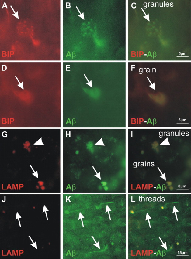
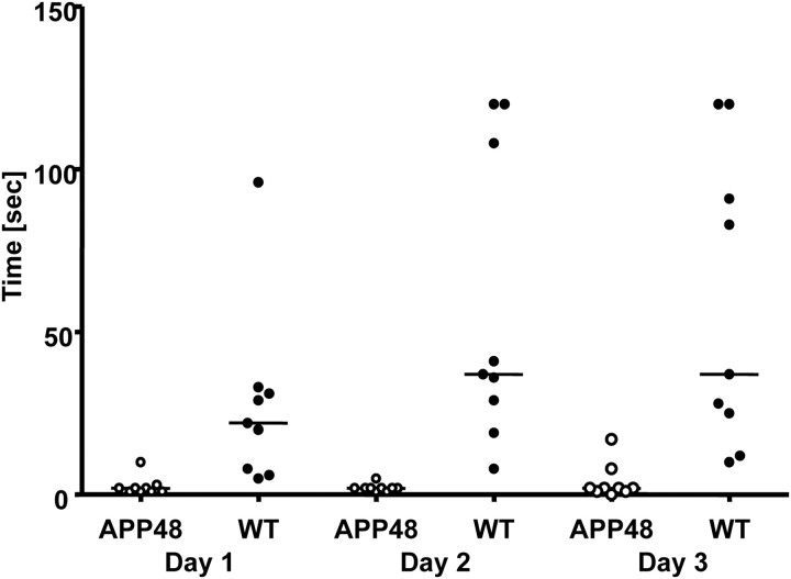
An early role of amyloid-β peptide (Aβ) aggregation in Alzheimer's disease pathogenesis is well established. However, the contribution of intracellular or extracellular forms of Aβ to the neurodegenerative process is a subject of considerable debate. We here describe transgenic mice expressing Aβ1-40 (APP47) and Aβ1-42 (APP48) with a cleaved signal sequence to insert both peptides during synthesis into the endoplasmic reticulum. Although lower in transgene mRNA, APP48 mice reach a higher brain Aβ concentration. The reduced solubility and increased aggregation of Aβ1-42 may impair its degradation. APP48 mice develop intracellular Aβ lesions in dendrites and lysosomes. The hippocampal neuron number is reduced already at young age. The brain weight decreases during aging in conjunction with severe white matter atrophy. The mice show a motor impairment. Only very few Aβ1-40 lesions are found in APP47 mice. Neither APP47 nor APP48 nor the bigenic mice develop extracellular amyloid plaques. While intracellular membrane expression of Aβ1-42 in APP48 mice does not lead to the AD-typical lesions, Aβ aggregates develop within cells accompanied by considerable neurodegeneration.


Figures

Figure 1.
Figure 1.
APP47 and APP48 transgenes and brain mRNA expression. A, Schematic representation of the APP47 and APP48 expression constructs. The box represents the translated sequence comprising the cleaved N-terminal signal sequence SPENK (signal peptide preproenkephalin) followed by Aβ1–40 or Aβ1–42. The gray C-terminal end denotes the hydrophobic amino acid stretch of Aβ, which is approximately one-half of the APP transmembrane region (located in the luminal leaflet of the membrane bilayer). B, In situ hybridization to locate the transgene expression in 2-month-old APP47 and APP48 mouse brain. C, Relative transgene mRNA levels in forebrain of female APP47, APP48, and APP47 × APP48 (47 × 48) mice as determined by quantitative PCR. Animals were 2 months of age. Differences between Aβ1–40 and Aβ1–42 mRNAs were statistically significant (Student's t test, two-tailed, p < 0.0001), whereas the same mRNAs did not differ significantly (p > 0.1) between single- and double-transgenic mice. Error bars indicate SEM.
Figure 2.
Figure 2.
Characterization of human Aβ40 and Aβ42 peptides in brain. A, Western blot of forebrain homogenates from representative Aβ-expressing mice at 2 months. Female mice are shown, but results were similar for males. Homogenates were dissolved in SDS-sample buffer and run on a SDS/urea gel to separate Aβ1–40 and Aβ1–42. The gel was overloaded to improve detection. Synthetic Aβ peptides spiked into a nontransgenic mouse brain homogenate are shown on the left. Note that Aβ1–40 and Aβ1–42 blot with different efficiency and cannot be compared directly. B, Formic acid-extracted total Aβ was quantified by an electrochemoluminescence assay (MSD). Significant differences (Student's t test, two-tailed) are indicated by asterisks: ***p < 0.001. One-way ANOVA and Tukey's test showed a significant difference between Aβ40 and Aβ42 in APP47 versus APP48 mice (p < 0.001) but not within the double-transgenic animals (47 × 48; Student's t test, two-tailed, p = 0.83). Error bars indicate SEM. C, MALDI spectra of immunoprecipitates with Aβ antibody 6E10 from brain extracts of APP47 and APP48 mice showed peaks at the expected molecular weight of Aβ1–40 and Aβ1–42, respectively. D, Forebrain homogenates of 2- and 21-month-old APP47, APP48, and APP47 × APP48 (47 × 48) mice were immunoprecipitated with Aβ antibody 6E10, separated on a SDS/urea gel, and detected by Western blotting using an N3pE (pyroglutamate; upper panel) or general (6E10; lower panel) Aβ antibody. The two faster migrating bands are indicated by arrowheads. Pyroglutamate Aβ was detected as a minor portion of the fastest band in aged double-transgenic mice only. The migration positions of synthetic standards are indicated on the right.
Figure 3.
Figure 3.
Age-dependent changes and solubility characteristics of brain Aβ in APP47 and APP48 mice. Formic acid-extracted total Aβ40 and Aβ42 in cerebral cortex was analyzed at the indicated ages using electrochemoluminescence assays. A, The Aβ42 concentration in APP48 brain significantly increased with age (Student's t test vs 1 month, two-tailed) as indicated by asterisks: *p < 0.05, **p < 0.01, ***p < 0.001. Linear regression analysis (regression ANOVA F(1,32) = 317.7, p < 0.001) indicated age as a strong and linear determinant of Aβ42. B, The Aβ40 concentration in APP47 mouse brain remained unchanged (p > 0.05, Student's t test, two-tailed, and linear regression). C, Compared with APP47, Aβ40 was considerably elevated in aged APP47 × APP48 mice, whereas Aβ42 was not consistently different from APP48 (Student's t test vs single-transgenic mice, two-tailed). Error bars indicate SEM. D, Forebrain homogenates of 1.5- and 21-month-old APP47, APP48, and APP47 × APP48 (bi) mice were sequentially extracted with 1% Triton X-100, 2% SDS, and 70% formic acid. Extracts were immunoprecipitated with antibody 4G8 and analyzed by Western blotting with antibody 6E10, both directed against Aβ. A gel without urea was used, which does not separate Aβ1–40 and Aβ1–42. The faster migrating band corresponds to the truncated Aβ isoforms separated on SDS/urea gels (see above). This band is primarily found in the Triton and SDS extracts from young mice. An extract from a young APP transgenic mouse (APP23) is shown for comparison.
Figure 4.
Figure 4.
Macroscopic analysis of brains from APP48 mice. The forebrain of APP48 (B) mice was ∼10% smaller than that of wild-type mice (A) as measured by the bi-hemispheric diameter, which is indicated as black bar below the brain section. The relative forebrain white matter volume was decreased in APP48 mice as seen morphologically in the central white matter and the corpus callosum (B, stars) and as documented by quantitative assessment (Student's t test, two-tailed, **p < 0.01; C). D, E, The spinal cord was also ∼10% smaller in APP48 mice (E) than in wild-type mice (D). However, morphological changes and especially white matter loss did not become obvious.
Figure 5.
Figure 5.
N3pEAβ staining in APP47 and APP48 mice. Neocortical sections from 18-month-old APP47 (A–C) and APP48 (D, E) mice were immunostained with antibodies specific for Aβ40 (A, D), Aβ42 (B, E), or N3pEAβ (C, F). As expected, APP47 brains were stained with Aβ40 but not Aβ42 antibodies, whereas APP48 reacted with Aβ42 but not Aβ40 antibodies. Pyroglutamate was found in neuropil Aβ grains and few dendritic Aβ treads but not in somatic Aβ granules of APP48 but not in APP47 mice. Scale bar, 15 μm.
Figure 6.
Figure 6.
Aβ lesions in APP48 mice. A, B, In the neocortex, Aβ changes were mainly located in layers II, III, V, and VI in 3- as well as in 18-month-old animals. No amyloid plaques were visible. C, At 3 months of age, Aβ antibodies predominantly stained grain-like structures, whereas thread-like material and somatic granules were less frequently found. D, In 18-month-old APP48 mice, thread-like lesions predominated, while grains and granules became less abundant (see Fig. 8 for quantification). E–G, Higher magnification of the three major Aβ accumulations in APP48 mice: dendritic Aβ threads (E) representing dendrites filled with Aβ; somatic Aβ granules (F), which are dot-like Aβ-positive structures within the perikaryon of neurons. They were distributed in the cell soma; Aβ grains (G) representing extraneuronal accumulations of Aβ. H, Double-label immunofluorescence for Aβ and MAP2 confirmed the dendritic localization of the Aβ threads. These three Aβ lesions are found throughout the gray matter of the CNS, although Aβ threads were missing in the spinal cord (I, J). I, At 5 months of age, Aβ grains were predominant (arrows). Only few interneurons with somatic granules were observed (black arrowhead). Motor neurons were free of Aβ (open arrowheads). J, At 19 months of age, somatic granules were also seen in motor neurons (arrowheads) and Aβ grains were less abundant (arrows).
Figure 7.
Figure 7.
Immuno-electron-microscopic localization of Aβ lesions and ultrastructural analyzes. A, Campbell–Switzer silver staining of dendritic Aβ threads indicated the fibrillar nature of the aggregates (arrows). B, At the immuno-electron-microscopic level, a distinct number of dendrites (example marked by the dashed line) in the neuropil of the frontocentral neocortex contained Aβ-positive material (arrows). Note that the axon did not contain Aβ. C, At higher magnification, the Aβ-positive dendritic material exhibited a fibrillar structure (arrows) consistent with the SDS resistance of an Aβ1–42 subpopulation. D, Immuno-electron microscopy showed a neuron with somatic Aβ granules. E, Higher magnification (area of the top two arrows in D) detected Aβ within lysosomes (arrows) and more rarely in the endoplasmic reticulum (arrowheads). F, Double-label immunofluorescence indicated that Aβ grains (labeled in green) were associated with CD45-positive microglial cells. G, Using immuno-electron microscopy, microglial cells were found, which exhibited lysosomal Aβ42-reactive material. H, Higher magnification of the lysosomal region outlined by arrows. I–K, Epon-embedded tissue exhibited a better structural resolution than the immuno-electron material. I, Neurons did not show obvious alterations of their subcellular organization. J, No specific changes were found in lysosomes (arrow) at higher magnification (I, frame). K, Dendrites and synapses appeared normal. Mitochondria (arrowheads) within the dendrites and axons showed no obvious changes (arrows indicate dendritic threads).
Figure 8.
Figure 8.
Localization of Aβ lesions at intracellular membrane compartments by double immunolabeling. Double-label immunohistochemistry for BIP (A, D), a marker of post-endoplasmatic reticulum compartments, and Aβ (B, E) showed colocalization of BIP and Aβ (C, F) in Aβ granules (A–C, arrow) and microglial Aβ grains (D–F, arrow). The lysosomal marker LAMP-1 (G) demonstrated a similar colocalization (I) with Aβ (H) in granules (arrowhead) and grains (arrow) as BIP, indicating their lysosomal location. Aβ labeling (K) of dendritic threads (J–L, arrows) did not colocalize (L) with the lysosomal marker LAMP-1 (J), further distinguishing Aβ threads from Aβ granules and grains.
Figure 9.
Figure 9.
Quantification of Aβ lesions and neuron numbers in APP48 mice. Dendritic Aβ threads (A), somatic Aβ granules (B), and microglia Aβ grains (C) were quantified in the frontocentral neocortex of 3- and 18-month-old APP48 mice. This analysis revealed an increase in the number of dendritic Aβ threads with age but a decrease of somatic Aβ granules and microglial Aβ grains. Stereology was used to quantify neurons in the frontal cortex (D) of 18-month-old APP48 mice compared with wild-type littermate controls, which did not show a difference (p = 0.613). The total number of neurons was reduced in hippocampus (E) at both 3 and 18 months of age. Significant differences are indicated (Student's t test, two-tailed, *p < 0.05).
Figure 10.
Figure 10.
Motor impairment of APP48 mice. APP48 mice (open circles) and littermate controls (closed circles) at the age of 5–7 months were evaluated in the Rotarod test on 3 consecutive days. The time on the rod is shown for each individual animal, and the median is indicated. In all three tests, APP48 mice stayed significantly less long on the rod than the controls (Mann–Whitney U test; trial 1, p < 0.001; trial 2, p < 0.0003; trial 3, p < 0.0007).

References

    1. Abramowski D, Wiederhold KH, Furrer U, Jaton AL, Neuenschwander A, Runser MJ, Danner S, Reichwald J, Ammaturo D, Staab D, Stoeckli M, Rueeger H, Neumann U, Staufenbiel M. Dynamics of A beta turnover and deposition in different beta-amyloid precursor protein transgenic mouse models following gamma-secretase inhibition. J Pharmacol Exp Ther. 2008;327:411–424. - PubMed
    1. Braak H. On the structure of the human archicortex. I. The cornu ammonis. A Golgi and pigmentarchitectonic study. Cell Tissue Res. 1974;152:349–383. - PubMed
    1. Braak H, Braak E. Demonstration of amyloid deposits and neurofibrillary changes in whole brain sections. Brain Pathol. 1991;1:213–216. - PubMed
    1. Calhoun ME, Wiederhold KH, Abramowski D, Phinney AL, Probst A, Sturchler-Pierrat C, Staufenbiel M, Sommer B, Jucker M. Neuron loss in APP transgenic mice. Nature. 1998;395:755–756. - PubMed
    1. Calhoun ME, Burgermeister P, Phinney AL, Stalder M, Tolnay M, Wiederhold KH, Abramowski D, Sturchler-Pierrat C, Sommer B, Staufenbiel M, Jucker M. Neuronal overexpression of mutant amyloid precursor protein results in prominent deposition of cerebrovascular amyloid. Proc Natl Acad Sci U S A. 1999;96:14088–14093. - PMC - PubMed

Publication types

MeSH terms