doi: 10.4103/1477-3163.90676.
Epub 2011 Dec 8.
Paget's disease of the breast
Affiliations
- PMID: 22279416
- PMCID: PMC3263015
- DOI: 10.4103/1477-3163.90676
Item in Clipboard
Paget's disease of the breast
J Carcinog.
2011.
Abstract
Paget's disease of the breast is a rare type of cancer of the nipple-areola complex and that is often associated with an underlying in situ or invasive carcinoma. This article provides an overview and we review the main clinicopathological and therapeutic features of mammary Paget's disease.
Keywords: Breast; Paget's disease; nipple.
Figures
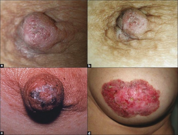
(a and b) Paget's disease of the nipple. The clinical appearance is usually a thickened, eczematoid crusted lesion with irregular borders. (c) Scaly, erythematous, crusty pigmentation and thickened plaques on the nipple, spreading to the surrounding areolar areas. (d) Advanced lesions show skin thickening, redness, erythema, erosion of the nipple and scaling around the nipple–areola
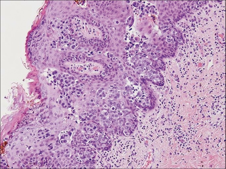
Paget's disease of the nipple. Nests and groups of malignant Paget's cells predominantly involving the lower layers of the epidermis. Epidermis may be eroded and hyperplastic (H and E, ×40)
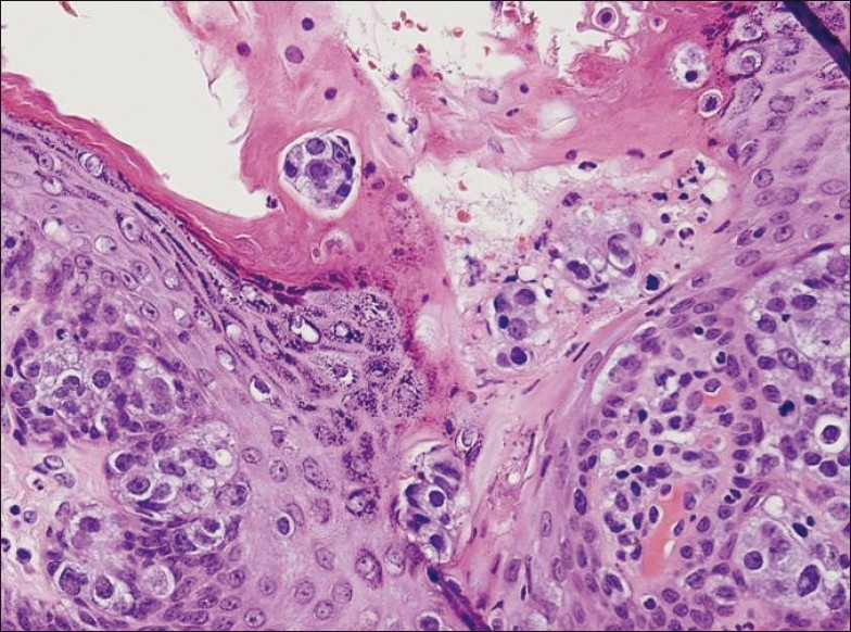
Paget's disease of the nipple with superficial ulceration. The tumor cells have abundant pale cytoplasm, pleomorphic and hyperchromatic nuclei with prominent nucleoli. Mitotic figures are seen (H and E, ×100)
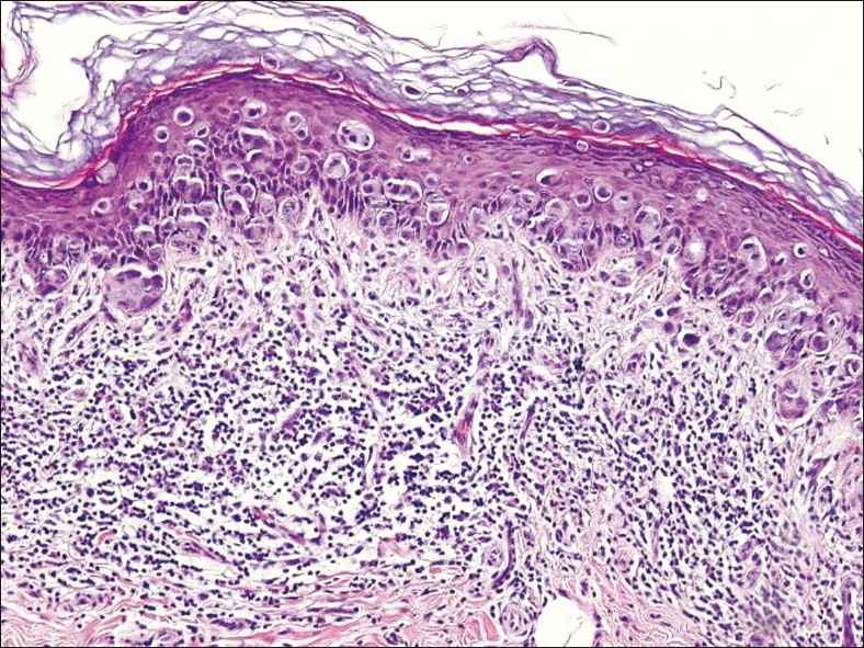
Carcinoma cells form a band in the deep epidermis and they are scattered individually throughout the squamous epithelium. The lacunar arrangement of carcinoma cells is commonly seen in Paget's disease. An extensive lymphocytic infiltrate with involvement largely concentrated in the deep epidermis (H and E, ×40)
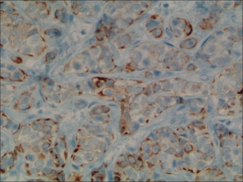
Paget's cells are highlighted by an immunostain for (CK7, ×200)
Similar articles
-
Paget's disease of the breast.Cancer Treat Rev. 2001 Feb;27(1):9-18. doi: 10.1053/ctrv.2000.0203. Cancer Treat Rev. 2001. PMID: 11237774 Review.
-
Paget's disease of the breast: a clinical perspective.Langenbecks Arch Surg. 2001 Nov;386(6):444-50. doi: 10.1007/s004230100250. Epub 2001 Oct 17. Langenbecks Arch Surg. 2001. PMID: 11735019 Review.
-
[Pathology of the nipple-areola complex : I. Paget's disease of the nipple, variants, and differential diagnoses].Pathologe. 2020 Jul;41(4):393-399. doi: 10.1007/s00292-020-00772-1. Pathologe. 2020. PMID: 32405655 Review. German.
-
Paget's disease of the nipple in a population based cohort.Breast Cancer Res Treat. 2008 Sep;111(2):313-9. doi: 10.1007/s10549-007-9783-5. Epub 2007 Oct 19. Breast Cancer Res Treat. 2008. PMID: 17952590
-
Mammary and extramammary Paget's disease.J Eur Acad Dermatol Venereol. 2007 May;21(5):581-90. doi: 10.1111/j.1468-3083.2007.02154.x. J Eur Acad Dermatol Venereol. 2007. PMID: 17447970 Review.
Cited by
-
Paget's Disease of Nipple in Male Breast with Cancer.J Clin Diagn Res. 2016 Feb;10(2):PD14-6. doi: 10.7860/JCDR/2016/17778.7217. Epub 2016 Feb 1. J Clin Diagn Res. 2016. PMID: 27042526 Free PMC article.
-
A novel web-based prognostic nomogram and the features influencing the curative effect of chemotherapy and radiotherapy for Paget's disease with invasive ductal carcinoma.Am J Cancer Res. 2023 Oct 15;13(10):4508-4530. eCollection 2023. Am J Cancer Res. 2023. PMID: 37970339 Free PMC article.
-
A tale of three common nipple diseases.Proc (Bayl Univ Med Cent). 2022 Feb 4;35(3):354-356. doi: 10.1080/08998280.2022.2027195. eCollection 2022. Proc (Bayl Univ Med Cent). 2022. PMID: 35518793 Free PMC article.
-
Clinicopathological characteristics and survival outcomes in Paget disease: a SEER population-based study.Cancer Med. 2018 Jun;7(6):2307-2318. doi: 10.1002/cam4.1475. Epub 2018 May 2. Cancer Med. 2018. PMID: 29722170 Free PMC article.
-
Managing Necrotizing Soft-Tissue Infection in Breast Cancer: A Case of Emergency Toilet Mastectomy.Am J Case Rep. 2025 May 12;26:e946669. doi: 10.12659/AJCR.946669. Am J Case Rep. 2025. PMID: 40350661 Free PMC article.
References
-
- Paget J. On the disease of the mammary areola preceding cancer of the mammary gland. St Bartholomews Hosp Rep. 1874;10:87–9.
-
- Tavassoli FA. Norwalk, Connecticut: AppletonandLange; 1999. Pathology of the breast; pp. 731–60.
-
- Sakorafas GH, Blanchard K, Sarr MG, Farley DR. Paget's disease of the breast. Cancer Treat Rev. 2001;27:9–18. - PubMed
-
- Kanitakis J. Mammary and extramammary Paget's disease. J Eur Acad Dermatol Venereol. 2007;21:581–90. - PubMed
-
- Martin VG, Pellettiere EV, Gress D, Miller AW. Paget's disease in an adolescent arising in a supernumerary nipple. J Cutan Pathol. 1994;21:283–6. - PubMed
LinkOut - more resources
Full Text Sources