. 2012;7(1):e29995.
doi: 10.1371/journal.pone.0029995. Epub 2012 Jan 18.

TRAPPC9 mediates the interaction between p150 and COPII vesicles at the target membrane

Affiliations

TRAPPC9 mediates the interaction between p150 and COPII vesicles at the target membrane

Min Zong et al. PLoS One. 2012.

Abstract

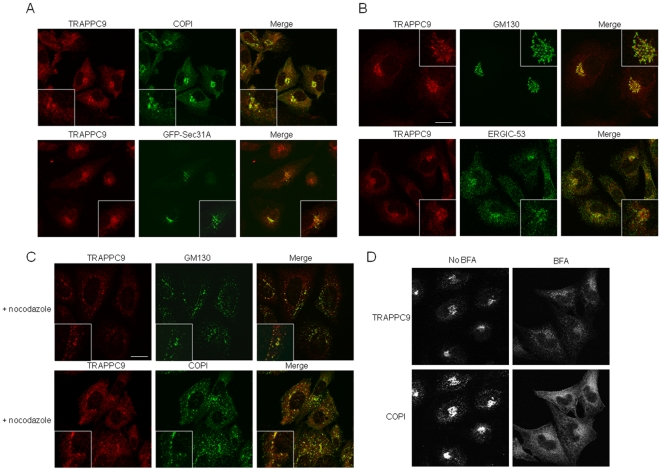
Background: The transport of endoplasmic reticulum (ER)-derived COPII vesicles toward the ER-Golgi intermediate compartment (ERGIC) requires cytoplasmic dynein and is dependent on microtubules. p150(Glued), a subunit of dynactin, has been implicated in the transport of COPII vesicles via its interaction with COPII coat components Sec23 and Sec24. However, whether and how COPII vesicle tether, TRAPP (Transport protein particle), plays a role in the interaction between COPII vesicles and microtubules is currently unknown.

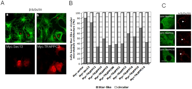
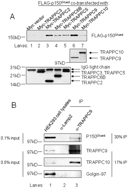
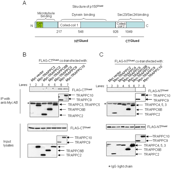
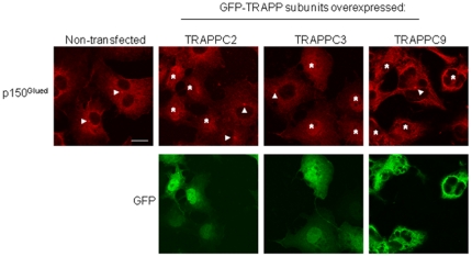
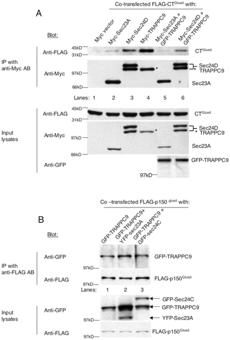
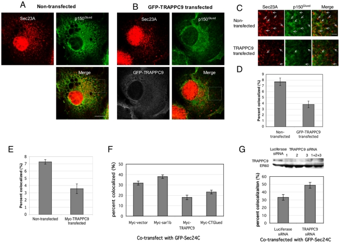
Principle findings: We address the functional relationship between COPII tether TRAPP and dynactin. Overexpressed TRAPP subunits interfered with microtubule architecture by competing p150(Glued) away from the MTOC. TRAPP subunit TRAPPC9 bound directly to p150(Glued) via the same carboxyl terminal domain of p150(Glued) that binds Sec23 and Sec24. TRAPPC9 also inhibited the interaction between p150(Glued) and Sec23/Sec24 both in vitro and in vivo, suggesting that TRAPPC9 serves to uncouple p150(Glued) from the COPII coat, and to relay the vesicle-dynactin interaction at the target membrane.

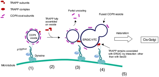
Conclusions: These findings provide a new perspective on the function of TRAPP as an adaptor between the ERGIC membrane and dynactin. By preserving the connection between dynactin and the tethered and/or fused vesicles, TRAPP allows nascent ERGIC to continue the movement along the microtubules as they mature into the cis-Golgi.


Conflict of interest statement

Competing Interests: The authors have declared that no competing interests exist.

Figures

Figure 1
Figure 1. TRAPPC9 is predominantly enriched in cis-Golgi and ERGIC.
CHO cells were stained with TRAPPC9 and various markers by indirect immunofluorescence. TRAPPC9 were detected by affinity purified rabbit IgG specific to TRAPPC9, followed by Alexa Fluor 568- conjugated goat anti rabbit secondary antibody. Organelle markers are detected by mouse monoclonal antibodies against the indicated proteins, followed by Alexa Fluor 488 conjugated goat anti- mouse secondary antibody. The extent of colocalization is presented in the merge images. The boxed areas are enlarged as insets. (A). TRAPPC9 (red) extensively colocalizes with COPI (green), which is enriched in the ERGIC and cis Golgi, and partially with transfected GFP-Sec31. (B). TRAPPC9 also extensively colocalizes with cis Golgi marker, GM130, and partially with ERGIC (ERGIC-53). (C). CHO cells treated with nocodazole for 1 h before fixed and stained with indicated antibodies. Small puncta derived from the dispersal of the Golgi membranes showed extensive overlap of TRAPPC9 signal (red) with GM130 (green, top panels) and COPI (green, bottom panels). (D). CHO cells treated with or without 10 µg/ml BFA for 1 hours before staining with antibodies against TRAPPC9 (top panels) or COPI (bottom panels). Scale bar = 20 µm.
Figure 2
Figure 2. Overexpression of TRAPP subunits disrupts the microtubule “star-like” astral architecture in COS cells.
(A) COS cells overexpressing the indicated Myc-tagged proteins are shown in the lower panels and the same cells are marked by red asterisks in the upper panels. Myc-tagged proteins were stained with Rhodamine-conjugated mouse anti-Myc antibody. Microtubules were stained with mouse antibody against β-tubulin, followed by Alexa Fluor 488 conjugated goat anti mouse secondary antibody. (B) Quantification of the status of microtubule architectures in COS cells overexpressing various TRAPP subunits. In all samples, at least 100 cells were counted. (C) TRAPP overexpression did not disrupt the MTOC. COS cells transfected with the indicated TRAPP subunits were stained with MTOC marker γ-tubulin (red). The integrity of the MTOC can be visualized by a single intense fluorescent dot (arrows). Scale bar = 20 µm (A);  = 10 µm (C).
Figure 3
Figure 3. p150Glued interacts with TRAPP.
(A) The indicated myc-tagged TRAPP subunits were overexpressed with FLAG-tagged p150Glued in COS cells. Co-immunoprecipitations were performed using antibody against c-Myc. The presence of FLAG-p150Glued in the pull-down is detected by immunoblotting with antibody against FLAG (top panel). The extent of Myc-tagged TRAPP subunits precipitated was determined in immunoblotting using antibody against c-Myc (middle and bottom panels). Approximately 1% of the input lysates and 25% of the immunoprecipitants were loaded. (B) HEK293 cell lysates were subjected to immunoprecipitation using antibody specific to TRAPPC9 or to LAMP2 (negative control). The presence of TRAPPC9 (top panel), p150Glued (second panel), and TRAPPC10 (third panel) in the immunoprecipitants were detected by immunoblotting using antibodies specific to these proteins. As negative control, Golgin-97 was not detected in the immunoprecipitants (bottom panel). For TRAPPC9, TRAPPC10 and Golgin-97, approximately 0.5% of the input lysates and 17% of the immunoprecipitants were loaded. For p150Glued, approximately 0.1% of the input lysates and 30% of the immunoprecipitants were loaded.
Figure 4
Figure 4. TRAPPC9 interacts with CTGlued.
(A) Schematic diagram of p150Glued structure. NTGlued and CTGlued were subjected to co-immunoprecipitation experiments. FLAG-tagged CTGlued (B) and FLAG-tagged NTGlued (C) were co-transfected with various Myc-tagged TRAPP subunits for immunoprecipitation. The extent of Myc-TRAPP subunit precipitation was determined by immunoblotting with anti-Myc antibody (middle panels). The presence of co-precipitated FLAG-CTGlued or FLAG-NTGlued was determined by anti-FLAG antibody (top panels). The level of protein expression in input lysates was determined by anti-FLAG or anti-Myc antibody (bottom panels). The precipitated anti-Myc IgG light chain was marked as asterisk in the immunoprecipitation samples. Approximately 1% of the input lysates and 25% of the immunoprecipitants were loaded.
Figure 5
Figure 5. Overexpression of TRAPP subunits disrupts the MTOC localization of endogenous p150Glued.
COS cells transfected with the indicated GFP tagged TRAPP subunits (bottom panels) were stained with endogenous p150Glued. The presence of p150Glued signal at the MTOC can be observed in the non-transfected cells (Arrowheads). In cells transfected with the indicated TRAPP subunits, (asterisks, top panels), the signal of p150Glued at the MTOC disappeared. Scale bar = 20 µm.
Figure 6
Figure 6. TRAPPC9 competes with Sec23 and Sec24 for binding to p150Glued.
(A) Myc-Sec23 (lanes 2 and 5) or -Sec24 (lanes 3 and 6) were co-transfected FLAG-CTGlued for co-immunoprecipitation experiments. The ability of TRAPPC9 to inhibit the interaction between Sec23 or Sec24 with CTGlued was determined by the presence of GFP-TRAPPC9 (lanes 5–6). The amount of FLAG-CTGlued co-precipitated was determined by immunoblotting with anti-FLAG antibody (top panel). The amount of Myc-tagged protein precipitated was determined by blotting with anti-Myc antibody (second panel). Levels of protein expression in the transfected cell lysates were determined by blotting the input lysates with the indicated antibodies (bottom three panels). (B) Transfected Sec23 or Sec24 lost the ability to bind to p150Glued in the presence of co-transfected TRAPPC9. Transfected FLAG-p150Glued was precipitated by antibody against FLAG (second panel). The presence of co-precipitated YFP-Sec23A, GFP-Sec24C or GFP-TRAPPC9 was detected by antibody against GFP (top panel). Only TRAPPC9, but not Sec23 or Sec24, could be co-precipitated with FLAG-p150Glued. The levels of protein expression in the transfected cell lysates were determined by blotting the input lysates with the indicated antibodies (bottom two panels). In both experiments shown in (A) and (B), approximately 1% of input lysates and 25% of the immunoprecipitants were loaded.
Figure 7
Figure 7. Overexpression of TRAPPC9 reduces the association of p150Glued and ER exit sites in vivo.
Non-transfected (A) or GFP-TRAPPC9-transfected (B) COS cells, were treated with nocodazole before staining with p150Glued and Sec23A. p150Glued signal was scanned by confocal microscopy at 633 nm and pseudo-colored in green. Sec23A was scanned at 543 nm and visualized in red; GFP-TRAPPC9 signal was scanned at 488 nm and visualized in white. Scale bar = 20 µm. The boxed areas are enlarged and presented in (C). Fluorescent dots of p150Glued and Sec23A that are colocalized are indicated by arrows. Scale bar = 5 µm. (D) Quantifications of the colocalized signals in (C) as percentages of Sec23A-positive fluorescent dots that colocalized with p150Glued-positive dots. In non-transfected cells, 7.72% (S.E.M. = 0.648%) of the Sec23A dots were found to colocalize with p150Glued. A total of 890 Sec23A fluorescent dots in 8 cells were counted. In GFP-TRAPPC9 transfected cells, 3.83% (S.E.M. = 0.577%) Sec23A dots colocalized with p150Glued. 493 dots in 7 cells were analyzed. (E) Myc-TRAPPC9, instead of GFP-TRAPPC9, was transfected into COS cells. Sec23A was labeled with AF488-conjugated secondary antibody, p150Glued was labeled with AF633-conjugated secondary antibody, and Myc-TRAPPC9 was labeled with TRITC-conjugated mouse anti-Myc antibody. In 11 non-transfected cells analyzed, a total of 1571 Sec23A-positive dots were analyzed and 7.27% (S.E.M. = 0.313%) colocalized with p150Glued-positive dots. In comparison, 3.56% (S.E.M. = 0.665%) colocalization was observed in Myc-TRAPPC9 transfected cells. 1087 Sec23A-positive dots in 10 transfected cells were analyzed. (F) Hela cells co-transfected with GFP-Sec24C and the indicated sequences in Myc-tagged vector. Endogenous p150Glued was labeled with AF633-conjugated secondary antibody and the overexpressed Myc-tagged proteins were labeled with rhodamine-conjugated mouse anti-c-Myc IgG. Colocalized signals between p150Glued and GFP-Sec24C were counted as above. Fluorescence signals from at least six cells were counted, with totally 262 dots or more for each sample. (G) HEK293 cells were transfected with Luciferase siRNA or three combined TRAPPC9 siRNAs for 72 hrs. Three TRAPPC9-specific siRNAs efficiently depleted the target protein as shown by immunoblotting (top panels). TRAPPC9 depletion significantly increased the co-localization of GFP-Sec24C and p150Glued from 33.15% (S.E.M. = 0.369%) to 48.74% (S.E.M. = 0.22%). In this experiment, 473 fluorescent dots in 10 cells (control luciferase knockdown), and 494 dots in 11 cells (TRAPPC9 knockdown) were analyzed. Error bars = S.E.M.
Figure 8
Figure 8. Schematic diagram depicting how TRAPP and p150Glued coordinate the movement and tethering of a COPII vesicle is tethered at the ERGIC.
(1). Dyneine-powered movement of a fully coated COPII vesicle along microtubule is mediated by an interaction between p150Glued and Sec23/Sec24. (2). TRAPP subunits assemble onto the vesicle to form a fully functional TRAPP complex during the movement. (3). When the vesicle reaches the ERGIC, COPII coat will be partially shed to facilitate vesicle fusion but the interaction between p150Glued and the vesicle is now mediated by the TRAPPC9-p150Glued interaction. (4). TRAPP is anchored on the target membrane by a currently unknown mechanism. (5). The vesicle may fuse with the ERGIC but the TRAPPC9-p150Glued interaction continues to help move the ERGIC towards the cis-Golgi.

Similar articles

Cited by

References

    1. Wehland J, Henkart M, Klausner R, Sandoval IV. Role of microtubules in the distribution of the Golgi apparatus: effect of taxol and microinjected anti-alpha-tubulin antibodies. Proceedings of the National Academy of Sciences of the United States of America. 1983;80:4286–4290. - PMC - PubMed
    1. Rogalski AA, Bergmann JE, Singer SJ. Effect of microtubule assembly status on the intracellular processing and surface expression of an integral protein of the plasma membrane. The Journal of Cell Biology. 1984;99:1101–1109. - PMC - PubMed
    1. Rogalski AA, Singer SJ. Associations of elements of the Golgi apparatus with microtubules. J Cell Biol. 1984;99:1092–1100. - PMC - PubMed
    1. Corthesy-Theulaz I, Pauloin A, Rfeffer SR. Cytoplasmic dynein participates in the centrosomal localization of the Golgi complex. J Cell Biol. 1992;118:1333–1345. - PMC - PubMed
    1. Roghi C, Allan VJ. Dynamic association of cytoplasmic dynein heavy chain 1a with the Golgi apparatus and intermediate compartment. J Cell Sci. 1999;112:4673–4685. - PubMed

Publication types

MeSH terms