. 2012;7(1):e30341.
doi: 10.1371/journal.pone.0030341. Epub 2012 Jan 18.

CNS penetration of intrathecal-lumbar idursulfase in the monkey, dog and mouse: implications for neurological outcomes of lysosomal storage disorder

Affiliations

CNS penetration of intrathecal-lumbar idursulfase in the monkey, dog and mouse: implications for neurological outcomes of lysosomal storage disorder

Pericles Calias et al. PLoS One. 2012.

Abstract

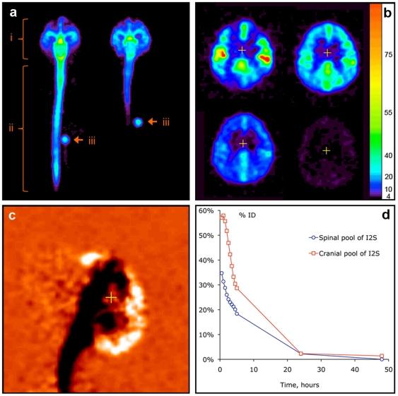
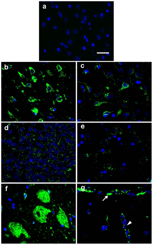
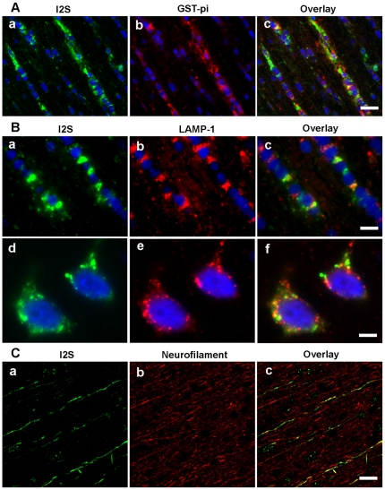
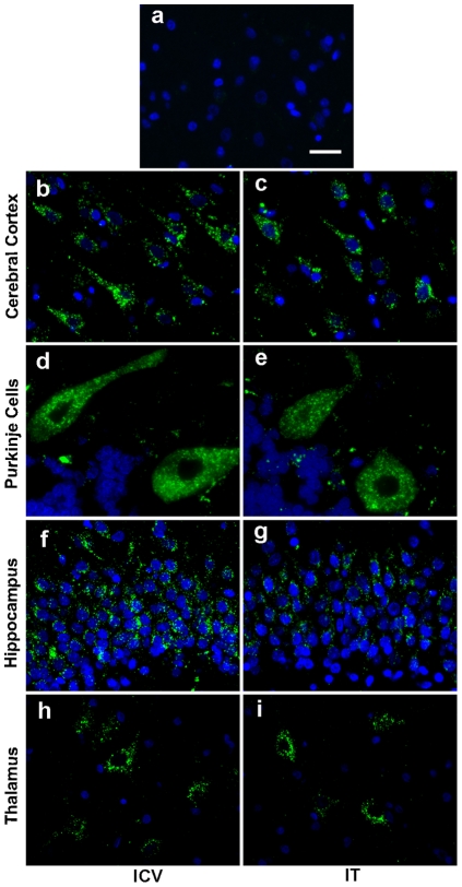
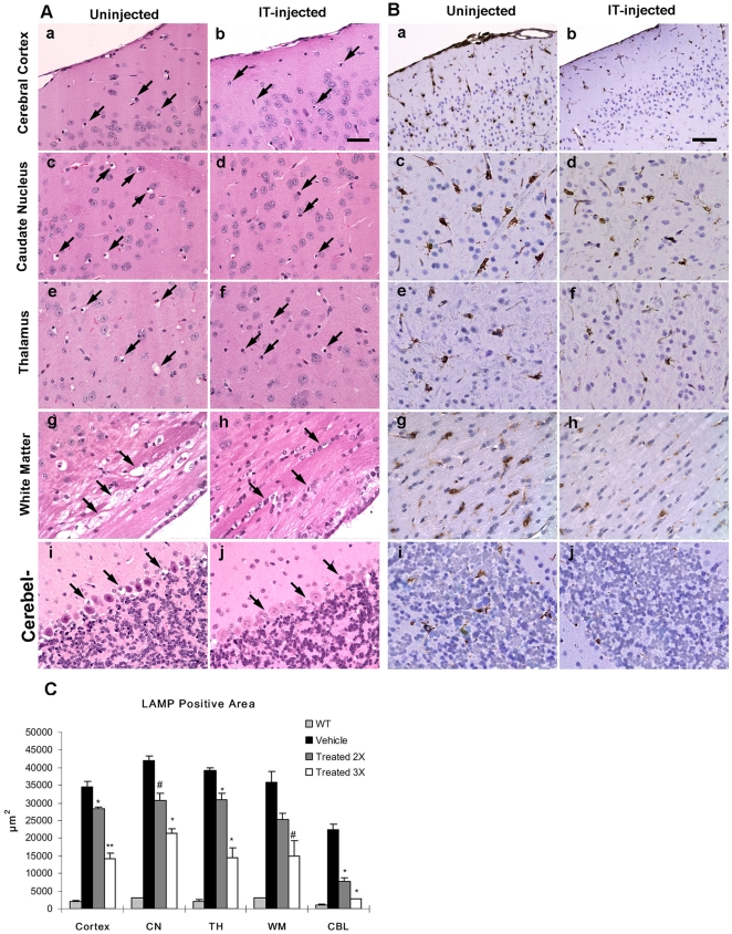
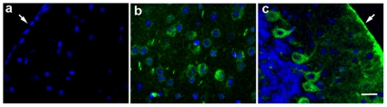
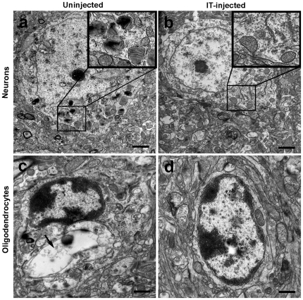
A major challenge for the treatment of many central nervous system (CNS) disorders is the lack of convenient and effective methods for delivering biological agents to the brain. Mucopolysaccharidosis II (Hunter syndrome) is a rare inherited lysosomal storage disorder resulting from a deficiency of iduronate-2-sulfatase (I2S). I2S is a large, highly glycosylated enzyme. Intravenous administration is not likely to be an effective therapy for disease-related neurological outcomes that require enzyme access to the brain cells, in particular neurons and oligodendrocytes. We demonstrate that intracerebroventricular and lumbar intrathecal administration of recombinant I2S in dogs and nonhuman primates resulted in widespread enzyme distribution in the brain parenchyma, including remarkable deposition in the lysosomes of both neurons and oligodendrocytes. Lumbar intrathecal administration also resulted in enzyme delivery to the spinal cord, whereas little enzyme was detected there after intraventricular administration. Mucopolysaccharidosis II model is available in mice. Lumbar administration of recombinant I2S to enzyme deficient animals reduced the storage of glycosaminoglycans in both superficial and deep brain tissues, with concurrent morphological improvements. The observed patterns of enzyme transport from cerebrospinal fluid to the CNS tissues and the resultant biological activity (a) warrant further investigation of intrathecal delivery of I2S via lumbar catheter as an experimental treatment for the neurological symptoms of Hunter syndrome and (b) may have broader implications for CNS treatment with biopharmaceuticals.


Conflict of interest statement

Competing Interests: The authors have read the journal's policy and have the following conflicts: PC, JP, YH, MA, NL, NS, JL, JLP and MWH are full-time employees of Shire Human Genetic Therapies, Inc. MP, VB and AJF have no competing interests to declare. This does not alter the authors′ adherence to all the PLoS ONE policies on sharing data and materials.

Figures

Figure 1
Figure 1. In vivo distribution of 124I -labeled I2S (3 mg/animal) in cynomolgus monkeys by PET.
(A) Distribution of I2S administered through the lumbar (left) and ICV (right) catheters 30 minutes after the administration as demonstrated by a projection PET image (sum of all slices). Relative linear color scale. (B) The distribution of I2S in the brain at 0.5, 2.5, 5 and 24 hours after lumbar administration; PET image, 1.2 mm slice through the corpus callosum region in the plane parallel to the occipital bone. The color scale is calibrated in µg/ml of I2S. (C) Changes in the cerebral I2S distribution between 0.5 and 5 hours after lumbar administration shown in monochrome linear color scale. The image was obtained by subtraction of the quantitative data matrix obtained at 5 hours from the one obtained at 0.5 hours. Neutral orange color represents no change. Clearance of I2S from the CSF is seen as black, and accumulation in the parenchyma and arachnoid as white color. (D) An example of single-animal dynamics of I2S clearance from the leptomeningeal compartment and CNS.
Figure 2
Figure 2. Cellular uptake of I2S in the neurons and vascular cells of the brain of monkeys.
(A) I2S immunohistochemical staining of the cerebral cortex of vehicle control monkeys was negative. Representative images showing I2S following 6 monthly IT injections of 100 mg/dose detected in the neurons by immunohistochemistry in the cerebral cortex (B), thalamus (C), hippocampus (D), caudate nucleus (E) and spinal cord (F). (G) Meningeal cells (arrow) covering the surface of the cerebral cortex and perivascular cells surrounding the blood vessel (V) were also positive for I2S. Green, I2S; blue, 4′-6-diamidino-2-phenylindole (DAPI)-stained nuclei. Scale bar: 25 µm.
Figure 3
Figure 3. I2S is detected within the lysosomes of oligodendrocytes and in the axons of white matter.
(A) Representative images showing I2S uptake in oligodendrocytes in the white matter of the 100 mg/dose IT-injected monkeys as demonstrated by colocalization of I2S (a, green) with glutathione-S-transferase-pi, an oligodendrocyte marker (b, red) and the overlay image (c). (B) I2S was located within lysosomes of the oligodendrocytes as demonstrated by colocalization of I2S (a, green) with lysosomal associated membrane protein-1 LAMP-1 (b, red) and the overlay image (c). I2S is located within the lysosomes of neurons, as demonstrated by colocalization of I2S (d, green) with LAMP-1 (e, red) and the overlay image (f). (C) I2S was also detected in some axons in the white matter as demonstrated by colocalization of I2S (a, green) with neurofilament, an axonal marker (b, red) and the overlay image (c). Scale bars: A, 25 µm; B, a–c, 10 µm, d–f, 5 µm; C, 30 µm.
Figure 4
Figure 4. Widespread distribution in the brains of dogs following either intracerebroventricular (ICV) or intrathecal (IT) administration.
(A) I2S immunohistochemical staining of the cerebral cortex of vehicle control dogs was negative. Representative fluorescent immunohistochemical images showed uptake of I2S in the neurons of both ICV-dosed (C,D,F,H) and IT-dosed (C,E,G,I) dogs. I2S was detected in neurons in the deep internal layer of the cerebral cortex (B,C), Purkinje cells of the cerebellum (D,E) and neurons in the hippocampus (F,G) and thalamus (H,I). Green, I2S; blue, DAPI-stained nuclei. Scale bar: 25 µm.
Figure 5
Figure 5. Reversal of pathology in I2S knockout (mucopolysaccharidosis II) mice after three IT-lumbar injections of I2S.
(A) Hematoxylin and eosin-stained brain tissues of uninjected (left panels) and injected (right panels) mice showed numerous cellular storage vacuoles (arrows) in the uninjected brain that were markedly reduced in injected mice in the cerebral cortex (a,b), caudate nucleus (c,d), thalamus (e,f), white matter (g,h), and cerebellum (i,j). Scale bar: 25 µm. (B) As demonstrated by immunohistochemical staining of lysosomal-associated membrane-1 (LAMP-1), there was a marked reduction of LAMP-1 immunoreactivity in the brains after three IT injections of I2S (right panels) compared with uninjected mice (left panels). There was a decrease in the number of LAMP-1 positive cells and lighter staining intensity in the cerebral cortex (a,b), caudate nucleus (c,d), thalamus (e,f), white matter (g,h), and cerebellum (i,j). Scale bar: 25 µm. (C) A comparison of the mean LAMP-1 positive area between uninjected and I2S (two or three IT injections) injected wild-type (WT) mice in the cerebral cortex (cortex), caudate nucleus (CP), thalamus (TH), white matter (WM) and cerebellum (CBL). Data are represented as the mean ± s.d. # P<0.05; * P<0.01; ** P<0.001.
Figure 6
Figure 6. Cellular uptake of I2S in IT-injected (three injections) I2S knockout mice.
(A) I2S immunohistochemical staining of the cerebral cortex of untreated control mice was negative. In IT-injected mice, I2S positive staining was found in neurons of the cerebral cortex (B) and Purkinje cells of the cerebellum (C). Meningeal cells (arrows) were also I2S positive in IT-injected animals. Scale bar: 25 µm.
Figure 7
Figure 7. Electron micrographs of brain cells in uninjected and IT-injected (3 doses) I2S knockout mice.
Pathological improvements occurred at the ultrastructural level. (A) Neurons of uninjected mice had lamellated inclusions, zebra body-like structures and vacuoles containing granular storage material (insert), which was reduced in I2S injected mice (B). Oligodendrocytes of uninjected mice showed large electron-lucent storage vacuoles (arrow; C) while oligodendrocytes of I2S-injected mice had minimal vacuolation (D).

References

    1. Abbott NJ, Patabendige AA, Dolman DE, Yusof SR, Begley DJ. Structure and function of the blood-brain barrier. Neurobiol Dis. 2010;37:13–25. - PubMed
    1. Brown PD, Davies SL, Speake T, Millar ID. Molecular mechanisms of cerebrospinal fluid production. Neuroscience. 2004;129:957–970. - PMC - PubMed
    1. Laterra J, Goldstein GW. Ventricular organization of the cerebrospinal fluid: blood-brain barrier, brain edema, and hydrocephalus. In: Kandel E, Schwartz JH, Jessel TM, editors. Principles of neural science. New York: McGraw-Hill; 2000. pp. 1288–1301.
    1. Pardridge WM. Blood-brain barrier drug targeting: the future of brain drug development. Mol Interv. 2003;3:90–105, 151. - PubMed
    1. Boado RJ, Pardridge WM. Genetic engineering of IgG-glucuronidase fusion proteins. J. Drug Target. 2010;18:205–211. - PMC - PubMed

Publication types

MeSH terms

LinkOut - more resources