. 2012;7(1):e30358.
doi: 10.1371/journal.pone.0030358. Epub 2012 Jan 18.

A novel Atoh1 "self-terminating" mouse model reveals the necessity of proper Atoh1 level and duration for hair cell differentiation and viability

Affiliations

A novel Atoh1 "self-terminating" mouse model reveals the necessity of proper Atoh1 level and duration for hair cell differentiation and viability

Ning Pan et al. PLoS One. 2012.

Abstract

Atonal homolog1 (Atoh1) is a bHLH transcription factor essential for inner ear hair cell differentiation. Targeted expression of Atoh1 at various stages in development can result in hair cell differentiation in the ear. However, the level and duration of Atoh1 expression required for proper hair cell differentiation and maintenance remain unknown. We generated an Atoh1 conditional knockout (CKO) mouse line using Tg(Atoh1-cre), in which the cre expression is driven by an Atoh1 enhancer element that is regulated by Atoh1 protein to "self-terminate" its expression. The mutant mice show transient, limited expression of Atoh1 in all hair cells in the ear. In the organ of Corti, reduction and delayed deletion of Atoh1 result in progressive loss of almost all the inner hair cells and the majority of the outer hair cells within three weeks after birth. The remaining cells express hair cell marker Myo7a and attract nerve fibers, but do not differentiate normal stereocilia bundles. Some Myo7a-positive cells persist in the cochlea into adult stages in the position of outer hair cells, flanked by a single row of pillar cells and two to three rows of disorganized Deiters cells. Gene expression analyses of Atoh1, Barhl1 and Pou4f3, genes required for survival and maturation of hair cells, reveal earlier and higher expression levels in the inner compared to the outer hair cells. Our data show that Atoh1 is crucial for hair cell mechanotransduction development, viability, and maintenance and also suggest that Atoh1 expression level and duration may play a role in inner vs. outer hair cell development. These genetically engineered Atoh1 CKO mice provide a novel model for establishing critical conditions needed to regenerate viable and functional hair cells with Atoh1 therapy.


Conflict of interest statement

Competing Interests: The authors have declared that no competing interests exist.

Figures

Figure 1
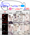
Figure 1. Atoh1-cre is expressed in hair cells and causes transient limited expression of Atoh1 in CKO ears.
(a) This diagram shows that Atoh1-cre is driven by an Atoh1 enhancer element (En) which is activated when endogenous Atoh1 protein binds. After the Cre protein is expressed, it excises the genomic Atoh1 region flanked by loxP sites, resulting in conditional deletion of Atoh1 after a transient expression of mRNA and protein. (b–c’) Immunohistochemistry of Cre protein (red) at E18.5 confirms that Cre is expressed in all hair cells in the control ear. The expression in the CKO mutant cochlea is limited to one to two rows of cells in the organ of Corti and becomes discontinuous with gaps (arrows in c’) in the base. (d–k’) In situ hybridization of Atoh1 shows its expression in the control and CKO mutant ears. The expression initiates in vestibular sensory epithelia (d) followed by progressive expression from the base to the apex in the cochlea (f, h, j). In the CKO ears Atoh1 expression is less profound and later is eliminated in all sensory epithelia (e, g, i, k). The loss of Atoh1 in the CKO cochlea also progresses from base to apex. At P4, only a small number of cells at the apical tip show Atoh1 expression (k, k’). All control and mutant ears of the same stages are from littermates processed simultaneously as left and right ears for identification. Images were electronically adjusted to the same orientation. AC, anterior crista; Co, cochlea; HC, horizontal crista; OC, organ of Corti; PC, posterior crista; S, saccule; U, utricle. Bar indicates 100 µm.
Figure 2
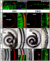
Figure 2. Conditional deletion of Atoh1 results in death of organ of Corti cells and patchy Myo7a-positive presumptive hair cells which are innervated by many nerve fibers.
(a–b) Cell death assay using immunohistochemistry of activated Caspase 3 (red) and PSVue staining (blue) shows that many cells undergo cell death in E15.5 and E16.5 CKO ears. Whole mount cochlea is shown with the lateral wall to the top and the modiolus to the bottom. Dying cells are seen in the region equivalent to the organ of Corti based on their location in close proximity to the nerve fibers shown by Tubulin staining (green in a). When combined with Atoh1 in situ hybridization (false-colored in green in b), many Caspase 3-positive dying cells are found in between the Atoh1-expressing cells (b). (c–d’) Myo7a and Tubulin immunostaining shows presumptive hair cells and their innervation in E18.5 cochlea. In contrast to the stereotyped one row of inner and three rows of outer hair cells seen in control cochlea (c’), the Myo7a-positive hair cells are reduced to only one to two rows in the CKO cochlea (d’). They are continuous in the apex but become patchy in the base with gaps in between (d, d’). The remaining hair cells apparently attract most of the nerve fibers that innervate the mutant ear. (e–f’) Double in situ hybridization for Bmp4 and Fgf10 shows the boundaries of the organ of Corti. Bmp4 is expressed in Claudius cells lateral to the organ of Corti. Fgf10 is expressed in the greater epithelial ridge and defines the medial boundary. In the CKO cochlea, the two expression regions are in parallel in the apex (f) like the control (e). However, the Fgf10 expression is discontinuous at the base and the Bmp4 expression is expanded towards the gaps in the Fgf10 expression (f, f’). (g–h”) Immunostaining of Myo7a is combined with the Bmp4/Fgf10 double in situ hybridization (false-colored in green in g’” and h”) and shows that the Myo7a-positive hair cells are located between Bmp4 and Fgf10 expression regions. In the base of the CKO cochlea, the gaps between Myo7a-positive cells correlate with the area where Bmp4 expression is expanded towards GER (arrows in h’ and h”). Cl, Claudius cells; GER, greater epithelial ridge; He, Hensen’s cells; OC, organ of Corti; SG, spiral ganglion. Bar indicates 10 µm in a, b and 100 µm in c–h”.
Figure 3
Figure 3. The remaining hair cells in the Atoh1 CKO ears develop abnormal stereocilia.
(a–f”) Immunohistochemistry of Espin (red) shows the stereocilia bundles in the hair cells in P3 ears. The control hair cells have well-ordered stereocilia in both inner and outer hair cells (a, c, e). In green, the Tubulin antibodies label nerve fibers, supporting cells and the kinocilia located off-center of the stereocilia bundle (arrowheads in e’, e”). In the CKO cochlea, the Espin-positive cells are reduced to only one to two rows and become discontinuous towards the base (b, d). The stereocilia bundles are misshaped with kinocilia at abnormal positions (arrows in f’, f”). e and f show all the Espin-positive cells by collapsing z-stack confocal images while e’, e”, f’ and f” are single optical slices. (g–h’) SEM images of P7 control and CKO cochlea hair cells. High magnification images of several CKO hair cells (h’) shows aberrant stereocilia compared to the control (g’). Note that one inner hair cell-like cell has unusually thick stereocilia (arrow in h’). (i–l) A series of images taken from base to the apex of the CKO organ of Corti show that hair cells are progressively lost along the length of the cochlea. There are only small patches of hair cells in the base, while two to three rows of outer hair cells and one row of inner hair cells (arrows in l) remain in the apical tip. However, the stereocilia of all these hair cells are abnormal compared to littermate control (g). IHC, inner hair cells; O1, O2, O3, three rows of outer hair cells; PC, pillar cells. Bar indicates 100 µm in a-d’, 10 µm in e-g, h, i-l, and 1 µm in g’, h’.
Figure 4
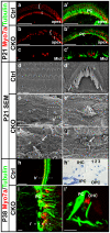
Figure 4. Some Myo7a-positive cells persist in adult Atoh1 CKO ears and display abnormal stereocilia.
(a–c’) In P21 control cochlea, one row of inner hair cells and three rows of outer hair cells are labeled by Myo7a (red) and the supporting cells are labeled by Tubulin (green). The pillar cells between the inner and outer hair cells are most strongly labeled. In the CKO cochlea, a few inner hair cells, which are medial to the strongly Tubulin-labeled pillar cells, can only be found at the apical tip (b, b’). The outer hair cells are reduced to one to two rows in the apex but become scattered in the middle turn and are surrounded or next to the remaining Tubulin-positive supporting cells. (d–g’) In contrast to the highly organized stereocilia found in all hair cells throughout the control cochlea, only few cells in the CKO cochlea are found to be hair cell-like, with stereocilia-like structures (e’, arrows in f, g). They are surrounded by, next to, or completely isolated from supporting cell-like cells. (h–i’) Immunohistochemistry of Myo7a and Tubulin shows hair cells and supporting cells in P38 ears. The control ear shows characteristic cellular distribution throughout the cochlea (h, h’). However, only a few Myo7a-positive cells remain in the CKO cochlea apex (i). The section shows that this hair cell is lateral to a Tubulin-labeled pillar cell-like cell and thus in the position of the first row of outer hair cells (i’). ‘[‘ and ‘{‘ in a and b indicate the location of inner and outer hair cells respectively. The dotted lines in h and i indicate the approximate position of the sections shown in h’ and i’. 1, 2, 3, three rows of outer hair cells; IHC, inner hair cells; IPC, inner pillar cell, OPC; outer pillar cell; PC, pillar cells. Bar indicates 100 µm in a–b’, h, 10 µm in c, c’, d, e, h’, i, i’, and 1 µm in d’ e’, f, g.
Figure 5
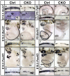
Figure 5. Atoh1, Fgf8 and Pou4f3, and to some extent Barhl1, are differentially expressed in inner and outer hair cells.
(a–b’) Atoh1 in situ hybridization in E14.5 ears shows its early expression in the cochlea. Both whole mount (a) and sections (a’) of control cochlea show a gradient of Atoh1 expression, with stronger signal in the inner hair cells and lower level in the outer hair cells. The tissue in the sections is stained (purple) and the Atoh1 in situ signal is dark blue. Note in unstained sections, low levels of Atoh1 signal can also be seen in several cells around the hair cells, possibly the supporting cells (insert in a’). In CKO cochlea, the Atoh1 signal appears sporadic in single or two rows of hair cells (b, b’). (c–d) Fgf8 is specifically expressed in the inner hair cells in the entire P0 control cochlea but only in the apex of the CKO cochlea. (e–f) Neurod1 is expressed and acts downstream of Atoh1 in the hair cells. In situ hybridization shows there is a complete loss of Neurod1 in P0 CKO cochlea. (g–j’) Barhl1, an Atoh1 downstream gene, follows its base to apex expression in the hair cells in the ear. At the leading end in E16.5 control cochlea, Barhl1 is first upregulated in the inner hair cells (g’). The expression is greatly reduced and becomes intermittent in the mutant ear (h, h’). At P0, all hair cells express Barhl1 in the control ear, but the expression is further reduced in the CKO cochlea. (k–n’) In situ hybridization of Pou4f3 shows its expression in hair cells. In E14.5 control cochlea, the expression starts from the base and the inner hair cells. Only a few outer hair cells express Pou4f3 at this stage (k’). At E18.5, the expression levels in all hair cells are similar. In CKO ears at both stages, the expression of Pou4f3 is greatly reduced, except for the apex. AC, anterior crista; Co, cochlea; GER, greater epithelia ridge; HC, horizontal crista; IHC, inner hair cells; OC, organ of Corti; PC, posterior crista; S, saccule; U, utricle. Bar indicates 10 µm in a-b’ and 100 µm in c–n’.
Figure 6
Figure 6. A working hypothesis shows the differential expression of Atoh1 in inner hair cells (IHC) and outer hair cells (OHC).
Based on our in situ hybridization data, Atoh1 is expressed earlier and at a higher level in IHCs (red line) than OHCs (green line), first in the base (top) followed by the apex (bottom). At early postnatal stages, Atoh1 is downregulated to below the detection threshold of in situ hybridization, but a low level of Atoh1 expression (grey lines) likely is sustained and can be detected by Atoh1-LacZ as late as P275 . In the CKO mutant cochlea, the Atoh1 expression is reduced to a range of different levels shortly after its upregulation and is then eliminated after a few days (dashed lines). Some cells at the apex express Atoh1 at a level close to control hair cells and can survive to adult stage.

References

    1. Izumikawa M, Batts SA, Miyazawa T, Swiderski DL, Raphael Y. Response of the flat cochlear epithelium to forced expression of Atoh1. Hear Res. 2008;240:52–56. - PMC - PubMed
    1. Izumikawa M, Minoda R, Kawamoto K, Abrashkin KA, Swiderski DL, et al. Auditory hair cell replacement and hearing improvement by Atoh1 gene therapy in deaf mammals. Nat Med. 2005;11:271–276. - PubMed
    1. Praetorius M, Hsu C, Baker K, Brough DE, Plinkert P, et al. Adenovector-mediated hair cell regeneration is affected by promoter type. Acta Otolaryngol. 2010;130:215–222. - PMC - PubMed
    1. Oshima K, Shin K, Diensthuber M, Peng AW, Ricci AJ, et al. Mechanosensitive hair cell-like cells from embryonic and induced pluripotent stem cells. Cell. 2010;141:704–716. - PMC - PubMed
    1. Hildebrand MS, Newton SS, Gubbels SP, Sheffield AM, Kochhar A, et al. Advances in molecular and cellular therapies for hearing loss. Mol Ther. 2008;16:224–236. - PubMed

Publication types

MeSH terms