Comparative Study
. 2012 Mar 9;53(3):1188-94.
doi: 10.1167/iovs.11-8408. Print 2012 Mar.

The posterior location of the dilator muscle induces anterior iris bowing during dilation, even in the absence of pupillary block

Affiliations
Comparative Study

The posterior location of the dilator muscle induces anterior iris bowing during dilation, even in the absence of pupillary block

Rouzbeh Amini et al. Invest Ophthalmol Vis Sci. .

Abstract

Purpose: To examine the effect of the posterior location of the dilator on iris anterior curvature during dilation.

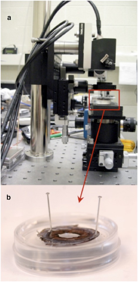
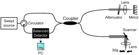
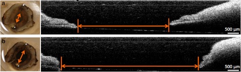
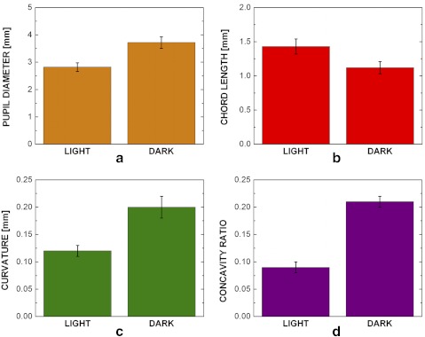
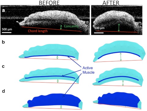
Methods: An in vivo human study, an ex vivo porcine experiment, and an in silico computational model were performed in parallel. Iris anterior curvature was measured in vivo before and after dilation by time-domain slit lamp optical coherence tomography (SL-OCT). All patients (n = 7) had undergone laser peripheral iridotomy to eliminate any pupillary block due to primary angle-closure glaucoma. In the ex vivo experiments, isolated porcine irides (n = 30) were secured at the periphery and immersed in an oxygenated Krebs-Ringer buffer. Dilation was induced pharmaceutically by the addition of 2.5% phenylephrine and 1% tropicamide. An in-house optical coherence tomography (OCT) system was used to obtain iris images before and after dilation. A finite element model was also developed based on typical geometry of the iris from the initial OCT image. The iris was modeled as a neo-Hookean solid, and the active muscle component was applied only to the region specified as the dilator.

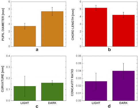
Results: An increase in curvature and a decrease in chord length after dilation were observed in both experiments. In both the in vivo and ex vivo experiments, the curvature-to-chord length ratio increased significantly during dilation. Computer simulations agreed well with the experimental results only when the proper anatomic position of dilator was used.

Conclusions: The posterior location of the dilator contributes to the anterior iris bowing via a nonpupillary block dependent mechanism.


Figures

Figure 1.
Figure 1.
Ex vivo experimental setup. Experimental setup of the in-house (a) OCT imaging system with the (b) iris sample pinned in the Petri dish.
Figure 2.
Figure 2.
The OCT system used to image the iris during dilation.
Figure 3.
Figure 3.
(a) OCT image of the iris before the dilator is activated. Iris chord length (A–B) is defined as the distance from the tip of the iris to the periphery; iris curvature (C–D) is defined as the longest distance between the iris chord and posterior epithelium. (b) Finite element model based on the OCT image of the iris before dilation. The region modeled as the active dilator muscle is marked with the darker color.
Figure 4.
Figure 4.
Histologic image of the pupillary and midperipheral portions of the porcine iris. Monoclonal anti-human α-smooth muscle actin stain was used to differentiate the muscular tissues, including the sphincter (S) and dilator (D). The pigment epithelium anterior border layer (ABL); stroma (ST), which is a loosely arranged collagen network; and another thick layer of pigment epithelial (PE) cells on the posterior surface of the iris is also identifiable. Magnification of the midperiphery regions illustrates that dilator muscle lies on the posterior iris surface and is very thin compared with the sphincter muscle.
Figure 5.
Figure 5.
In vivo ANA patients' iris chord length, curvature, and concavity ratio results. (a) The pupil diameter increased (P < 0.0001), (b) the iris chord length decreased (P < 0.0001), (c) the iris curvature increased (P = 0.481), and (d) the concavity ratio increased (P = 0.004) after dilation in the dark (bars, 95% confidence interval; n = 7).
Figure 6.
Figure 6.
Ex vivo pupil dilation results. Images taken before (a) and after (b) the addition of 40 μL of 2.5% phenylephrine and 40 μL of 1% tropicamide to the bath solution. On the left is a top-down view of an iris with the anterior surface facing up using a digital camera and in the middle is the cross section of the iris detected by the OCT imaging system. The pupil diameter (arrows) is shown to visibly increase after the addition of the drugs.
Figure 7.
Figure 7.
Ex vivo iris pupil diameter, chord length, curvature, and concavity ratio results. All measurements showed a significant difference before and after the addition of the dilation drugs. The pupil diameter (a) increased significantly (P < 0.001) after the addition of the drugs, the chord length (b) decreased (P < 0.0001), the curvature (c) increased (P < 0.002), and the (d) concavity ratio (P < 0.0001) increased (bars, 95% confidence interval, n = 30).
Figure 8.
Figure 8.
Iris chord length and curvature before (left) and after (right) dilation in (a) a typical ex vivo experiment, (b) an anatomic realistic model of the iris with the dilator muscle in the posterior, (c) an artificial model in which the dilator was positioned anteriorly, and (d) an artificial model in which the dilator was thickened throughout the entire iris contour.
Figure 9.
Figure 9.
Iris concavity ratio before and after dilation in a typical experiment and three models based on the geometry of the experiment. In the realistic model (Fig. 8b), the iris curvature was similar to that of the experiment. Anteriorly positioning (Fig. 8c) or thickening of the dilator muscle (Fig. 8d) in the artificial models led to a predicted curvature inconsistent with the experimental data.

References

    1. Mandell MA, Pavlin CJ, Weisbrod DJ, Simpson ER. Anterior chamber depth in plateau iris syndrome and pupillary block as measured by ultrasound biomicroscopy. Am J Ophthalmol. 2003;136:900–903 - PubMed
    1. Nonaka A, Iwawaki T, Kikuchi M, Fujihara M, Nishida A, Kurimoto Y. Quantitative evaluation of iris convexity in primary angle closure. Am J Ophthalmol. 2007;143:695–697 - PubMed
    1. Ritch LR. Angle-closure glaucoma: mechanisms and epidemiology. The Glaucomas; St. Louis: Mosby; 1996:chap 37
    1. Woo EK, Pavlin CJ, Slomovic A, Taback N, Buys YM. Ultrasound biomicroscopic quantitative analysis of light-dark changes associated with pupillary block. Am J Ophthalmol. 1999;127:43–47 - PubMed
    1. Huang EC, Barocas VH. Active iris mechanics and pupillary block: steady-state analysis and comparison with anatomical risk factors. Ann Biomed Eng. 2004;32:1276–1285 - PubMed

Publication types

MeSH terms