

A new, pachymetry-based approach for diagnostic cutoffs for normal, suspect and keratoconic cornea
- PMID: 22281864
- PMCID: PMC3351046
- DOI: 10.1038/eye.2011.365
A new, pachymetry-based approach for diagnostic cutoffs for normal, suspect and keratoconic cornea
Abstract
Purpose: To analyze whether an association exists between keratometric and pachymetric changes in the cornea, and whether it can be used to create pachymetric cutoff criteria secondary to keratometric criteria.
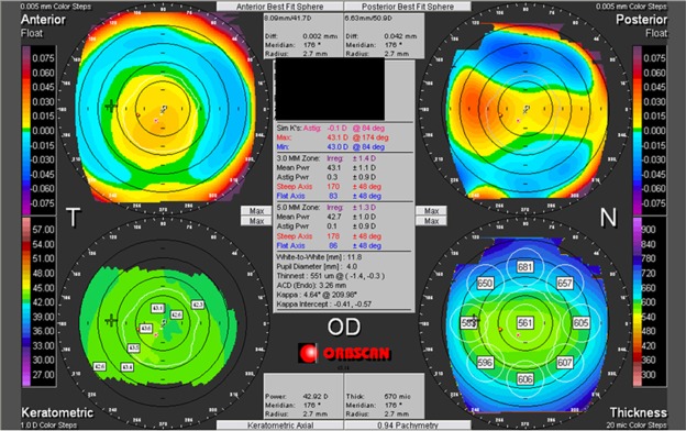
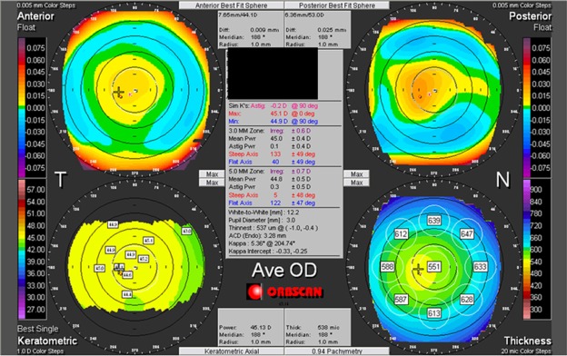
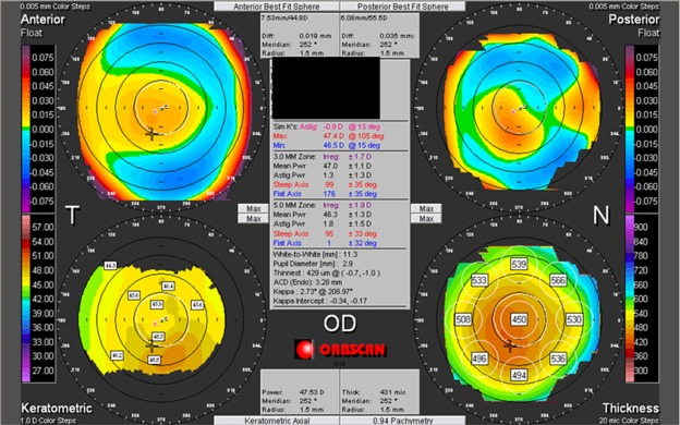
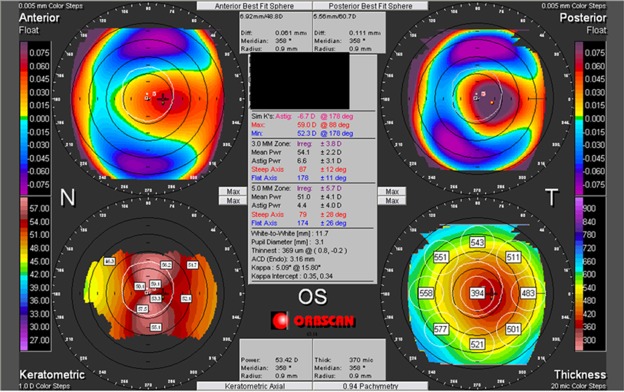
Methods: In this cross-sectional study, 1000 candidates presenting to the refractive surgery services of a tertiary care hospital underwent bilateral Orbscan IIz (Bausch and Lomb) assessment along with other ophthalmic evaluation.
Results: Stepwise regression analysis-based models showed that simulated keratometry (simK) astigmatism was significantly predicted by the minimum corneal thickness (MCT) and difference between central and MCT (δCT), mean SimK by the MCT and δCT, and maximum keratometry in the central 10-mm zone by the MCT and δCT (P<0.001). The mean MCT values were 542.5 ± 39.6, 539.9 ± 39.2, 524.2 ± 49.5, and 449.3 ± 73.7 μm for flatter normal (<44 D), steeper normal (≥ 44 D), keratoconus suspect and keratoconic eyes, respectively (P<0.001). The mean differences between central corneal thickness and MCT (δCT) were 12.2 ± 7.1 μm, 12.4 ± 7.4 μm, 14.4 ± 8.9 μm and 23.2 ± 10.1 μm for the flatter normal, steeper normal, keratoconus suspect, and keratoconic eyes, respectively (P<0.001). Mean and 2SD cutoff were used to suggest that a cornea having MCT< 461 μm or δCT>27 μm has only a 2.5% chance of being normal and not a keratoconus suspect or worse.
Conclusion: Pachymetric diagnostic cutoffs can be used as adjuncts to the existing topographic criteria to screen keratoconus suspect and keratoconic eyes.
Figures
References
-
- Rabinowitz YS. Keratoconus. Surv Ophthalmol. 1998;42 (4:297–319. - PubMed
-
- Rabinowitz YS, Rasheed K. KISA% index: a quantitative videokeratography algorithm embodying minimal keratometric criteria for diagnosing keratoconus. J Cataract Refract Surg. 1999;25 (10:1327–1335. - PubMed
-
- Dastjerdi MH, Hashemi H. A quantitative corneal topography index for detection of keratoconus. J Refract Surg. 1998;14 (4:427–436. - PubMed
-
- Smolek MK, Klyce SD. Current keratoconus detection methods compared with a neural network approach. Invest Ophthalmol Vis Sci. 1997;38 (11:2290–2299. - PubMed
-
- Maeda N, Klyce SD, Smolek MK. Comparison of methods for detecting keratoconus using videokeratography. Arch Ophthalmol. 1995;113 (7:870–874. - PubMed
MeSH terms
LinkOut - more resources
Full Text Sources
