. 2012 Mar 2;110(5):739-48.
doi: 10.1161/CIRCRESAHA.111.258426. Epub 2012 Jan 26.

Selective class I histone deacetylase inhibition suppresses hypoxia-induced cardiopulmonary remodeling through an antiproliferative mechanism

Affiliations

Selective class I histone deacetylase inhibition suppresses hypoxia-induced cardiopulmonary remodeling through an antiproliferative mechanism

Maria A Cavasin et al. Circ Res. .

Abstract

Rationale: Histone deacetylase (HDAC) inhibitors are efficacious in models of hypertension-induced left ventricular heart failure. The consequences of HDAC inhibition in the context of pulmonary hypertension with associated right ventricular cardiac remodeling are poorly understood.

Objective: This study was performed to assess the utility of selective small-molecule inhibitors of class I HDACs in a preclinical model of pulmonary hypertension.

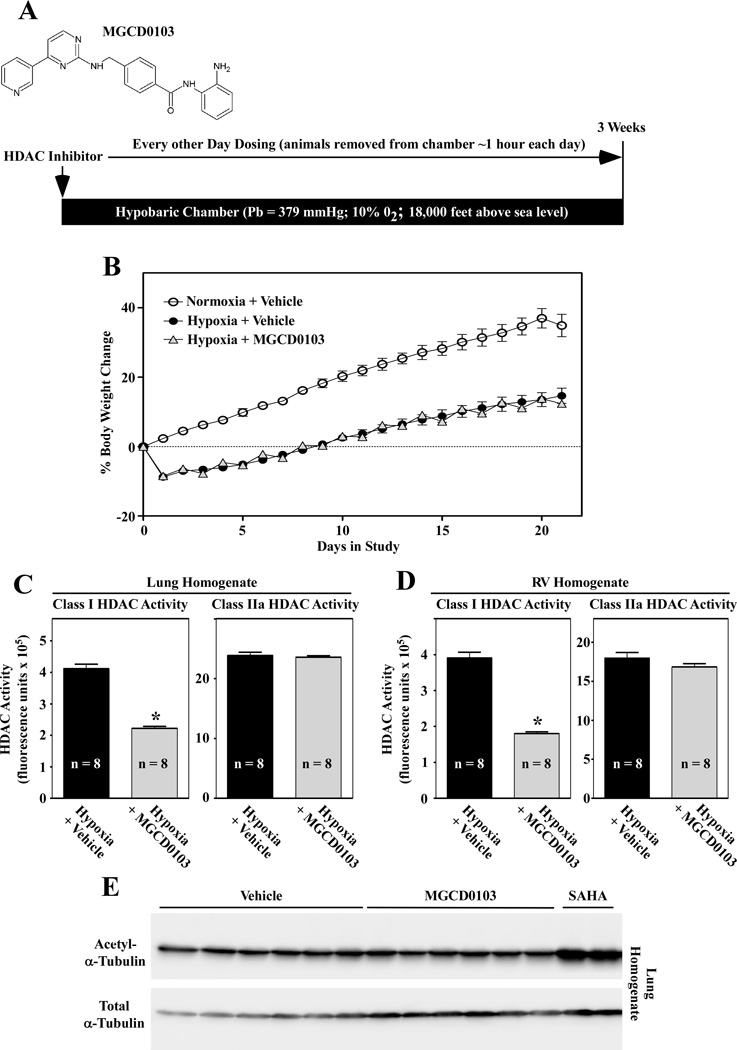
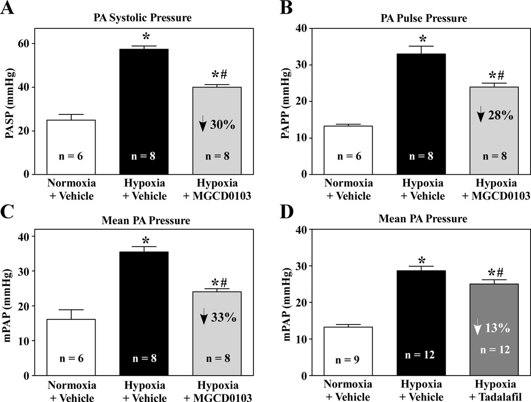
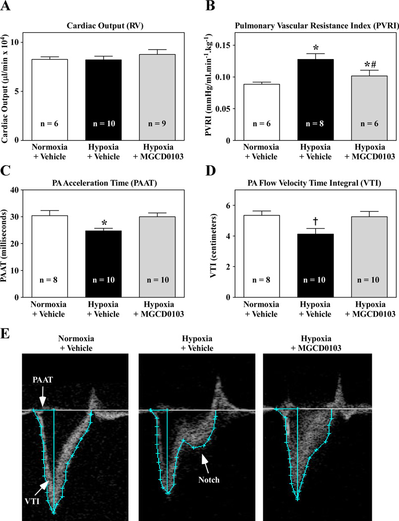
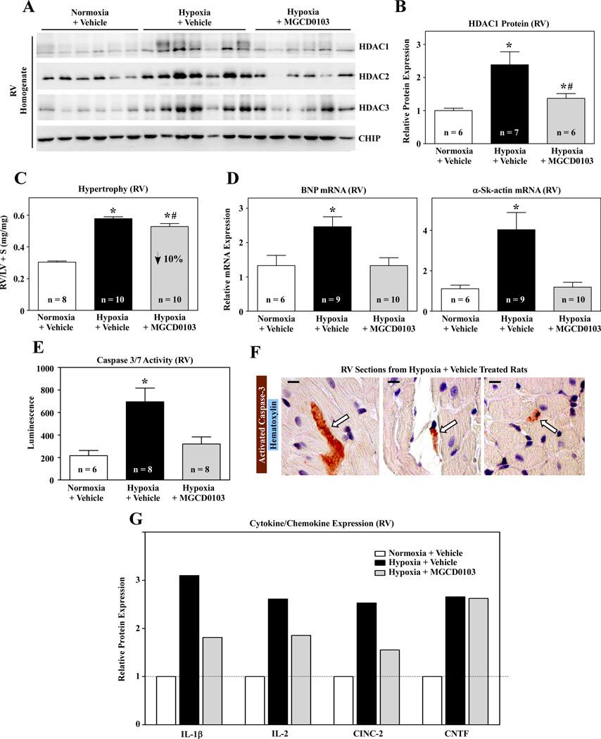
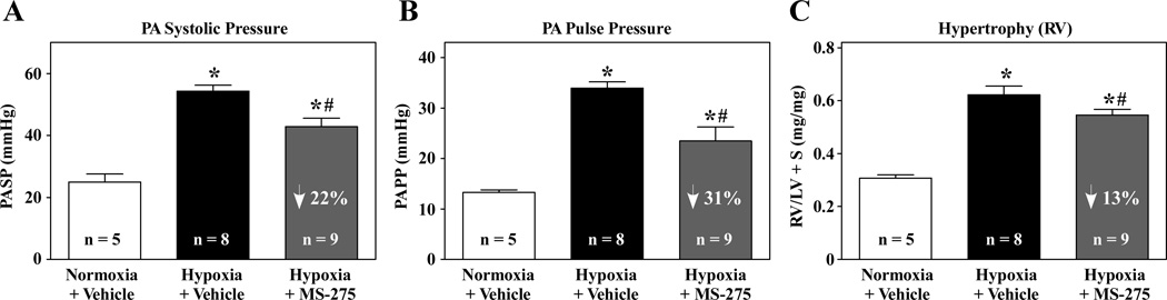
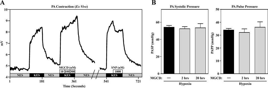
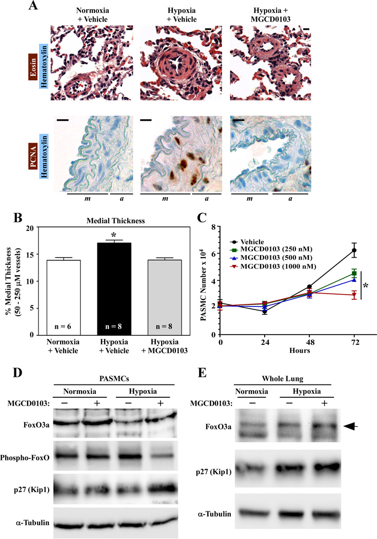
Methods and results: Rats were exposed to hypobaric hypoxia for 3 weeks in the absence or presence of a benzamide HDAC inhibitor, MGCD0103, which selectively inhibits class I HDACs 1, 2, and 3. The compound reduced pulmonary arterial pressure more dramatically than tadalafil, a standard-of-care therapy for human pulmonary hypertension that functions as a vasodilator. MGCD0103 improved pulmonary artery acceleration time and reduced systolic notching of the pulmonary artery flow envelope, which suggests a positive impact of the HDAC inhibitor on pulmonary vascular remodeling and stiffening. Similar results were obtained with an independent class I HDAC-selective inhibitor, MS-275. Reduced pulmonary arterial pressure in MGCD0103-treated animals was associated with blunted pulmonary arterial wall thickening because of suppression of smooth muscle cell proliferation. Right ventricular function was maintained in MGCD0103-treated animals. Although the class I HDAC inhibitor only modestly reduced right ventricular hypertrophy, it had multiple beneficial effects on the right ventricle, which included suppression of pathological gene expression, inhibition of proapoptotic caspase activity, and repression of proinflammatory protein expression.

Conclusions: By targeting distinct pathogenic mechanisms, isoform-selective HDAC inhibitors have potential as novel therapeutics for pulmonary hypertension that will complement vasodilator standards of care.


Conflict of interest statement

Disclosures

No conflicts of interest exist for the authors.

Figures

Figure 1
Figure 1. Class I HDAC inhibition in a hypoxia model of pulmonary hypertension
A, Rats were housed in hypobaric chambers and were injected i.p. with the class I HDAC inhibitor, MGCD0103 (10 mg/kg), every other day for three weeks. On days when compound was not delivered, animals were treated with vehicle control. Normoxic and hypoxic control rats were dosed with compound vehicle on a daily basis. B, Rats were weighed daily. C and D, Class I and class IIa HDAC enzymatic activity was quantified in lung and RV homogenates. E, Lung homogenates were immunoblotted for acetyl- and total α-tubulin. Lung lysates from an independent study with the pan-HDAC inhibitor, SAHA, were used as controls.
Figure 2
Figure 2. Class I HDAC inhibition suppresses hypoxia-dependent pulmonary hypertension
A – C, Pulmonary arterial pressure (PAP) was measured in normoxic and hypoxic rats treated with vehicle or MGCD0103 for 3 weeks. PA systolic pressure (PASP); PA pulse pressure (PAPP); mean PAP, mPAP. D, mPAP values from an independent study of tadalafil are shown. Tadalafil was dosed daily by oral gavage at a concentration of 10 mg/kg.
Figure 3
Figure 3. Improved hemodynamics in class I HDAC inhibitor-treated animals
A, Rats were dosed i.p. with compound vehicle or MGCD0103 (10 mg/kg) for 3 weeks. RV cardiac output was determined using an invasive pressure-volume catheter. B, Cardiac output was used to calculate total pulmonary vascular resistance. C and D, Pulmonary artery acceleration time (PAAT) and velocity time integral (VTI) were quantified using Doppler images from normoxic and hypoxic rats treated with vehicle or MGCD0103. The reduction in VTI in the hypoxia + vehicle group was significant by t-test but not ANOVA. E, Doppler images of transpulmonary blood flow. Regions of the images used to calculate PAAT and VTI are shown. Systolic notching of PA blood flow in a hypoxic rat treated with vehicle is indicated. For all graphs, values represent mean +SEM. *P<0.05 vs. normoxia (ANOVA); #P<0.05 vs. hypoxia plus vehicle (ANOVA); P<0.05 vs. normoxia (t-test).
Figure 4
Figure 4. Class I HDAC inhibition suppresses multiple pathological pathways in the RV
A, Immunoblotting of class I HDAC proteins in RV lysates. C-terminus Hsp70 interacting protein (CHIP) was immunoblotted as a control. B, HDAC1 levels were quantified using a digital imager. C, RV hypertrophy was assessed by weighing ventricular chambers at the time of necropsy. D, Quantitative PCR analysis of RV brain natriuretic peptide (BNP) and alpha-skeletal actin (α-Sk-actin) mRNA levels. E, Caspase activity was measured in RV homogenates using a fluorescent substrate that is cleaved by caspase-3 and -7. For B – E, Values presented are mean +SEM. *P<0.05 vs. normoxia; #P<0.05 vs. hypoxia plus vehicle. F, RV sections from hypoxic rats stained for cleaved (active) caspase-3. Arrows indicate caspase-positive cells. Caspase-positive cells were not detected in RVs from normoxic controls. Scale bar = 10 µm. G, Cytokine protein levels in RV homogenates. For each group, RV protein from four independent animals was pooled prior to analysis.
Figure 5
Figure 5. A second class I HDAC inhibitor suppresses hypoxia-dependent pulmonary hypertension and RV hypertrophy
Rats were housed in hypobaric chambers and were injected with the class I HDAC inhibitor, MS-275 (3 mg/kg), every other day for three weeks. On days when compound was not delivered, animals were treated with vehicle control. Normoxic and hypoxic control rats were dosed with compound vehicle on a daily basis. MS-275 reduced PASP (A), PAPP (B) and RV hypertrophy (C). For all graphs, values represent mean +SEM. *P<0.05 vs. normoxia (ANOVA); #P<0.05 vs. hypoxia plus vehicle (ANOVA).
Figure 6
Figure 6. MGCD0103 is not an acute vasodilator
A, Rat pulmonary artery strips (1.5 mm X 200 µm) were hung on a “bubble plate” between two tungsten wires. One wire was fixed and the other attached to a force transducer. Intact strips were stimulated with high potassium (KES). MGCD0103 had no effect on the potassium-mediated contraction at any concentration (10–300 nM). However, when strips were washed with normal extracellular solution (NES) and contracted again with potassium, sodium nitroprusside (SNP, 1 µM) effectively relaxed the vessel. B, Rats were maintained in hypobaric chambers for 3 weeks. On the final day of the study, animals received a single injection of MGDC0103 (10 mg/kg) 2 or 20 hours prior to measuring PAP. Acute administration of MGCD0103 failed to lower hypoxia-induced increases in PASP or PAPP.
Figure 7
Figure 7. Class I HDAC inhibition suppresses hypoxia-induced pulmonary arterial medial thickening
A, Images of hematoxylin and eosin- (upper panel) and proliferating cell nuclear antigen (PCNA)-stained lung sections. PCNA-stained sections were counterstained with hematoxylin. Scale bar = 10 µm. B, Medial thickness of pulmonary arteries between 50 and 250 µm in diameter. Values are presented as mean +SEM. *P<0.05 vs. normoxia; #P<0.05 vs. hypoxia plus vehicle. C, Rat PASMCs were cultured in the presence of FBS (10%) with the indicated concentrations of MGCD0103. D, Rat PASMCs were grown under normoxic or hypoxic conditions for 48 hours in the absence or presence of MGCD0103 (500 nM). Protein lysates from these cells were immunoblotted with antibodies for FoxO3a, phospho-FoxO3a (P-Thr-32), p27 or α-tubulin. E, Homogenates of lungs (n = 4 lungs/condition, pooled) were immunoblotted for FoxO3a, p27 or α-tubulin.

Similar articles

Cited by

References

    1. McLaughlin VV, Archer SL, Badesch DB, Barst RJ, Farber HW, Lindner JR, Mathier MA, McGoon MD, Park MH, Rosenson RS, Rubin LJ, Tapson VF, Varga J. ACCF/AHA 2009 expert consensus document on pulmonary hypertension a report of the American College of Cardiology Foundation Task Force on Expert Consensus Documents and the American Heart Association developed in collaboration with the American College of Chest Physicians; American Thoracic Society, Inc. and the Pulmonary Hypertension Association. J Am Coll Cardiol. 2009;53:1573–1619. - PubMed
    1. Humbert M, Morrell NW, Archer SL, Stenmark KR, MacLean MR, Lang IM, Christman BW, Weir EK, Eickelberg O, Voelkel NF, Rabinovitch M. Cellular and molecular pathobiology of pulmonary arterial hypertension. J Am Coll Cardiol. 2004;43:13S–24S. - PubMed
    1. D'Alonzo GE, Barst RJ, Ayres SM, Bergofsky EH, Brundage BH, Detre KM, Fishman AP, Goldring RM, Groves BM, Kernis JT. Survival in patients with primary pulmonary hypertension. Results from a national prospective registry. Ann Intern Med. 1991;115:343–349. - PubMed
    1. Simon MA, Pinsky MR. Right ventricular dysfunction and failure in chronic pressure overload. Cardiol Res Pract. 2011;2011:568095. - PMC - PubMed
    1. Walker LA, Buttrick PM. The right ventricle: biologic insights and response to disease. Curr Cardiol Rev. 2009;5:22–28. - PMC - PubMed

Publication types

MeSH terms