

Fast T1 mapping determined using incomplete inversion recovery look-locker 3D balanced SSFP acquisition and a simple two-parameter model fit
- PMID: 22282318
- PMCID: PMC3343218
- DOI: 10.1002/jmri.23576
Fast T1 mapping determined using incomplete inversion recovery look-locker 3D balanced SSFP acquisition and a simple two-parameter model fit
Abstract
Purpose: To evaluate a fast T1 mapping technique using incomplete inversion recovery 3D balanced steady-state free precession acquisition along with a two-parameter model fit.
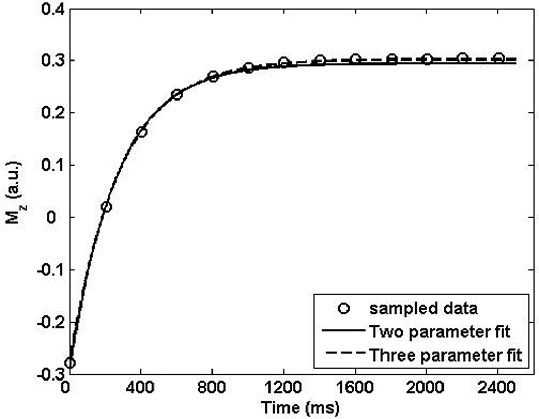
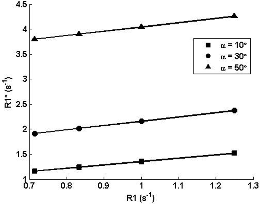
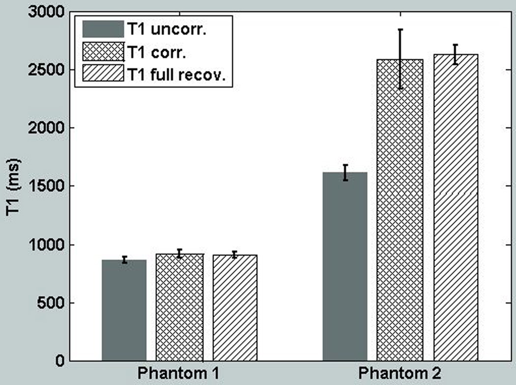
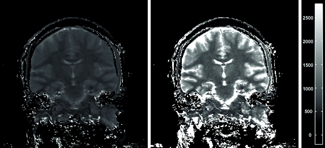
Materials and methods: Using Bloch simulations, we explored the two-parameter model fit for data acquired using such an acquisition scheme. The parameter space over which the fit holds good was determined through simulations. A linear correction was derived for the R1* (1/T1*) values so determined. Two phantoms and six volunteers were scanned using the described technique. Comparison scans using full recovery as well as gold standard inversion recovery spin echo were also performed.
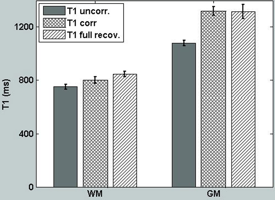
Results: The two-parameter fit works exceedingly well over a large parameter space. T1 values in the phantoms showed an error of 4.9% and 39% before correction and 0.9% and 1.6% after correction. For the six volunteers, error in T1 value was 5.3% for white matter (WM) and 2.4% for gray matter (GM) after correction, while it was 11.2% and 18.2% before correction.
Conclusion: The work presented here allows for T1 map determination with higher resolution and shorter acquisition time than previously possible. The technique is especially well suited for GM/WM T1 mapping.
Copyright © 2012 Wiley Periodicals, Inc.
Figures


Similar articles
-
Modulated repetition time look-locker (MORTLL): a method for rapid high resolution three-dimensional T1 mapping.J Magn Reson Imaging. 2009 Sep;30(3):640-8. doi: 10.1002/jmri.21842. J Magn Reson Imaging. 2009. PMID: 19630081 Free PMC article.
-
Free-breathing 3D whole-heart joint T1/T2 mapping and water/fat imaging at 0.55 T.Magn Reson Med. 2024 Oct;92(4):1511-1524. doi: 10.1002/mrm.30139. Epub 2024 Jun 13. Magn Reson Med. 2024. PMID: 38872384
-
Comparison of spoiled gradient echo and steady-state free-precession imaging for native myocardial T1 mapping using the slice-interleaved T1 mapping (STONE) sequence.NMR Biomed. 2016 Oct;29(10):1486-96. doi: 10.1002/nbm.3598. NMR Biomed. 2016. PMID: 27658506 Free PMC article.
-
Non-contrast-enhanced MR portography with time-spatial labeling inversion pulses: comparison of imaging with three-dimensional half-fourier fast spin-echo and true steady-state free-precession sequences.J Magn Reson Imaging. 2009 May;29(5):1140-6. doi: 10.1002/jmri.21753. J Magn Reson Imaging. 2009. PMID: 19388119
-
Simultaneous T1 and T2 quantification of the myocardium using cardiac balanced-SSFP inversion recovery with interleaved sampling acquisition (CABIRIA).Magn Reson Med. 2015 Aug;74(2):365-71. doi: 10.1002/mrm.25402. Epub 2014 Aug 11. Magn Reson Med. 2015. PMID: 25113911
Cited by
-
Automatic Brain Tissue and Lesion Segmentation and Multi-Parametric Mapping of Contrast-Enhancing Gliomas without the Injection of Contrast Agents: A Preliminary Study.Cancers (Basel). 2024 Apr 17;16(8):1524. doi: 10.3390/cancers16081524. Cancers (Basel). 2024. PMID: 38672606 Free PMC article.
-
First clinical application of a novel T1 mapping of the whole brain.Neuroradiol J. 2022 Dec;35(6):684-691. doi: 10.1177/19714009221084244. Epub 2022 Apr 21. Neuroradiol J. 2022. PMID: 35446175 Free PMC article.
References
-
- Haase A. Snapshot FLASH MRI. Applications to T1, T2, and chemical-shift imaging. Magn Reson Med. 1990;13(1):77–89. - PubMed
-
- Deichmann R, Haase A. Quantification of T1 Values by Snapshot-Flash Nmr Imaging. Journal of Magnetic Resonance. 1992;96(3):608–612.
-
- Scheffler K, Hennig J. T-1 quantification with inversion recovery TrueFISP. Magnet Reson Med. 2001;45(4):720–723. - PubMed
-
- Schmitt P, Griswold MA, Jakob PM, et al. Inversion recovery TrueFISP: Quantification of T-1, T-2, and spin density. Magnet Reson Med. 2004;51(4):661–667. - PubMed
-
- Messroghli DR, Radjenovic A, Kozerke S, Higgins DM, Sivananthan MU, Ridgway JP. Modified Look-Locker inversion recovery (MOLLI) for high-resolution T-1 mapping of the heart. Magnet Reson Med. 2004;52(1):141–146. - PubMed
Publication types
MeSH terms
Grants and funding
LinkOut - more resources
Full Text Sources
Other Literature Sources
Medical
