. 2012 Apr;32(4):1045-53.
doi: 10.1161/ATVBAHA.111.244210. Epub 2012 Jan 26.

Flow cytometric identification and functional characterization of immature and mature circulating endothelial cells

Affiliations

Flow cytometric identification and functional characterization of immature and mature circulating endothelial cells

Julie A Mund et al. Arterioscler Thromb Vasc Biol. 2012 Apr.

Abstract

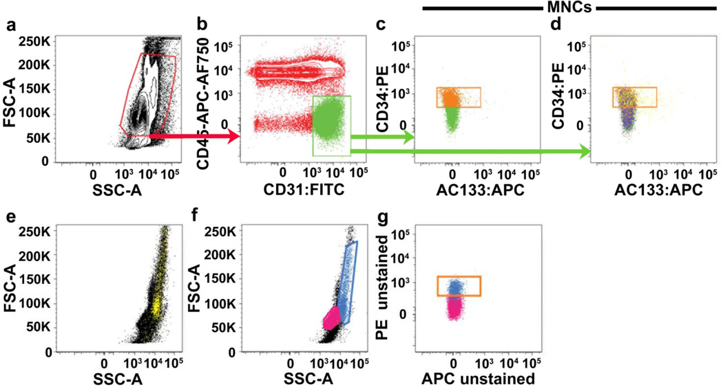
Objective: We sought to identify and characterize 2 distinct populations of bona fide circulating endothelial cells, including the endothelial colony-forming cell (ECFC), by polychromatic flow cytometry (PFC), colony assays, immunomagnetic selection, and electron microscopy.

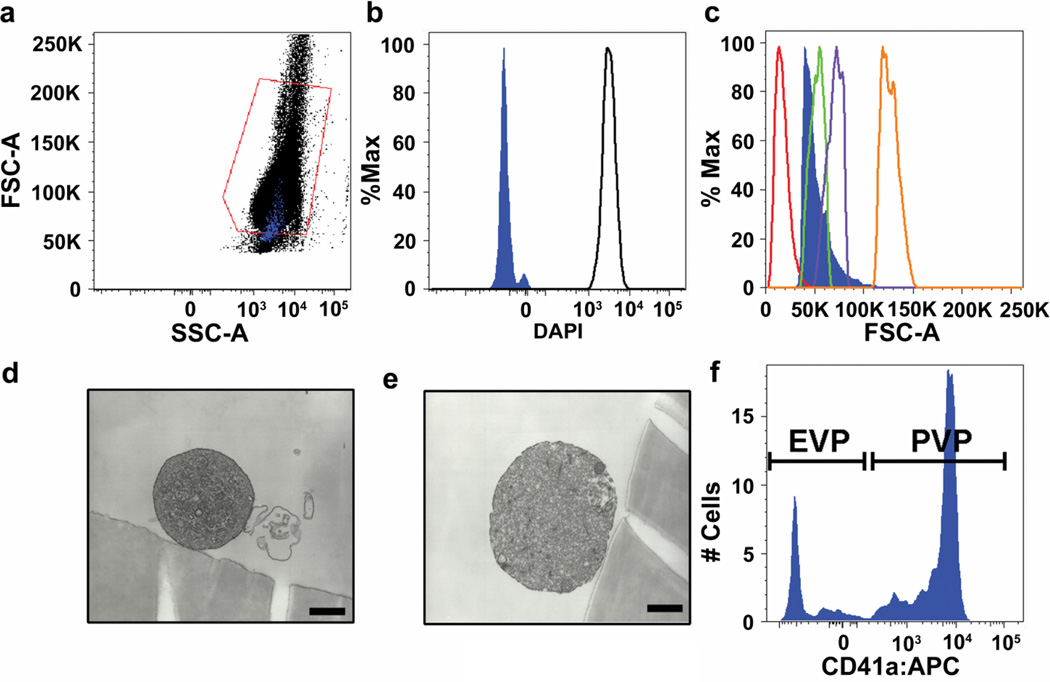
Methods and results: Mononuclear cells from human umbilical cord blood and peripheral blood were analyzed using our recently published PFC protocol. A population of cells containing both ECFCs and mature circulating endothelial cells was determined by varying expressions of CD34, CD31, and CD146 but not AC133 and CD45. After immunomagnetic separation, these cells failed to form hematopoietic colonies, yet clonogenic endothelial colonies with proliferative potential were obtained, thus verifying their identity as ECFCs. The frequency of ECFCs were increased in cord blood and were extremely rare in the peripheral blood of healthy adults. We also detected another mature endothelial cell population in the circulation that was apoptotic. Finally, when comparing this new protocol with a prior method, we determined that the present protocol identifies circulating endothelial cells, whereas the earlier protocol identified extracellular vesicles.

Conclusions: Two populations of circulating endothelial cells, including the functionally characterized ECFC, are now identifiable in human cord blood and peripheral blood by PFC.


Conflict of interest statement

DISCLOSURES

All authors declare no conflict of interest.

Figures

Figure 1
Figure 1. Threshold gating of mononuclear cells based on forward scatter and side scatter does not remove red blood cells, dead cell and monocyte contamination
Representative PFC analysis of a CPT mononuclear cell preparation of peripheral blood either (a–e) stained with the six-antibody/viability marker panel (as defined in Methods), which includes glyA (red blood cell marker), CD14 (monocyte marker) and LIVE/DEAD® (cell viability marker) or (f–g) completely unstained. Mononuclear cells (red gate in a) are initially gated on a FSC/SSC plot in an attempt to exclude red blood cells, dead cells and debris, and sub-gated onto a bivariant antigen plot (b) for identification of CD31brightCD45 cells (green gate). CD31brightCD45 cells are further sub-gated to identify the CD34+AC133 subset (orange gate in c and d). In (d), glyA+ red blood cells and/or dead/apoptotic cells (purple events) and CD14+ monocytes (yellow events), contained within the initial mononuclear cell gate, are mapped onto the bi-variant plot and can clearly be seen distributed throughout the CD34+AC133 gate (orange gate). Monocytes are difficult to exclude based on their scatter profile. CD14+ monocytes (yellow cells mapped in e) can be seen distributed throughout the mononuclear cells when back-gated onto a forward scatter/side scatter plot (e). In analysis of unstained samples (f–g), cells that have the scatter profile of monocytes (blue gate in f and blue cells mapped onto g), are highly auto-fluorescent and contaminate the orange gate used for frequency analysis of CD34+AC133 cells. Lymphocytes are gated and mapped in pink for reference (f–g). Similar results were seen in 9 other samples from different donors.
Figure 2
Figure 2. Frequency analysis of CD31brightCD34+CD45AC133 cells
Two strategies for frequency analysis of CD31brightCD34+CD45AC133 cells from peripheral blood stained with the six-antibody/viability marker panel are shown. In the first strategy (a–d), conventional flow cytometry techniques were utilized. In the second strategy (e–i), PFC techniques were utilized, including the removal of monocytes, red blood cells and dead cells that lead to a more uniform population with a tighter expression profile.
Figure 3
Figure 3. Comparison of Conventional Flow Cytometry to Polychromatic Flow Cytometry
Figure 4
Figure 4. Characterization of CD31brightCD34+CD45AC133 cells
CD14glyALIVE/DEAD®− CD31brightCD34+CD45AC133 events (as identified in Figure 1e–i) (solid blue in b) stain negatively for the nuclear dye DAPI. (c) CD14glyALIVE/DEAD®−CD31brightCD34+CD45AC133 events (solid blue in c) are in the range of 1–5µm in size. (d–e) Representative electron microscopy photomicrographs of sorted CD14glyALIVE/DEAD®−CD31brightCD34+CD45AC133 events. Scale bars represent 500nm. (f) CD41a+ events are platelet-derived microvesicles (PMV) and CD41a events are endothelial-derived microvesicles (EMV).
Figure 5
Figure 5. Prospective identification and isolation of circulating ECFCs by PFC and Immunomagnetic Selection
Representative PFC analysis of (a) adult peripheral blood mononuclear cells and (b) cord blood mononuclear cells. Putative CD34+CD45 ECFCs are gated in green. (c–e) Cells (b gate) were analyzed for expression of (c) CD31, (d) CD146, and (e) CD105. (f–g) PFC analysis of cord blood mononuclear cells (f) prior to and (g) following isolation of CD146+CD45 cells via MACS. (h) Representative photomicrograph of an ECFC colony derived from the CD146+CD45 fraction of cord blood mononuclear cells 6 days after culture in endothelial-specific media. Scale bar represents 200µm. (i) Low (×1,900) and (j) high (×13,000) magnification electron microscopy of sorted cord blood LIVE/DEAD®−CD14glyACD31brightCD34+CD45 AC133 cells, is consistent with endothelial morphology.
Figure 6
Figure 6. Photomicrograph of human endothelial cells expressing CD31 forming blood vessels that contain mouse erythrocytes
(a) Photomicrograph (original X60) of cellularized gels containing red blood cell perfused blood vessels stained with hematoxylin and eosin. (b) The vast majority of perfused blood vessel are lined by human endothelial cells (derived from the implanted ECFC) that express CD31 (brown stain). The scale bar represents 100µ. Similar results were seen in 4 control and immunomagnetic selected -isolated ECFC cultures.

Comment in

References

    1. Mund JA, Ingram DA, Yoder MC, Case J. Endothelial progenitor cells and cardiovascular cell-based therapies. Cytotherapy. 2009;11:103–113. - PubMed
    1. Ingram DA, Mead LE, Tanaka H, Meade V, Fenoglio A, Mortell K, Pollok K, Ferkowicz MJ, Gilley D, Yoder MC. Identification of a novel hierarchy of endothelial progenitor cells utilizing human peripheral and umbilical cord blood. Blood. 2004;104:2752–2760. - PubMed
    1. Asahara T, Murohara T, Sullivan A, Silver M, van der Zee R, Li T, Witzenbichler B, Schatteman G, Isner JM. Isolation of putative progenitor endothelial cells for angiogenesis. Science. 1997;275:964–967. - PubMed
    1. Urbich C, Dimmeler S. Endothelial progenitor cells: characterization and role in vascular biology. Circ Res. 2004;95:343–353. - PubMed
    1. Bertolini F, Shaked Y, Mancuso P, Kerbel RS. The multifaceted circulating endothelial cell in cancer: towards marker and target identification. Nat Rev Cancer. 2006;6:835–845. - PubMed

Publication types

MeSH terms