

Susceptibility-weighted imaging in patients with pyogenic brain abscesses at 1.5T: characteristics of the abscess capsule
- PMID: 22282449
- PMCID: PMC7968824
- DOI: 10.3174/ajnr.A2866
Susceptibility-weighted imaging in patients with pyogenic brain abscesses at 1.5T: characteristics of the abscess capsule
Abstract
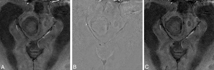
Background and purpose: SWI is a high-resolution 3D, fully velocity-compensated gradient-echo sequence that uses both magnitude and phase data. The purpose of this study was to investigate the phase behavior of the capsule of pyogenic brain abscesses with noncontrast SWI.
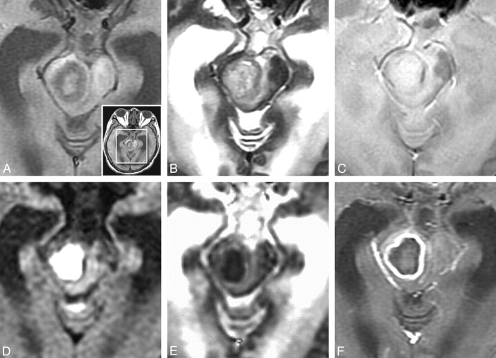
Materials and methods: Fourteen patients with pyogenic brain abscesses were studied at 1.5T. In all of the patients, SWI images were obtained and reviewed in addition to conventional MR images. Phase values within the abscess capsule were measured and compared with those from the abscess cavities and contralateral normal white matter using 1-way repeated measures ANOVA with post hoc Bonferroni analysis.
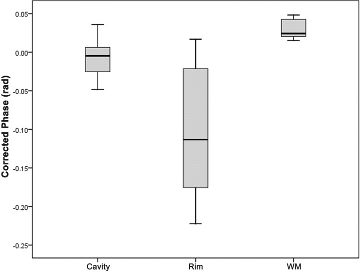
Results: SWI phase images showed mild hypointesity in 6 patients, isointensity in 3 patients, and mixed iso- to mild hypointensity in 5 patients. The means of phase in the cavity, rim of abscesses, and contralateral normal white matter were -7.552 × 10(-3) ± 0.024, -0.105 ± 0.080, and +0.029 ± 0.011 radians, respectively. Post hoc comparisons showed significant differences between any pair of the 3 regions (abscess cavity, rim capsule, and normal white matter) in SWI (all Ps < .005).
Conclusions: SWI phase imaging shows evidence of paramagnetic substances in agreement with the presence of free radicals from phagocytosis. SWI may provide additional information valuable in the characterization of pyogenic brain abscesses.
Figures
References
-
- Reichenbach JR, Venkatesan R, Schillinger DJ, et al. . Small vessels in the human brain: MR venography with deoxyhemoglobin as an intrinsic contrast agent. Radiology 1997;204:272–77 - PubMed
-
- Reichenbach JR, Haacke EM. High-resolution BOLD venographic imaging: a window into brain function. NMR Biomed 2001;14:453–67 - PubMed
-
- Sehgal V, Delproposto Z, Haacke EM, et al. . Clinical applications of neuroimaging with susceptibility-weighted imaging. J Magn Reson Imaging 2005;22:439–50 - PubMed
-
- Essig M, Reichenbach JR, Schad L, et al. . High resolution MR-venography of cerebral arteriovenous malformations. Radiologe 2001;41:288–95 - PubMed
Publication types
MeSH terms
LinkOut - more resources
Full Text Sources
Medical