

doi: 10.1126/science.1209678.
Epub 2012 Jan 26.
Epithelial nitration by a peroxidase/NOX5 system mediates mosquito antiplasmodial immunity
Affiliations
- PMID: 22282475
- PMCID: PMC3444286
- DOI: 10.1126/science.1209678
Item in Clipboard
Epithelial nitration by a peroxidase/NOX5 system mediates mosquito antiplasmodial immunity
Science.
.
Abstract
Plasmodium ookinetes traverse midgut epithelial cells before they encounter the complement system in the mosquito hemolymph. We identified a heme peroxidase (HPX2) and NADPH oxidase 5 (NOX5) as critical mediators of midgut epithelial nitration and antiplasmodial immunity that enhance nitric oxide toxicity in Anopheles gambiae. We show that the two immune mechanisms that target ookinetes-epithelial nitration and thioester-containing protein 1 (TEP1)-mediated lysis-work sequentially, and we propose that epithelial nitration works as an opsonization-like system that promotes activation of the mosquito complement cascade.
Figures
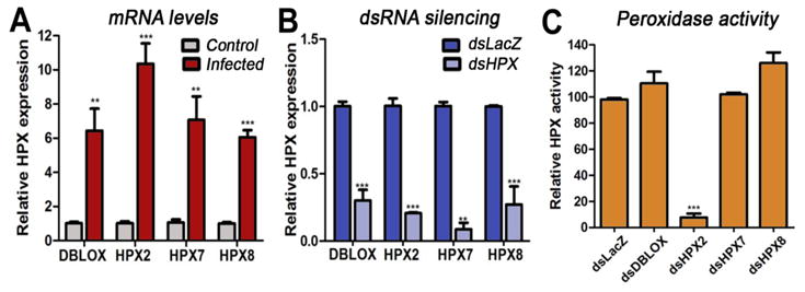
Expression and activity of midgut heme peroxidases (HPXs). (A) mRNA expression of double peroxidase (DBLOX), HPX2, HPX7, and HPX8 in the midguts of mosquitoes fed on a healthy (C, control) or a Plasmodium-infected (I, infected) mouse 24 h post-feeding (hpf); and (B) in the midguts of infected mosquitoes injected with dsLacZ or with dsRNA from each of the four HPXs 24 hpf. (C) Effect of silencing each HPX on Plasmodium-induced midgut peroxidase activity (mean ± SEM).
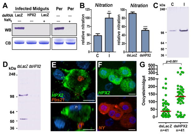
Localization of HPX2 and effect of HPX2 silencing on nitration and Plasmodium infection. (A) In vitro bovine serum albumin (BSA) nitration by a commercial peroxidase (Per) or by peroxidase from Plasmodium-infected midguts. Inhibition of peroxidase activity by addition of sodium azide (NaN3) or silencing HPX2 prevent in vitro nitration. Western blot (WB) with an anti-nitrotyrosine antibody and Coomassie blue (CB) staining of BSA. (B) Effect of Plasmodium infection (left panel) and of silencing HPX2 (right panel) in Plasmodium-infected mosquitoes on in vivo midgut nitration (mean ± SEM). Western blot detection of HPX2 protein in midgut homogenates from (C) mosquitoes fed on a healthy (C, control) or a Plasmodium-infected (I, infected) mouse 24 h post feeding (hpf) and (D) Plasmodium-infected mosquitoes injected with dsLacZ control or dsHPX2 collected 24 hpf. (E, F) Immunofluorescent staining of Plasmodium-infected midguts 24 hpf. (E) Ookinetes stained with anti-Pbs21 antibodies (red) and HPX2 (green). Lower panel shows side view; n, nuclei of apoptotic cell (blue). (F) HPX2 (upper panel) and nitrotyrosine (lower panel) expression in Plasmodium-infected midguts m (bar = 10 μm). (G) Effect of HPX2 silencing on Plasmodium infection 7 days post feeding. Each circle represents the number of parasites in individual midguts (median = red line).
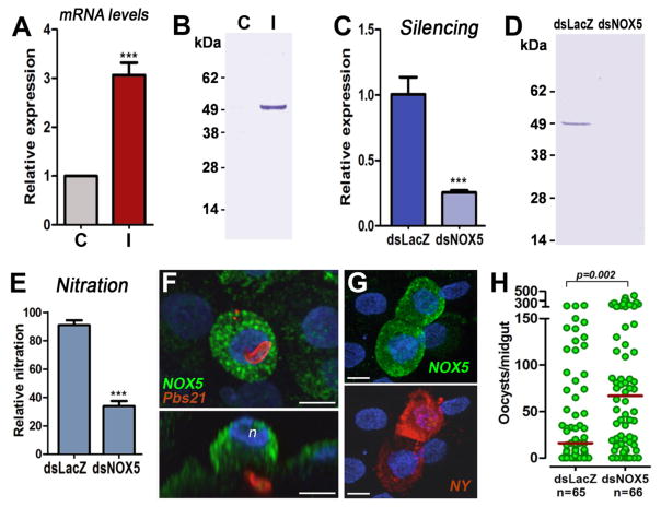
Expression and localization of NOX5 and effect of NOX5 silencing on Plasmodium infection. (A) Midgut NOX5 mRNA and (B) protein expression 24 h post-feeding (hpf) in mosquitoes fed on a healthy (C, control) or a Plasmodium-infected (I, infected) mouse. (C) Midgut NOX5 mRNA and (D) protein expression 24 hpf on mosquitoes injected with dsLacZ or dsNOX5. (E) Effect of NOX5 silencing on in vivo midgut protein nitration (mean ± SEM). (F, G) Immunofluorescence staining of midguts 24 hpf on a Plasmodium-infected mouse. (F) Ookinetes stained with Pbs21 (red) and NOX5 (green). Lower panel, side view; n, nuclei of apoptotic cell (blue). (G) Nitrotyrosine (red) and NOX5 (green) expression in cells of Plasmodium-infected midguts undergoing apoptosis (bar = 10 μm). (H) Effect of NOX5 silencing on Plasmodium infection 7 dpf. Each circle represents the number of parasites in individual midguts (median = red line).
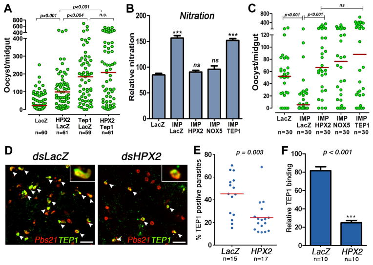
Midgut nitration and complement activation. (A) Effect of double silencing heme peroxidase 2 (HPX2) and thioester-containing protein 1 (TEP1) on Plasmodium infection 7 days post feeding (dpf). (B) Effect of silencing IMPer or co-silencing NADPH 5 (NOX5), HPX2, or TEP1 on in vivo protein nitration and (C) Plasmodium infection 7 dpf. Each circle represents the number of parasites in an individual midgut; the horizontal lines indicate medians. Effect of HPX2 silencing on (D) TEP1 staining (green) on ookinetes labeled with anti-Pbs21 antibody (red), (E) percentage of parasites on individual midguts that stain positive for TEP1, and (F) Plasmodium-induced TEP1 binding (difference in binding to infected and uninfected midguts) 28–30 hours post-feeding. Arrowheads indicate examples of TEP1-positive parasites.
References
Publication types
MeSH terms
Substances
Grants and funding
LinkOut - more resources
Full Text Sources
Other Literature Sources
Research Materials
