Comparative Study
. 2012 Apr;23(4):629-40.
doi: 10.1681/ASN.2011050449. Epub 2012 Jan 26.

Subtotal ablation of parietal epithelial cells induces crescent formation

Affiliations
Comparative Study

Subtotal ablation of parietal epithelial cells induces crescent formation

Eva-Maria Sicking et al. J Am Soc Nephrol. 2012 Apr.

Abstract

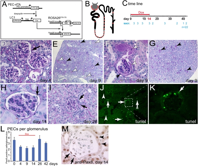
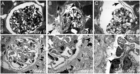
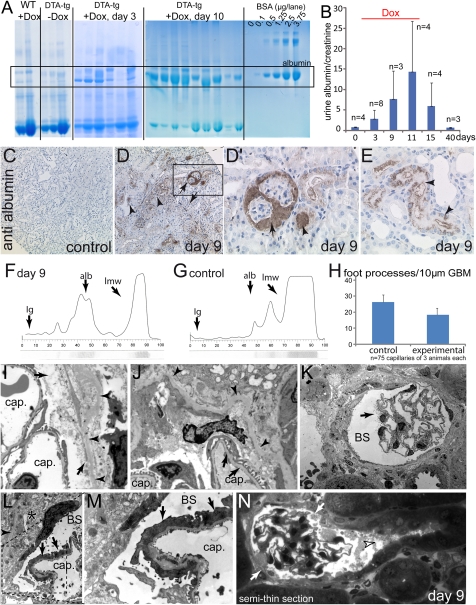
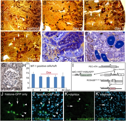
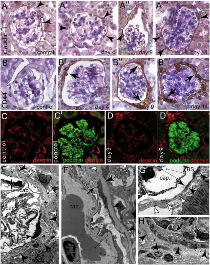
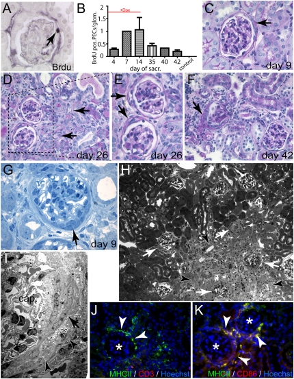
Parietal epithelial cells (PECs) of the renal glomerulus contribute to the formation of both cellular crescents in rapidly progressive GN and sclerotic lesions in FSGS. Subtotal transgenic ablation of podocytes induces FSGS but the effect of specific ablation of PECs is unknown. Here, we established an inducible transgenic mouse to allow subtotal ablation of PECs. Proteinuria developed during doxycycline-induced cellular ablation but fully reversed 26 days after termination of doxycycline administration. The ablation of PECs was focal, with only 30% of glomeruli exhibiting histologic changes; however, the number of PECs was reduced up to 90% within affected glomeruli. Ultrastructural analysis revealed disruption of PEC plasma membranes with cytoplasm shedding into Bowman's space. Podocytes showed focal foot process effacement, which was the most likely cause for transient proteinuria. After >9 days of cellular ablation, the remaining PECs formed cellular extensions to cover the denuded Bowman's capsule and expressed the activation marker CD44 de novo. The induced proliferation of PECs persisted throughout the observation period, resulting in the formation of typical cellular crescents with periglomerular infiltrate, albeit without accompanying proteinuria. In summary, subtotal ablation of PECs leads the remaining PECs to react with cellular activation and proliferation, which ultimately forms cellular crescents.


Figures

Figure 1.
Figure 1.
Transgenic model for inducible ablation of PECs. (A) Schematic of the transgenes. The reverse tetracycline-inducible transactivator (PEC-rtTA) is expressed in PECs. Upon administration of doxycycline, Cre recombinase is transiently expressed from the second transgene (LC1). This activates expression of an attenuated DTA (DTA176). (B) Schematic of expected cellular ablation within the nephron. The transgenic PEC-rtTA is active in PECs, the loop of Henle, and the thick ascending limp (as indicated with an X). In addition, single scattered cells are targeted within the proximal tubule and the collecting duct. (C) Study timeline. Animals were treated with doxycycline for 14 days and killed at the indicated time points (numerals indicate the number of sacrificed animals per time point). (D through I) PAS-stained paraffin section of the kidney at different time points. (D) On day 4, no histomorphological abnormalities were observed in PECs (arrow) or elsewhere within the kidney (female). (E) On day 9, protein was observed within the Bowman’s space of a fraction of the glomeruli (black arrowheads). Protein-filled tubuli are marked by white arrowheads. (F) Protein has leaked into the primary urine (white arrowheads). Parts of the inner aspect of Bowman’s capsule are covered by fuzzy material (black arrowhead). (G) Within the renal papilla, a fraction of the tubules contain protein (arrowheads; mice in E through G are male). (H) On day 14, PECs appear partly detached (arrow, female). (I) Late time points are characterized by extracapillary proliferations (i.e., cellular crescents, arrows, 10-week-old female mice). (J) TUNEL assay 9 days after induction. A fraction of the glomeruli contain TUNEL-positive PECs (arrows), whereas other glomeruli do not show any TUNEL-positive cells (arrowheads). Scattered tubular TUNEL-positive cells could be observed (arrow with tails). (K) Higher magnification of inset in J. Cells lining the inner aspect of Bowman’s capsule contained DNA fragments. Negative control not shown. (L) Pax8-positive cells quantified per glomerulus at different time points (n=3 per time point, 50 glomeruli per mouse). Overall, approximately 30% of all PECs were ablated. (M) Representative image of a glomerulus. Pax8-positive oval nuclei of cells with a flat cytoplasm were counted as PECs. Tubular cells lining Bowman’s capsule were not counted as PECs (arrowheads, male). Dox, doxycycline; sacr, sacrificed.
Figure 2.
Figure 2.
Ultrastructural analysis of subtotal PEC ablation. (A) Normal glomerulus 9 days after induction. (B) Cellular debris (black arrowhead) and protein within Bowman’s space on day 9. Remnants of PECs line Bowman’s capsule (white arrowheads), also within an adjacent glomerulus (arrow). (C) PECs have partially detached from Bowman’s capsule (arrow) (A through C, semi-thin sections). (D and D′) TEM images of a glomerulus 9 days after induction of PEC ablation. A viable PEC is marked by a white arrow. The remaining PECs are disintegrating (black arrowheads; D′ shows a higher magnification of the inset in D). (E) In this higher magnification, the apical plasma membrane of the PEC has disappeared. The cellular contents including the organelles are in direct contact with Bowman’s space (arrowhead). Bowman’s capsule (i.e., the PEC basement membrane) is wrinkled but still intact (white arrow, all animals aged 8 weeks; female mice are shown in A, C, and D, and male mice are shown in B and E).
Figure 3.
Figure 3.
Ablation of PECs induces acute permeability defects of the glomerular filtration barrier. (A) Five microliters of urine from different experimental animals was run on SDS-PAGE and stained with Coomassie. Significant progressive albuminuria was observed exclusively in mice subjected to acute PEC ablation (see BSA standards for comparison). (B) The albumin/creatinine ratios were determined in 17-hour urine collections at the time points indicated. Progressive albuminuria was observed during the PEC ablation period (doxycycline administration), which was fully reversible when doxycycline was discontinued. (C–E) Immunohistologic staining for mouse albumin (hematoxylin counterstain) in control and experimental animals on day 9 (male). (D and D′) Significant staining was observed in Bowman’s space and associated tubuli (arrows). Significantly more albumin reabsorption droplets were detected in other tubules (arrowheads). (F and G) SDS-PAGE with subsequent silver stain (lower part) and densitometric analysis (graph) of a proteinuric animal (day 9) and a healthy control animal. The expected position of Ig, albumin (alb), and low molecular weight proteins (lmw, which are abundant in mice) are indicated. (H) The number of foot processes was evaluated in 25 capillary circumferences per length of GBM of three experimental or control animals each (n=75). (I and J) Normal foot processes on capillary loops (cap) within the immediate vicinity of disintegrating PECs (arrows). The PEC basement membrane is wrinkled but intact (arrowheads). (K) Focal microvillous transformation of a single podocyte (arrow) projecting into Bowman’s space (BS). (L and M) Focal effacement of podocyte foot processes (arrows) on a segment of a capillary loop. An electron-dense viable PEC can be seen extending onto the cellular debris (asterisk). The intact Bowman’s capsule is marked by arrowheads (G shows a higher magnification of the inset in F). (H) Representative example of semi-thin serial sections of an affected glomerulus (see Supplemental Figure 2 for the entire series). Bowman’s space is filled with cellular debris (arrowhead); however, the tubular outflow is not blocked. PECs are partially disintegrated (arrows) (mice in I–N are male).
Figure 4.
Figure 4.
No evidence for tubular obstruction or ectopic cell ablation in the glomerulus. (A–F) To test if tubular obstruction occurs as a result of PEC ablation, experimental animals were injected with gold-labeled BSA 15 minutes before sacrifice (A–D, 9-µm thick sections). In control animals, gold-labeled BSA reabsorption occurred in the proximal part of proximal tubules throughout the entire cortex (A, arrows). (B) The same was true for experimental animals 4 days after induction with doxycycline. (C) On day 9, gold-labeled BSA was observed as a brown color in proximal tubules throughout the entire cortex similar to controls (black arrows). Because of proteinuria, gold-labeled BSA was also observed as a gray color in other parts of the tubular system (white arrows). (D) As a positive control for focal tubular obstruction by protein casts, a Thy1.1 transgenic mouse is shown 1 day after injection of Thy1.1 antiserum, which induces nephrotic-range proteinuria within minutes. Reabsorption of gold-labeled BSA can only be seen in some proximal tubuli (black arrow), whereas other tubuli are unstained (white arrows). (E) On semi-thin sections, gold-labeled BSA was still present within the capillary lumen (arrowheads) and reabsorbed in proximal tubuli (arrows). (F) In the Thy1.1 transgenic mouse with tubular obstruction, gold-labeled BSA–containing tubules (white arrow) were found next to protein-filled tubules without any gold-labeled BSA but instead with signs of cellular degeneration (black arrow). Some tubular lumina were blocked by protein casts (pc). All animals in A–F were 16-week-old male mice. (G and H) Absolute podocyte number per glomerular cross-section (n=150 in three animals of each time point as indicated; G, female). (I) To test whether leaky expression of the PEC-rtTA transgene occurs, an additional transgene tetO7-HIST1H2BJ/GFP was introduced that expresses histone-eGFP in a doxycycline-inducible fashion. (J and J′) When doxycycline was administered in triple transgenic mice that only lacked the ROSADTA176 transgene (suicide transgene), histone-eGFP was expressed specifically in almost all PECs (arrowheads) and no trace was observed elsewhere in the glomerulus (arrow). (K and K′) When doxycycline was administered in quadruple transgenic mice, histone-eGFP labeled PECs were observed much less frequently (arrowhead, 5 days after induction). No labeling of other glomerular cells, specifically of podocytes was observed (mice in J–K′ are male).
Figure 5.
Figure 5.
Activation of PECs after subtotal ablation. Immunohistologic stainings for PEC marker claudin-1 (A–A′′′) or PEC activation marker CD44 (B–B′′′) combined with PAS counterstainings were performed at different time points as indicated. In controls, PECs stained only for the PEC marker claudin-1, confirming that PECs are not activated under physiologic conditions (A and B, arrowheads). Upon induction of PEC ablation, PECs (arrowheads) expressed CD44 progressively (arrows) (mice in A′, A′′, B, and B′ are female; mice in A, A′′′, B′′, and B′′′ are male). (C and D) To analyze podocytes, double stainings for podocyte-marker podocin (green) and podocyte activation marker desmin (red) were performed in control animals and 9 days after induction. In both groups, a similar expression pattern was observed. Podocytes stained positive for podocin. Desmin was expressed predominantly within the mesangium but also in endothelial cells (female). (E) Viable electron-dense PECs (arrows) extended long thin processes across areas of Bowman’s capsule covered by cellular debris (asterisks). In addition, focal podocyte foot process effacement can be seen (thin arrows). Bowman’s capsule remains intact (arrowheads) even in areas with no viable PECs (white arrowheads). (F and G) Higher magnification of a cellular extension of a PEC (arrow points to the tip of the extension, white arrowhead point to the denuded area of Bowman’s capsule). Thin arrows point to focal podocyte foot process effacement (cap, capillary lumen; BS, Bowman’s space). (H) An accumulation of PECs (P) is shown within cellular debris. Arrowheads point to Bowman’s capsule. A cellular extension is seen emerging from the right lining the inner aspect of Bowman’s capsule and forming a multilayered crescent (arrow).
Figure 6.
Figure 6.
Proliferation of PECs results in formation of cellular crescents. (A and B) To determine if PECs engage in cellular proliferation, the number of BrdU-positive PECs per glomerulus was determined using immunohistochemistry at the indicated time points (A, arrow indicates a BrdU-positive PEC, female). Proliferation of PECs increased progressively during the administration of doxycycline and remained elevated even after 40 days. (C–F) PAS-stained paraffin sections at different time points. At early time points, cellular crescents can be seen in glomeruli without significant tubular damage (C, arrow). At later time points, glomeruli with cellular crescents are found next to or within areas of tubular degeneration (D, arrows; E shows a higher magnification of inset in D). Cellular crescents show the typical morphology of multilayered extracapillary proliferations (E and F, arrows; mice in C through F are female). (G) Semi-thin section of a cellular crescent (arrow) with the typical morphology. No capillary necrosis could be seen (male). (H) Semi-thin section of an area with significant tubular degeneration (arrowheads) and associated glomeruli with cellular crescents (white arrows). (I) TEM of a cellular crescent with multiple layers of PECs (arrow), cellular debris (asterisk), and an intact Bowman’s capsule (arrowheads). The capillary tuft (cap.) shows normal capillary loops. (J and K) Glomeruli with extracapillary proliferations (asterisk) recruited a typical periglomerular infiltrate of MHCII-positive, CD3-negative, CD86-positive cells, representing activated dendritic cells (arrowheads).

References

    1. Moeller MJ, Soofi A, Hartmann I, Le Hir M, Wiggins R, Kriz W, Holzman LB: Podocytes populate cellular crescents in a murine model of inflammatory glomerulonephritis. J Am Soc Nephrol 15: 61–67, 2004 - PubMed
    1. Ding M, Cui S, Li C, Jothy S, Haase V, Steer BM, Marsden PA, Pippin J, Shankland S, Rastaldi MP, Cohen CD, Kretzler M, Quaggin SE: Loss of the tumor suppressor Vhlh leads to upregulation of Cxcr4 and rapidly progressive glomerulonephritis in mice. Nat Med 12: 1081–1087, 2006 - PubMed
    1. Smeets B, Uhlig S, Fuss A, Mooren F, Wetzels JF, Floege J, Moeller MJ: Tracing the origin of glomerular extracapillary lesions from parietal epithelial cells. J Am Soc Nephrol 20: 2604–2615, 2009 - PMC - PubMed
    1. Smeets B, Kuppe C, Sicking EM, Fuss A, Jirak P, van Kuppelvelt TH, Endlich KH, Wetzels JF, Grone HJ, Floege J, Moeller MJ: Parietal epithelial cells participate in the formation of sclerotic lesions in focal and segmental glomerulosclerosis (FSGS). J Am Soc Nephrol 22: 1262-1274, 2011 - PMC - PubMed
    1. Appel D, Kershaw DB, Smeets B, Yuan G, Fuss A, Frye B, Elger M, Kriz W, Floege J, Moeller MJ: Recruitment of podocytes from glomerular parietal epithelial cells. J Am Soc Nephrol 20: 333–343, 2009 - PMC - PubMed

Publication types

MeSH terms

LinkOut - more resources