. 2012 Mar 15;72(6):1384-94.
doi: 10.1158/0008-5472.CAN-11-2905. Epub 2012 Jan 26.

Myeloid progenitor cells in the premetastatic lung promote metastases by inducing mesenchymal to epithelial transition

Affiliations

Myeloid progenitor cells in the premetastatic lung promote metastases by inducing mesenchymal to epithelial transition

Dingcheng Gao et al. Cancer Res. .

Abstract

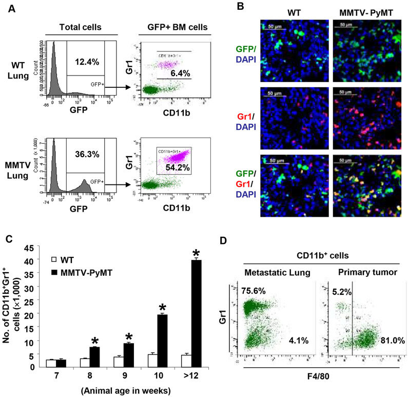
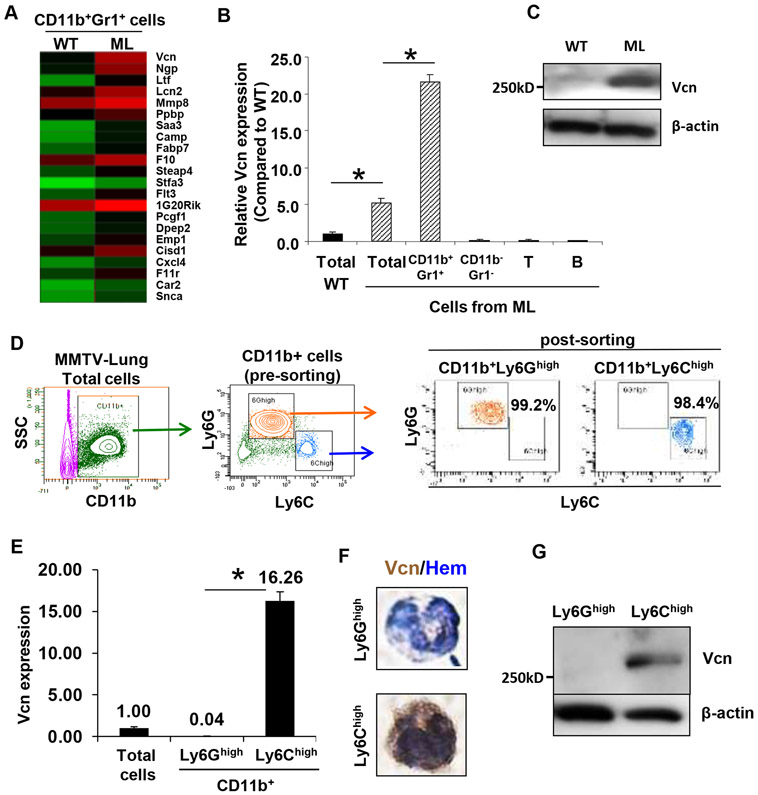
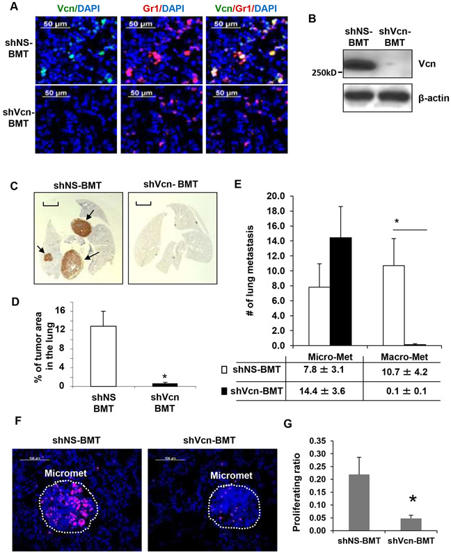
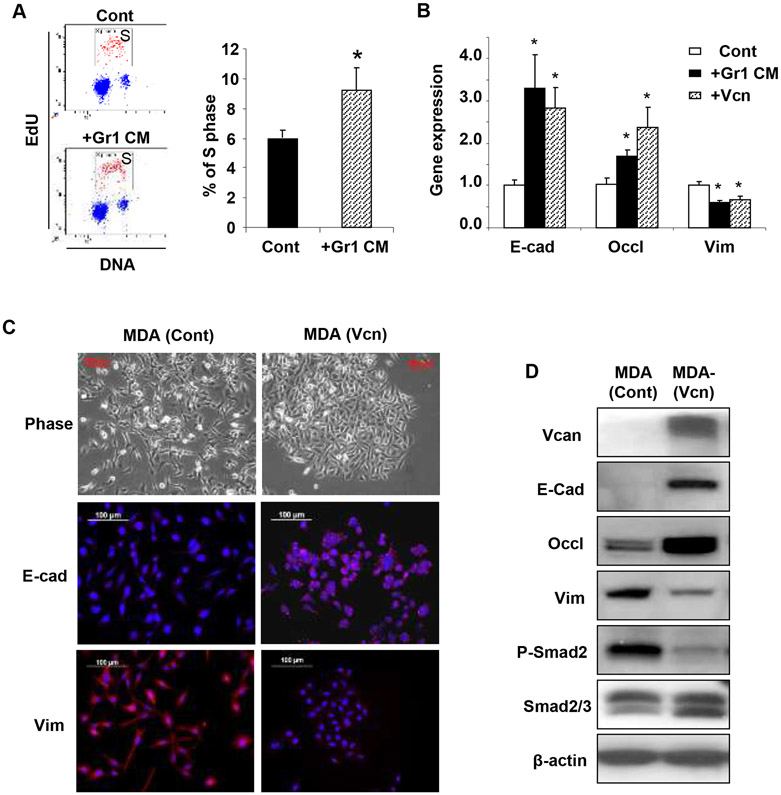
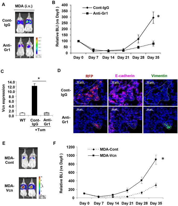
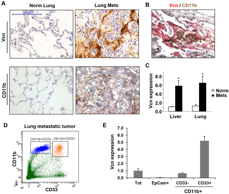
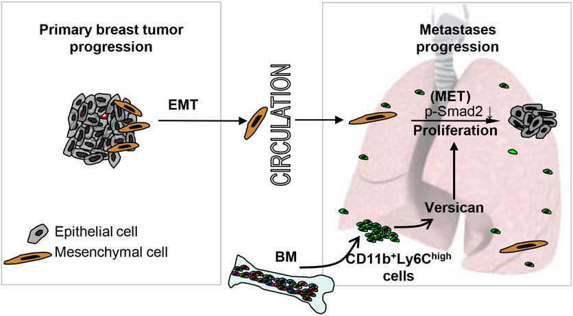
Tumors systemically initiate metastatic niches in distant target metastatic organs. These niches, composed of bone marrow-derived hematopoietic cells, provide permissive conditions for future metastases. However, the mechanisms by which these cells mediate outgrowth of metastatic tumor cells are not completely known. Using mouse models of spontaneous breast cancer, we show enhanced recruitment of bone marrow-derived CD11b(+)Gr1(+) myeloid progenitor cells in the premetastatic lungs. Gene expression profiling revealed that the myeloid cells from metastatic lungs express versican, an extracellular matrix proteoglycan. Notably, versican in metastatic lungs was mainly contributed by the CD11b(+)Ly6C(high) monocytic fraction of the myeloid cells and not the tumor cells or other stromal cells. Versican knockdown in the bone marrow significantly impaired lung metastases in vivo, without impacting their recruitment to the lungs or altering the immune microenvironment. Versican stimulated mesenchymal to epithelial transition of metastatic tumor cells by attenuating phospho-Smad2 levels, which resulted in elevated cell proliferation and accelerated metastases. Analysis of clinical specimens showed elevated versican expression within the metastatic lung of patients with breast cancer. Together, our findings suggest that selectively targeting tumor-elicited myeloid cells or versican represents a potential therapeutic strategy for combating metastatic disease.


Figures

Figure 1.
Figure 1.. Myeloid cells are recruited to the lung in MMTV-PyMT mice.
A, Flow cytometry plots showing increased recruitment of BM-derived (GFP+) CD11b+Gr1+ myeloid cells in the lungs of MMTV-PyMT mice (12 weeks old) compared to wild type (WT) mice. Representative plots were derived from three independent experiments. B, Immunostaining showing increased recruitment of BM-derived GFP+Gr1+ cells in the lungs of MMTV-PyMT mice compared to WT mice. C, Kinetic analysis of the recruitment of CD11b+Gr1+ myeloid cells in the lungs of MMTV-PyMT mice (age in weeks). The numbers of CD11b+Gr1+ cells were normalized to 1×10⁵ total lung cells analyzed per animal. Mean±s.d. *p<0.01 as compared to WT mice of same age. D, Flow cytometry analysis showing the majority of myeloid cells are Gr1+F4/80 in the metastatic lung, but are Gr1F4/80+ macrophages in the primary tumors.
Figure 2.
Figure 2.. Versican is expressed by CD11b+Ly6Chigh myeloid cells in the metastatic lung.
A, Heat map obtained from gene expression profiling of CD11b+Gr1+ cells sorted from WT lungs and metastatic lungs (ML). Each row represents a differentially upregulated gene (≥ 2 fold) in ML samples, and each column represents data from one comparison (average of three biological replicates). Red, high expression; Green, low expression. B, RT-PCR showing versican expression in Gr1+ myeloid cells, Gr1 stromal cells, T cells (CD3+) and B cells (B220+) sorted from WT or ML (representative data from two individual experiments). Versican expression was normalized to the internal control (GAPDH). The relative expression level is shown as compared to WT lung, *p<0.01 as indicated. C, Western blot showing versican levels in the lungs from MMTV-PyMT mice and control mice. D, Left panels, Flow cytometry sorting of CD11b+Ly6Ghigh and CD11b+Ly6Chigh cells from the lungs of MMTV-PyMT mice. Right panels, Flow cytometry showing purity of the post-sorted cells. E, RT-PCR of versican expression in CD11b+Ly6Ghigh and CD11b+Ly6Chigh cells and total cells from metastatic lungs. GAPDH was used as internal control. Representative data is from three independent experiments. *p<0.01. F, IHC of versican on CD11b+Ly6Ghigh and CD11b+Ly6Chigh cells. Hematoxylin (Hem) was used to determine the morphology of the nucleus. G, Western blot showing versican in sorted CD11b+Ly6Ghigh and CD11b+Ly6Chigh cells from metastatic lungs of MMTV-PyMT mice.
Figure 3.
Figure 3.. Versican deficiency in myeloid cells impairs macrometastases in MMTV-PyMT mice.
A, Representative microscopy images showing versican (green) deficiency in Gr1+(red) cells in the lungs of shVcn-BMT MMTV-PyMT mice compared to shNS-BMT mice (10-weeks old). B, Western blots showing versican in the lungs from shNS-BMT and shVcn-BMT MMTV-PyMT mice. C, Representative lung images (stained with anti-PyMT antibody) of shNS-BMT and shVcn-BMT MMTV-PyMT mice (15-weeks old). Arrows mark pulmonary metastases. Scale bar, 2mm. D, Quantitation of the area of metastases in shNS-BMT and shVcn-BMT MMTV-PyMT mice (15-weeks old, n=7-9 per group, *p<0.01 as compared to shNS-BMT group). E, Quantitation of the number of metastases in shNS-BMT and shVcn-BMT MMTV-PyMT mice. The average number of micrometastasis (<1mm in diameter) and macrometastasis (>1mm in diameter) were counted from at least 5 sections from individual animals, n = 7-9, *p<0.01 as compared to shNS-BMT group. F, Staining of Ki67 (magenta) showing proliferating cells in the micrometastases in lungs of shNS-BMT mice as compared with that from shVcn-BMT mice. G, Quantification of the proliferation ratio in micrometastases showing less proliferating cells in lesions from shVcn-BMT mice compared to shNS-BMT mice. n=10, *p<0.01.
Figure 4.
Figure 4.. Versican enhances proliferation and induces MET in metastatic tumor cells.
A, Flow cytometry plots depicting cell cycle analysis of metastatic breast cancer MDA-MB-231 cells treated with Gr1+ CM or control media (Cont). Percentage of S phase cells are indicated. Representative plots are from 3 experiments. B, Quantitative RT-PCR analysis of epithelial/mesenchymal marker expression in MDA-MB-231 cells treated with Gr1 CM, or biochemically purified versican (+Vcn, 2.5 μg/mL). n=3, *p<0.01 as compared to untreated cells (Cont). C, Microscopy images of MDA-control cells and MDA-versican cells (MDA-Vcn) depicting morphology (Phase) and expression of epithelial/mesenchymal markers. E-cad,E-cadherin; Vim,vimentin. D, Western blot analysis of versican (>250 KDa), EMT markers, phospho (p)-Smad2, and total Smad2/3 expression in MDA (Vcn) cells and MDA (Cont) cells. Representative data from three experiments.
Figure 5.
Figure 5.. Versican deficiency impairs MET-mediated lung metastases in vivo.
A, BLI images showing that depletion of versican-producing Gr1+ cells by anti-Gr1 antibody treatment inhibited lung metastases formed by MDA-MB-231 cells compared to IgG treated controls. Scale bar depicts the photon flux (photons per second). B, Quantification of pulmonary metastases by BLI at days 0,7,14,21,28,35 after inoculation. The relative BLIs are normalized using the values from day 0, n=10. *p<0.01. C, Myeloid cells harvested from WT and control antibody treated (Cont-IgG) or anti-Gr1 antibody treated (Anti-Gr1) tumor-bearing animals. Versican expression was analyzed by RT-PCR (n=3 in each group). GAPDH was used as internal control for RT-PCR. *p<0.01 as indicated. D, Images showing expression of E-cadherin (magenta) and vimentin (green) in pulmonary metastases formed by MDA-MB-231 cells in mice treated with control IgG or anti-Gr1 antibodies. Tumor cells were detected by the intrinsic RFP signal (red). The metastatic lesions are shown within the dotted line. Note that lung epithelial cells surrounding the metastases also stain for E-cadherin. E, Versican promotes lung metastasis in vivo. Representative bioluminiscence images showing accelerated metastases of MDA-Vcn cells in the lung after tail vein injection as compared to MDA-Cont cells (n=5). F, Quantification of pulmonary metastases by BLI at days 0,7,14,21,28,35 after inoculation. The relative BLIs are normalized using the values from day 0, n=5, *P<0.01.
Figure 6.
Figure 6.. Versican expression in the metastatic tumors of breast cancer patients.
A, Representative IHC images of lungs from normal healthy subjects (Normal Lung, n=5) and breast cancer patients with metastases (Lung Mets, n=11) showing stromal versican expression. Scale bar,200μm. B, IHC of lung metastases from a breast cancer patient showing colocalization of recruited CD11b+ (brown) myeloid cells with versican (red). C, Quantification of versican expression by RT-PCR in lung metastases (n=6) and liver metastases (n=11) of breast cancer patients as compared to healthy normal tissues (n=5, n=4 respectively). D, Flow cytometry showing that CD11b+ cells in the human metastatic lungs are comprised of CD11b+CD33+ and CD11b+CD33 populations. E, RT-PCR of versican expression in sorted total cells (Tot), tumor cells (EpCam+), CD11b+CD33+ myeloid cells, and CD11b+CD33 cells in a breast cancer patient with lung metastases.
Figure 7.
Figure 7.. Schematic depicting the contribution of BM-derived myeloid cells to the formation of lung metastases from breast tumor.
EMT,epithelial to mesenchymal transition; MET,mesenchymal to epithelial transition; BM,bone marrow; p-Smad2, phospho-Smad2.

References

    1. Townson JL, Chambers AF. Dormancy of solitary metastatic cells. Cell Cycle. 2006;5:1744–50. - PubMed
    1. Naumov GN, Bender E, Zurakowski D, Kang SY, Sampson D, Flynn E, et al. A model of human tumor dormancy: an angiogenic switch from the nonangiogenic phenotype. J Natl Cancer Inst. 2006;98:316–25. - PubMed
    1. Gupta GP, Massague J. Cancer metastasis: building a framework. Cell. 2006;127:679–95. - PubMed
    1. Joyce JA, Pollard JW. Microenvironmental regulation of metastasis. Nat Rev Cancer. 2009;9:239–52. - PMC - PubMed
    1. Polyak K, Weinberg RA. Transitions between epithelial and mesenchymal states: acquisition of malignant and stem cell traits. Nat Rev Cancer. 2009;9:265–73. - PubMed

Publication types

MeSH terms