. 2012 Mar 15;72(6):1529-37.
doi: 10.1158/0008-5472.CAN-11-2602. Epub 2012 Jan 26.

PTP1B is an androgen receptor-regulated phosphatase that promotes the progression of prostate cancer

Affiliations

PTP1B is an androgen receptor-regulated phosphatase that promotes the progression of prostate cancer

Laurent Lessard et al. Cancer Res. .

Abstract

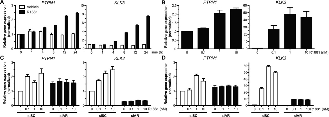
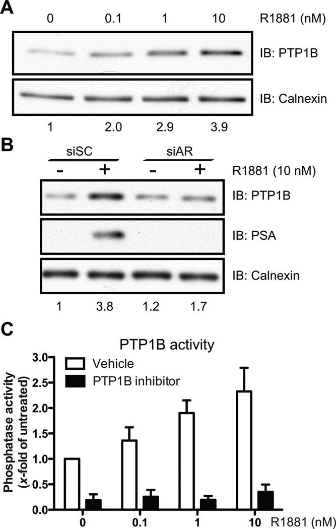
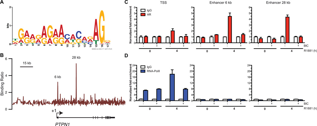
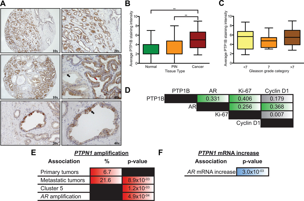
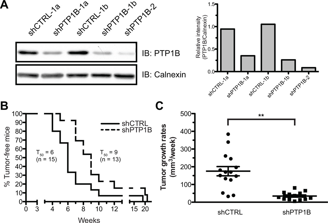
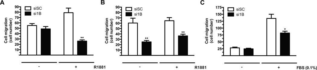
The androgen receptor (AR) signaling axis plays a key role in the pathogenesis of prostate cancer. In this study, we found that the protein tyrosine phosphatase PTP1B, a well-established regulator of metabolic signaling, was induced after androgen stimulation of AR-expressing prostate cancer cells. PTP1B induction by androgen occurred at the mRNA and protein levels to increase PTP1B activity. High-resolution chromosome mapping revealed AR recruitment to two response elements within the first intron of the PTP1B encoding gene PTPN1, correlating with an AR-mediated increase in RNA polymerase II recruitment to the PTPN1 transcriptional start site. We found that PTPN1 and AR genes were coamplified in metastatic tumors and that PTPN1 amplification was associated with a subset of high-risk primary tumors. Functionally, PTP1B depletion delayed the growth of androgen-dependent human prostate tumors and impaired androgen-induced cell migration and invasion in vitro. However, PTP1B was also required for optimal cell migration of androgen-independent cells. Collectively, our results established the AR as a transcriptional regulator of PTPN1 transcription and implicated PTP1B in a tumor-promoting role in prostate cancer. Our findings support the preclinical testing of PTP1B inhibitors for prostate cancer treatment.


Figures

Figure 1
Figure 1
PTP1B mRNA expression is induced upon R1881 treatment. (A and B) PTPN1 expression in LNCaP cells was quantified as described in Materials and Methods after treatment with R1881 (10nM or the specified concentration) or the vehicle. PTPN1 mRNA levels were significantly increased in a (A) time- and (B) dose-dependent (24hrs treatment) manner (Kruskal-Wallis test, p<0.01 and p<0.05 respectively; representative experiment, N=3, +/− SEM). Relative PSA gene (KLK3) expression levels were monitored as treatment control. (C and D) PTPN1 mRNA levels were quantified after 24hrs of R1881 treatment in cells transfected with siRNAs against the AR (siAR) or control sequences (siSC). The PTPN1 mRNA increase in siSC transfected (C) C4-2 or (D) LNCaP cells was significant (Kruskal-Wallis test, p<0.05 for both cell lines; representative experiment, N=3, +/− SEM). No R1881-dependent induction was observed in siAR conditions.
Figure 2
Figure 2
PTP1B protein and phosphatase activity is increased following R1881 treatment. (A and B) Cell lysates from LNCaP cells treated for 36hrs with (A) different R1881 concentrations or (B) with 10nM R1881 after transfection with siSC or siAR were resolved by SDS/PAGE followed by Western immunoblotting against PTP1B, PSA, and Calnexin.. Numbers below represent PTP1B levels relative to Calnexin (Representative experiments, (A) N=3 and (B) N=2). (C) PTP1B phosphatase activity in LNCaP cells was assessed after 36hrs of R1881 treatment, and a dose-dependent increase in phosphatase activity was observed (Kruskal-Wallis test, p<0.05; N=3, +/− SEM). A PTP1B inhibitor (50 µM) was used to address the specificity of the assay.
Figure 3
Figure 3
PTPN1 is a direct target of the AR. (A) Sequence logo depicting a GRE sequence enriched in AR-bound segments identified by ChIP-on-chip obtained following a de novo motif discovery. (B) Binding profile of AR from the ChIP-on-chip performed with LNCaP cells on a high-resolution tiled array covering PTPN1 and its vicinity (N=2). (C and D) Standard ChIP in LNCaP cells shows that (C) AR is recruited to the 6 kb (Kruskal-Wallis test, p<0.05) and 28 kb enhancers (Kruskal-Wallis test, near to significance, p=0.075) upon R1881 treatment. We also observed a slight but non-significant increase in AR recruitment at the TSS (Kruskal-Wallis test, p<0.05). AR binding at these sites was abrogated when cells were pretreated with BIC (Representative experiment, N=2 +/− SEM). (D) RNAPol II occupancy at the TSS is increased after treatment with R1881 (Kruskal-Wallis test, p<0.05) but not when cells where pretreated with BIC (Representative experiment, N=2 +/− SEM).
Figure 4
Figure 4
PTP1B and AR are co-expressed and co-amplified in prostate tumors. (A) Immunohistochemical analyses of a prostate tumor tissue microarray (TMA) demonstrated a strong PTP1B expression in tumor tissue (upper panel), a strong PTP1B expression in tumor cells (middle panel, red arrow), and weak expression in normal glands (middle panel, black arrow). Punctate staining was often observed in normal gland (lower panels). (B and C) Expression of PTP1B is (B) elevated in cancer tissues compared to normal or PIN glands (Mann-Whitney, **p<0.01; Median +/− SD), (C) but does not vary according to the degree of histological differentiation (Gleason score). (D) Spearman correlations between PTP1B expression and the nuclear localization of AR, Ki-67, and cyclin D1 (green boxes represent p<0.01). (E) PTPN1 amplification is significantly associated with metastatic (N=37) and not primary prostate tumors (N=181). It significantly correlates with AR amplification (all samples, N=217) an event exclusively observed in metastatic samples. PTPN1 amplification is also associated with a subset of genomic alterations (Cluster 5) which displays early recurrence after radical prostatectomy (19). (F) PTPN1 mRNA expression in prostate cancer tumors positively correlates with AR mRNA expression (samples with available mRNA expression data: N=150).
Figure 5
Figure 5
PTP1B down-regulation delays tumor occurrence and decreases tumor growth rates in vivo. (A) Western immunoblotting showing PTP1B levels in stable shCTRL and shPTP1B clones (left panel) relative to Calnexin (right panel). For the xenograft assays, a total of 40 mice were injected with either shCTRL-1a (n=10), shCTRL-1b (n=10), shPTP1B-1a (n=10), and shPTP1B-2 (n=10) clones. For data analysis, shCTRL and shPTP1B mice were pooled together (B) Tumor development occurred in 15 shCTRL mice, and 13 shPTP1B animals. Kaplan-Meier analyses of tumor occurence shows that shPTP1B tumors at a median time of onset (TD50) of 9 weeks compared to 6 weeks for shCTRL clones (Log-rank test, p<0.05). (C) Weekly monitoring of tumor growth revealed that shPTP1B tumors growth significantly slower than the shCTRL tumors (Mann-Whitney test, p<0.01).
Figure 6
Figure 6
PTP1B down-regulation impedes cell migration in vitro. (A and B) (A) LNCaP and (B) C4-2 cells transiently transfected with an siRNA against PTP1B (si1B) or a non-targeting sequence (siSC) were androgen-deprived for 48hrs. Cells were then treated with R1881 or the vehicle for 36hrs before FBS-induced cell migration as described in Materials and Methods. (C) DU145 cells were transiently transfected with si1B or siSC for 72hrs before FBS-induced cell migration (Mann-Whitney test; * p<0.05, ** p<0.01 compared to the corresponding siSC; N=3 +/− SEM).

References

    1. Jemal A, Siegel R, Xu J, Ward E. Cancer statistics, 2010. CA Cancer J Clin. 2010;60:277–300. - PubMed
    1. Shen MM, Abate-Shen C. Molecular genetics of prostate cancer: new prospects for old challenges. Genes Dev. 2010;24:1967–2000. - PMC - PubMed
    1. Damber JE, Aus G. Prostate cancer. Lancet. 2008;371:1710–1721. - PubMed
    1. Chen CD, Welsbie DS, Tran C, Baek SH, Chen R, Vessella R, et al. Molecular determinants of resistance to antiandrogen therapy. Nat Med. 2004;10:33–39. - PubMed
    1. Gurel B, Iwata T, Koh CM, Yegnasubramanian S, Nelson WG, De Marzo AM. Molecular alterations in prostate cancer as diagnostic, prognostic, and therapeutic targets. Adv Anat Pathol. 2008;15:319–331. - PMC - PubMed

Publication types

MeSH terms