Review
. 2012 Jan;109(1-2):15-25; quiz 26.
doi: 10.3238/arztebl.2012.0015. Epub 2012 Jan 9.

The differential diagnosis and treatment of normal-pressure hydrocephalus

Affiliations
Review

The differential diagnosis and treatment of normal-pressure hydrocephalus

Michael Kiefer et al. Dtsch Arztebl Int. 2012 Jan.

Abstract

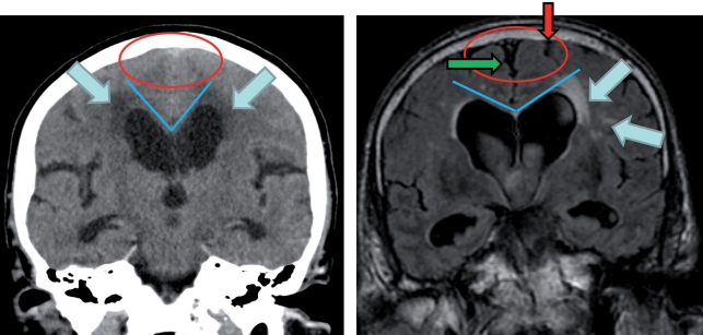
Background: Normal-pressure hydrocephalus (NPH) arises in adulthood and is characterized by a typical combination of clinical and radiological findings. The mean basal intracranial pressure is normal or mildly elevated. The typical signs of the disease are gait impairment, urinary incontinence, and dementia. The difficulty of distinguishing NPH from other neurodegenerative disorders is the likely reason why some 80% of cases remain unrecognized and untreated. According to current evidence, the spontaneous course of NPH ends, for the vast majority of patients, in dependence on nursing care.

Methods: This review article is based on relevant publications retrieved by a selective search in Medline and on national and international guidelines for the management of NPH.

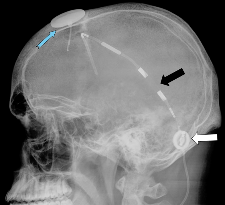
Results: Studies with a high evidence level are lacking; thus, the current state of knowledge about NPH is derived from studies of low or intermediate evidence levels, e.g., observational studies. Modern forms of treatment lead to clinical improvement in 70% to 90% of treated patients. The treatment of choice is the implantation of a ventriculoperitoneal shunt. The differential diagnosis is complicated by the fact that three-quarters of patients with NPH severe enough to require treatment also suffer from another neurodegenerative disorder. Therefore, the clinical findings and imaging studies often do not suffice to establish the indication for surgery. To do this, a further, semi-invasive diagnostic procedure is recommended. Current risk/benefit analyses indicate that shunt operations improve outcome compared to the spontaneous course of the disease.

Conclusion: Normal pressure hydrocephalus should always enter into the differential diagnosis of patients who present with its characteristic manifestations. If the diagnosis of NPH is confirmed, it should be treated at an early stage.


Figures

Figure 1
Figure 1
A current pathophysiological model of normal-pressure hydrocephalus According to the model, NPH results from low craniospinal compliance (intracranial reserve capacity) or low vascular compliance in the vessels of the circle of Willis. The model further postulates that the same causes can lessen cerebral blood flow and might also lead to Alzheimer’s disease. It would thus account for the frequent simultaneous occurrence of NPH, Alzheimer’s disease, and cerebral hypoperfusion.
Figure 2
Figure 2
Cranial CT of a patient with normal-pressure hydrocephalus before and after CSF shunting (upper and lower rows, respectively). Yellow arrows: There is hardly any change of ventricular size after shunting. The change may be very small or not noticeable at all at first glance. Blue arrows: After shunting, the CSF spaces over the convexity near the vertex are wider; this is the sole reliable sign of adequate CSF drainage through the shunt. Red arrows: The tip of the ventricular catheter lies in the right frontal horn. Photographs: Neuroradiology Dept., Universitätsklinikum des Saarlandes
Figure 3
Figure 3
Coronal head CT (left) and MRI (right) at the level of the posterior commissure: on the left image, the CSF spaces over the convexity near the vertex are narrowed (“tight convexity,” red circle), as are the medial cisterns (red circle)—these are typical findings of NPH. On the right image, however, the CSF spaces over the convexity near the vertex (red arrow) and the medial cisterns (green arrow) are widened, a finding consistent with brain atrophy. The blue lines in both images indicate the callosal angle: an angle less than 90° is typical of NPH (left image), while an angle greater than 90° is typical of brain atrophy (right image). The blue arrows indicate periventricular signal alterations. Their unilateral occurrence (right image) suggests that they are probably due to vascular encephalopathy. The abnormalities seen on the left image may well represent transependymal CSF diapedesis due to NPH Photographs: Neuroradiology Dept., Universitätsklinikum des Saarlandes
Figure 4
Figure 4
Typical cranial CT of a patient with iNPH. The CSF spaces near the vertex are narrow (blue arrows); the few wide sulci that are seen on the cerebral convexity (gray arrows) are all in the vicinity large, superficial arteries. Widening of the insular cisterns (red arrow) is a good indicator of iNPH that will respond to treatment Photographs: Neuroradiology Dept., Universitätsklinikum des Saarlandes
Figure 5
Figure 5
Diagnostic flowchart. If NPH is suspected on clinical and radiological grounds, diagnostic accuracy can be secured with invasive testing (CSF drainage or measurement of CSF dynamic variables such as compliance and resistance to outflow). Such tests are often needed for adequate confirmation of the indication for shunting, particularly in patients with iNPH. *1In the 10 m gait test, at least 20% improvement; in psychometric tests, at least 10% improvement. PVI, pressure-volume index; Rout, resistance to outflow
eFigure 1
eFigure 1
In this sagittal T2-weighted MR image of a patient with NPH, the flow-void phenomenon (blue arrows) reflects increased pendular flow of CSF in the cerebral aqueduct due to untreated hydrocephalus. Its presence supports the diagnosis, but its absence would not imply that no treatment is needed. The corpus callosum is typically thinned (red arrows). The disproportion between the third and fourth ventricles does not conclusively distinguish NPH from aqueductal stenosis. Photograph: Neuroradiology Dept., Universitätsklinikum des Saarlandes
eFigure 2
eFigure 2
Typical signal abnormalities (arrows) in which the extent of periventricular hypodensity is too great to be due to transependymal CSF seepage alone. Signal abnormalities extending this far out into the white matter must be presumed to be due mainly to microvascular changes. Photographs: Neuroradiology Dept., Universitätsklinikum des Saarlandes
eFigure 3
eFigure 3
This lateral skull film shows both a conventional differential-pressure valve (black arrow) and a gravity-controlled valve (white arrow). One can also see an ICP telemetry probe that has been implanted into the brain (blue arrow) for long-term ICP measurement, enabling optimal setting of the opening pressure of the adjustable G valve. Photograph: Neuroradiology Dept., Universitätsklinikum des Saarlandes

References

    1. Relkin N, Marmarou A, Klinge P, Bergsneider M, Black PM. Diagnosing idiopathic normal-pressure hydrocephalus. Neurosurgery. 2005;57:4–16. - PubMed
    1. Deutsche Gesellschaft für Neurologie. Leitlinie Normaldruckhydrocephalus. www.dgn.org/images/stories/dgn/leitlinien/LL2008/ll08kap_017.pdf.
    1. Ishikawa M, Hashimoto M, Kuwana N, et al. Guidelines for management of idiopathic normal pressure hydrocephalus. Neurol Med Chir (Tokyo) 2008;48(S1-23) - PubMed
    1. Ziegler U, Doblhammer G. Prevalence and incidence of dementia in Germany—a study based on data from the public sick funds in 2002. Gesundheitswesen. 2009;71:281–290. - PubMed
    1. Brean A, Fredo HL, Sollid S, Muller T, Sundstrom T, Eide PK. Five-year incidence of surgery for idiopathic normal pressure hydrocephalus in Norway. Acta Neurol Scand. 2009;120:314–316. - PubMed

MeSH terms