. 2012 Jan 27;335(6067):436-41.
doi: 10.1126/science.1213808.

Crystal structure of the human K2P TRAAK, a lipid- and mechano-sensitive K+ ion channel

Affiliations

Crystal structure of the human K2P TRAAK, a lipid- and mechano-sensitive K+ ion channel

Stephen G Brohawn et al. Science. .

Abstract

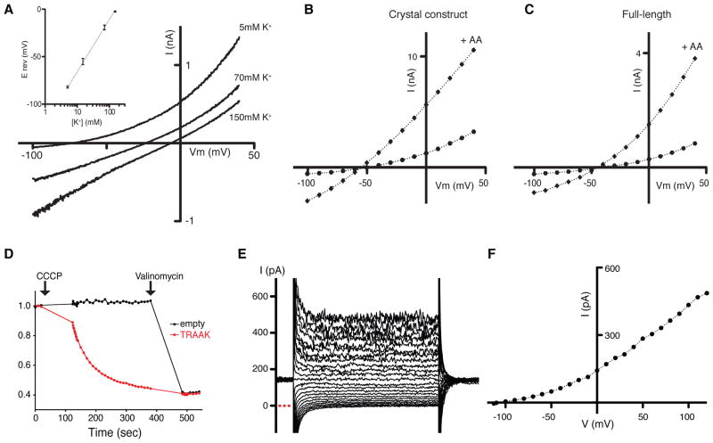
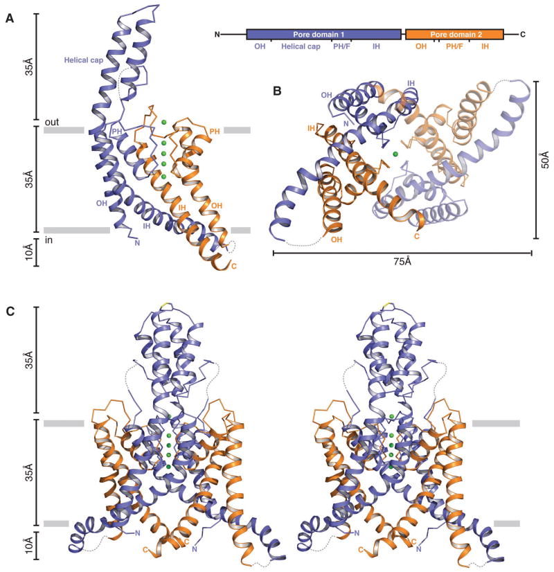
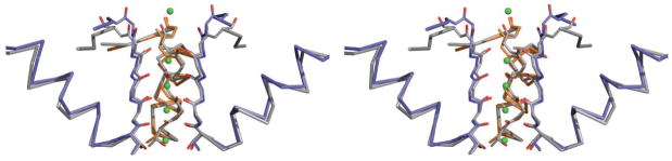
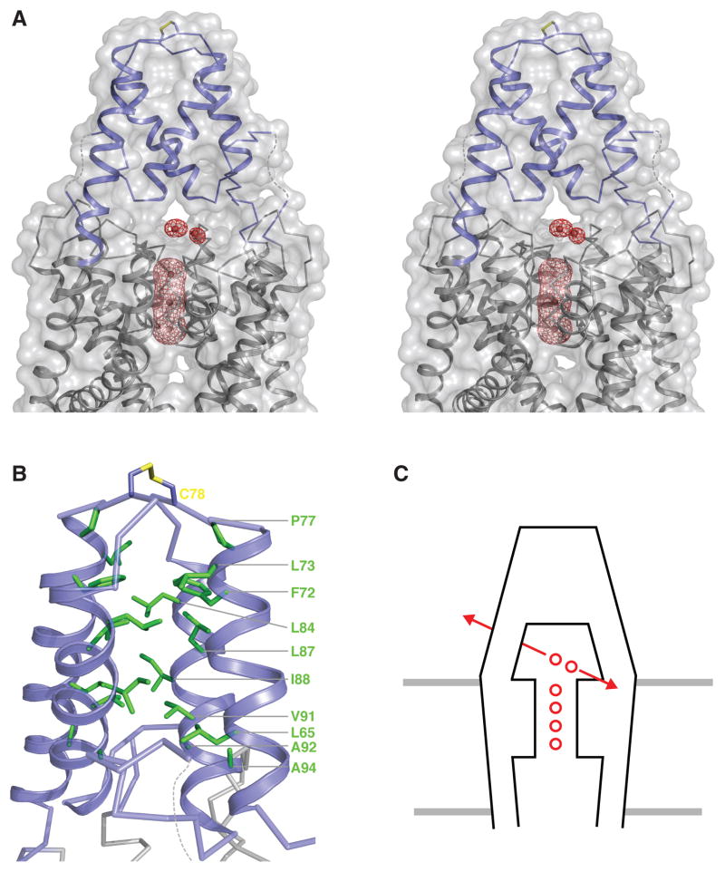
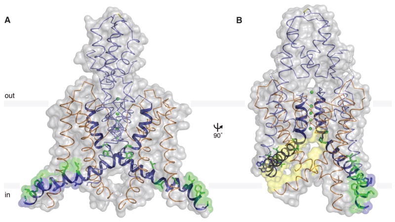
TRAAK channels, members of the two-pore domain K(+) (potassium ion) channel family K2P, are expressed almost exclusively in the nervous system and control the resting membrane potential. Their gating is sensitive to polyunsaturated fatty acids, mechanical deformation of the membrane, and temperature changes. Physiologically, these channels appear to control the noxious input threshold for temperature and pressure sensitivity in dorsal root ganglia neurons. We present the crystal structure of human TRAAK at a resolution of 3.8 angstroms. The channel comprises two protomers, each containing two distinct pore domains, which create a two-fold symmetric K(+) channel. The extracellular surface features a helical cap, 35 angstroms tall, that creates a bifurcated pore entryway and accounts for the insensitivity of two-pore domain K(+) channels to inhibitory toxins. Two diagonally opposed gate-forming inner helices form membrane-interacting structures that may underlie this channel's sensitivity to chemical and mechanical properties of the cell membrane.


Figures

Fig. 1
Fig. 1. Analysis of TRAAK channel activity
(A) Macroscopic currents from CHO cells expressing the crystal construct of TRAAK. (A) Current-voltage relationship during voltage ramp. Current was measured in whole cell mode during a voltage ramp from −100 mV to 40 mV from a holding potential of −80 mV in 800 msec. Curves recorded from cells in 5, 70, and 150 mM external [K+] are displayed. Reversal potential determined from voltage ramps recorded from cells in 5, 15, 70, and 150 mM external [K+] is plotted in the inset. (B) Arachidonic acid (AA) activation of the crystal construct of the TRAAK channel. (C) Arachidonic acid (AA) activation of the full-length channel. Current-voltage relationship is plotted from whole-cell recordings during voltage pulses from −100 to 40 mV from a holding potential of −80 mV before and after perfusion of arachidonic acid (+AA). (D) K+ flux assay in which K+ efflux drives the carbonyl cyanide m-chlorophenyl hydrazone (CCCP)-mediated uptake of protons, which are detected by fluorophore 9-amino-6-chloro-2-methoxyacridine (ACMA). Vesicles were loaded with 150 mM K+, 0 mM Na+ and assayed in buffer with 0 mM K+, 150 mM Na+. Relative fluorescence change recorded from TRAAK reconstituted (red) and empty (black) lipid vesicles is shown. Addition of the K+ ionophore valinomycin results in flux from crystal construct TRAAK reconstituted and empty vesicles. (E) Current recorded from TRAAK reconstituted into planar lipid bilayers under bi-ionic conditions. Current was recorded during voltage pulses from −120 mV to 120 mV from a holding potential of 0 mV with internal 150 mM K+, 0 mM Na+ and external 0 mM K+, 150 mM Na+ by electrophysiological convention. Zero current level is indicated with a dotted red line. (F) Current-voltage relationship plotted from data in (E).
Fig. 2
Fig. 2. Structure of TRAAK
(A) Ribbon representation of a single TRAAK protomer viewed from the membrane plane with the extracellular solution above (left). Approximate positions of the lipid bilayer boundaries are indicated as gray bars. Pore domain 1 is colored blue, pore domain 2 is colored orange, and potassium ions are shown as green spheres. Illustration of TRAAK protomer organization (right). Approximate boundaries of structural features are indicated in the illustration and labeled in the structure (N and C terminus, outer helix (OH), helical cap, pore helix (PH), selectivity filter (F), and inner helix (IH)). (B) A view of the TRAAK channel from the cytoplasmic solution. The protomer shown in (A) is rotated 90 both into the page and clockwise and the second protomer is half transparent. (C) Stereo view of TRAAK viewed from the membrane plane with the protomer shown in (A) rotated 90 . The disulfide bond bridging the apex of the helical cap is shown in stick representation with the cysteine sulfur colored yellow. Note that part of the cytoplasmic extension of protomer B (residues 180–187) is not present in the final TRAAK model due to weak electron density features. Here it is modeled from a superposition of the well-defined region in protomer A for visual clarity. Loops not modeled in the structure due to lack of interpretable electron density are drawn as dashed gray lines.
Fig. 3
Fig. 3. Convergent symmetry of the TRAAK pore helices and selectivity filter
Stereo view, from the membrane plane with extracellular solution above, shows a comparison between the TRAAK and KcsA (27) pore helices and selectivity filters. Pore helices and selectivity filter chains closest to the viewer are removed. Pore helices are shown as wires and selectivity filters as backbone sticks. TRAAK pore domain 1 is blue, TRAAK pore domain 2 is orange, and KcsA is gray with backbone carbonyl and threonine hydroxyl oxygen atoms from selectivity filter residues shown in red. K+ positions in TRAAK (green spheres) occupy canonical positions S0–S4 from the extracellular to intracellular side.
Fig. 4
Fig. 4. The TRAAK helical cap
(A) Stereo view of the helical cap viewed from the membrane plane with the extracellular solution above. TRAAK is shown as grey ribbons with the helical cap blue within a semi-transparent surface representation. A Tl+ anomalous-difference Fourier map (red mesh) calculated from 30–4.2 Å and contoured at 6σ is shown around Tl+ ions (red spheres). (B) The hydrophobic core of the helical cap. The helical cap is shown as a blue ribbon with hydrophobic residues (L65, F72, L73, P77, L84, L87, I88, V91, A92, A94) as green sticks. The C78 disulfide bond is shown as blue sticks with yellow sulfur atoms. (C) Cartoon depiction of the bifurcated ion pathway created by the TRAAK helical cap. View is analogous to (A). TRAAK (black) in a membrane (gray) coordinates four ions (red circles) in the selectivity filter and one ion directly above in site S0. A sixth ion is present in one of the two possible extracellular access/egress pathways (red arrows) that extend into the page and towards the viewer. The TRAAK helical cap blocks ion access from above and laterally in the image plane.
Fig. 5
Fig. 5. TRAAK channel inner helices and gating implications
(A) View from the membrane with extracellular solution above of the amphipathic segment of the pore domain 1 inner helix (as in Fig. 1C). TRAAK is shown in wire representation with inner helices from pore domain 1 as ribbons and key residues as sticks within a semi-transparent gray surface. Pore domain 1 is colored blue, pore domain 2 is colored orange, and K+ ions are shown as green spheres. The hinge glycine (G153), kink proline (P155), and hydrophobic face of the amphipathic segment (L168, L172, I176, I179, I182, F183, W186) are colored green. Basic residues conserved in TRAAK (R167, R173, H174, H178, K185) on the solution accessible face of the amphipathic helix are colored blue. (B) A view rotated 90 with respect to (A). The lipid bilayer accessible surface of the lateral opening into the TRAAK channel central cavity closest to the viewer is additionally colored yellow.

Comment in

References

    1. Hille B. Ion channels of excitable membranes. Sinauer Associates Inc; Sunderland, MA: 2001.
    1. Hodgkin AL, Huxley AF. Potassium leakage from an active nerve fibre. J Physiol. 1947;106:341. - PMC - PubMed
    1. Enyedi P, Czirjak G. Molecular background of leak K+ currents: two-pore domain potassium channels. Physiol Rev. 90:559. - PubMed
    1. Ketchum KA, Joiner WJ, Sellers AJ, Kaczmarek LK, Goldstein SA. A new family of outwardly rectifying potassium channel proteins with two pore domains in tandem. Nature. 1995;376:690. - PubMed
    1. Zhou XL, Vaillant B, Loukin SH, Kung C, Saimi Y. YKC1 encodes the depolarization-activated K+ channel in the plasma membrane of yeast. FEBS Lett. 1995;373:170. - PubMed

Publication types

MeSH terms

Associated data