. 2012 Feb 14;51(6):1188-98.
doi: 10.1021/bi200854q. Epub 2012 Feb 3.

Two ZnF-UBP domains in isopeptidase T (USP5)

Affiliations

Two ZnF-UBP domains in isopeptidase T (USP5)

George V Avvakumov et al. Biochemistry. .

Abstract

Human ubiquitin-specific cysteine protease 5 (USP5, also known as ISOT and isopeptidase T), an 835-residue multidomain enzyme, recycles ubiquitin by hydrolyzing isopeptide bonds in a variety of unanchored polyubiquitin substrates. Activation of the enzyme's hydrolytic activity toward ubiquitin-AMC (7-amino-4-methylcoumarin), a fluorogenic substrate, by the addition of free, unanchored monoubiquitin suggested an allosteric mechanism of activation by the ZnF-UBP domain (residues 163-291), which binds the substrate's unanchored diglycine carboxyl tail. By determining the structure of full-length USP5, we discovered the existence of a cryptic ZnF-UBP domain (residues 1-156), which was tightly bound to the catalytic core and was indispensable for catalytic activity. In contrast, the previously characterized ZnF-UBP domain did not contribute directly to the active site; a paucity of interactions suggested flexibility between these two domains consistent with an ability by the enzyme to hydrolyze a variety of different polyubiquitin chain linkages. Deletion of the known ZnF-UBP domain did not significantly affect rate of hydrolysis of ubiquitin-AMC and suggested that it is likely associated mainly with substrate targeting and specificity. Together, our findings show that USP5 uses multiple ZnF-UBP domains for substrate targeting and core catalytic function.


Conflict of interest statement

CONFLICT OF INTEREST

The authors declare that they have no conflict of interest.

Figures

Figure 1.
Figure 1.
USP5-Ub complex structure. Ribbon representation of the FL USP5 structure is shown with nUBP colored yellow, cUBP colored green, nUBA colored violet, and cUBA colored magenta. Ubiquitin, covalently attached to the catalytic cysteine, is colored orange. Peptide regions that were not modeled are represented as dashed lines. Secondary structure elements are labeled.
Figure 2.
Figure 2.
Biochemical assays. Mass-spectrometry results of reaction products (5 min at 23°C) using tetra-ubiquitin (A) and diubiquitin (B) isomers are shown. Substrates are listed above and enzyme variants used on the left. Positions of substrates and products are shown below. C) Shown are substrate-dependent initial rates determined using the fluorescence-based Ub-AMC assay, with Michaelis-Menten kinetic parameters listed in Table II.
Figure 3.
Figure 3.
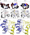
The nUBP domain. A) Cutaway view of an electrostatic surface representation (−70 to +70 kT/e) of the USP5 nUBP, cUBP, and HDAC6 UBP. Bound ubiquitin tails in cUBP and HDAC6 UBP are shown in yellow stick format. A close-up view of the corresponding ligand binding cleft is shown below the surface representation. Prominent UBP side chains are labeled and shown in stick format and their van der Waals radii are shown as dots. B) The Catalytic-nUBP interface is shown stereoscopically, with labeled domains as ribbons and interacting, labeled residues in stick format. Hydrogen and ionic bonds are shown as black dashed lines.
Figure 4.
Figure 4.
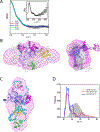
SAXS results for apo-USP5 in solution. A) Shown are representative fits of the curves from DAMMIN (red), GASBOR (blue), BUNCH (green), and EOM (cyan) to the experimental scattering profile of apo-USP5 (open black circle). The inset shows the Kratky plot (q2I(q) vs q) of the scattering profile of apo-USP5. B) Shown is the crystal structure fitted to the ab initio envelope generated by DAMMIN (magenta mesh). Also shown is a GASBOR dummy residue model (gray spheres) fitted to the DAMMIN envelope. C) A representative BUNCH model fitted to the DAMMIN envelope (magenta mesh) is shown; the linkers are displayed as cyan spheres. D) Distribution of the selected conformers (red to 20 conformers per ensemble and blue to 3 conformers per ensemble) and the initial pool of 10,000 random conformers (grey area) as a function of Rg is shown. The color code for individual domain is the same as those in Figure 1.

References

    1. Komander D (2009) The emerging complexity of protein ubiquitination, Biochem Soc Trans 37, 937–953. - PubMed
    1. Ikeda H, and Kerppola TK (2008) Lysosomal localization of ubiquitinated Jun requires multiple determinants in a lysine-27-linked polyubiquitin conjugate, Molecular biology of the cell 19, 4588–4601. - PMC - PubMed
    1. Peng J, Schwartz D, Elias JE, Thoreen CC, Cheng D, Marsischky G, Roelofs J, Finley D, and Gygi SP (2003) A proteomics approach to understanding protein ubiquitination, Nat Biotechnol 21, 921–926. - PubMed
    1. Bremm A, Freund SM, and Komander D (2010) Lys11-linked ubiquitin chains adopt compact conformations and are preferentially hydrolyzed by the deubiquitinase Cezanne, Nature structural & molecular biology 17, 939–947. - PMC - PubMed
    1. Fushman D, and Walker O (2010) Exploring the linkage dependence of polyubiquitin conformations using molecular modeling, Journal of molecular biology 395, 803–814. - PMC - PubMed

Publication types