. 2012 Jan 26;73(2):260-7.
doi: 10.1016/j.neuron.2011.11.020.

Postsynaptic complexin controls AMPA receptor exocytosis during LTP

Affiliations

Postsynaptic complexin controls AMPA receptor exocytosis during LTP

Mohiuddin Ahmad et al. Neuron. .

Abstract

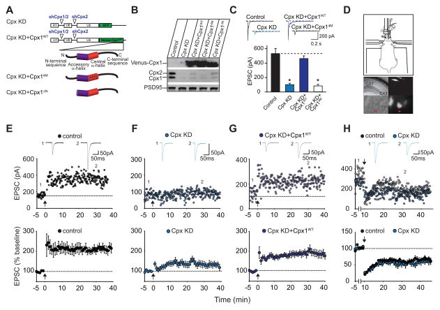
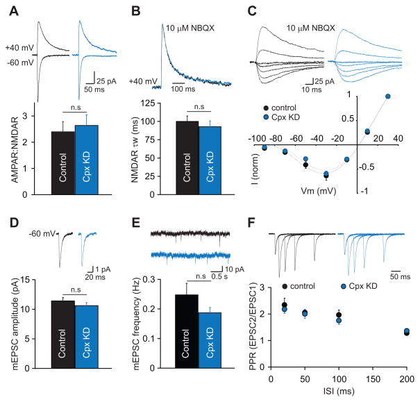
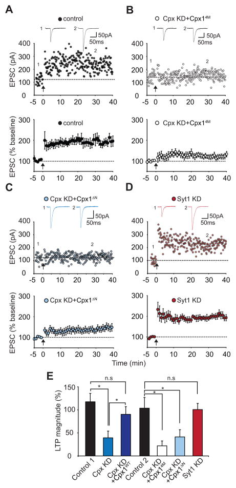
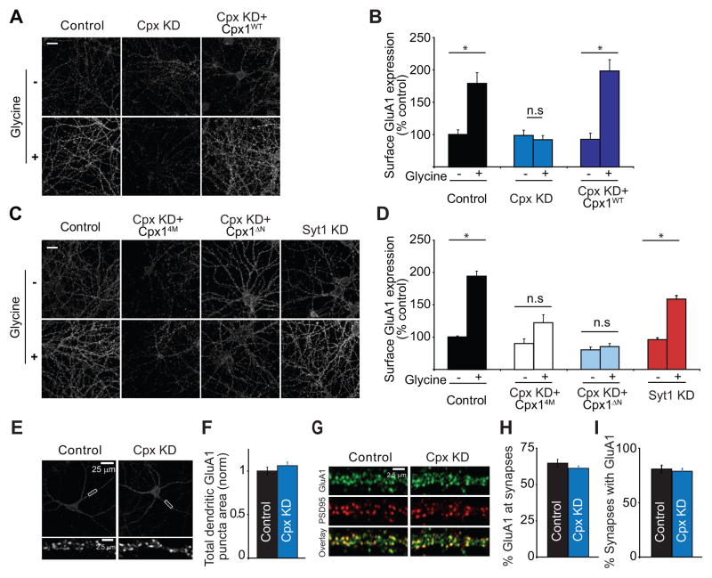
Long-term potentiation (LTP) is a compelling synaptic correlate of learning and memory. LTP induction requires NMDA receptor (NMDAR) activation, which triggers SNARE-dependent exocytosis of AMPA receptors (AMPARs). However, the molecular mechanisms mediating AMPAR exocytosis induced by NMDAR activation remain largely unknown. Here, we show that complexin, a protein that regulates neurotransmitter release via binding to SNARE complexes, is essential for AMPAR exocytosis during LTP but not for the constitutive AMPAR exocytosis that maintains basal synaptic strength. The regulated postsynaptic AMPAR exocytosis during LTP requires binding of complexin to SNARE complexes. In hippocampal neurons, presynaptic complexin acts together with synaptotagmin-1 to mediate neurotransmitter release. However, postsynaptic synaptotagmin-1 is not required for complexin-dependent AMPAR exocytosis during LTP. These results suggest a complexin-dependent molecular mechanism for regulating AMPAR delivery to synapses, a mechanism that is surprisingly similar to presynaptic exocytosis but controlled by regulators other than synaptotagmin-1.


Figures

Figure 1
Figure 1. Postsynaptic Knockdown of Complexin-1 and -2 (Cpx KD) In Vivo Blocks LTP
(A) Diagram of shRNA-expressing lentiviruses. (B) Western blots from cultured neurons showing efficacy of Cpx KD and its replacement by Venus-complexins. (C) Effects of Cpx KD and its replacement by Cpx1WT and Cpx14M on evoked AMPAR EPSCs in cultured neurons. Sample traces are shown above the bar graph. For each group, data are from 15 different neurons from 3 different cultures (Bar graphs represent means ± SEM; *p < 0.001). (D) Diagram (upper panel) showing mouse on stereotaxic apparatus for in vivo injection of lentiviruses. Low magnification DIC image of an acute hippocampal slice (middle left panel) and CA1 pyramidal cell layer (at 40X magnification, lower left panel) from injected animal. Same images (middle and lower right panels) showing GFP expression. (E) Sample experiment (top panel) and summary graph (lower panel) of LTP in control CA1 pyramidal cells recorded from slices prepared from animals injected with Cpx KD lentiviruses. In this and all subsequent figures: arrow indicates the time of tetanic stimulation; sample averaged EPSCs during the baseline (1) and 30 min post-LTP induction (2) are shown above the sample experiment. (F) Sample experiment (top panel) and summary graph (lower panel) of impaired LTP in Cpx KD cells. (G) Sample experiment (top panel) and summary graph (bottom panel) of LTP in Cpx KD cells also expressing recombinant full-length complexin-1 (Cpx KD+Cpx1WT). (H) Sample experiment (top panel) and summary graph (bottom panel) of LTD in control cells and Cpx KD cells. Bottom graphs in E–H represent means ± SEM.
Figure 2
Figure 2. Postsynaptic Knockdown of Complexin-1 and -2 (Cpx KD) In Vivo Does Not Alter Basal Synaptic Transmission
(A) The ratio of AMPAR- to NMDAR-mediated EPSCs is unchanged in Cpx KD cells. Representative EPSCs recorded at −60 mV and +40 mV are shown above the bar graph. (B) The weighted decay time constant of isolated NMDAR EPSCs at +40 mV is unchanged by postsynaptic Cpx KD. Scaled NMDAR EPSCs from two representative cells are shown above the bar graph. (C) Postsynaptic Cpx KD does not affect the I/V relationship of isolated NMDAR EPSCs. Representative traces from the two cells are shown above the graph. (D) The mean amplitude of mEPSCs is unaltered by postsynaptic Cpx KD. Averaged mEPSCs from representative cells are shown above the graph. (E) The mean frequency of mEPSCs is also unchanged by Cpx KD. Short traces of recordings from representative cells are shown above the graph. (F) Paired-pulse ratios of AMPAR EPSCs are unchanged in Cpx KD cells. EPSCs from two representative cells are shown above the graph. In all panels, bar graphs and individual points represent means ± SEM.
Figure 3
Figure 3. Postsynaptic Complexin Function in LTP Requires Its Interaction With the SNARE Complex and Its N-terminal Activation Domain
(A) Sample experiment (top panel) and summary graph (bottom panel) of LTP in control cells that were interleaved with the complexin replacement experiments. (B) Sample experiment (top panel) and summary graph (bottom panel) of impaired LTP in Cpx KD cells expressing the Cpx14M mutant (Cpx KD+Cpx14M). (C) Sample experiment (top panel) and summary graph (bottom panel) of impaired LTP in Cpx KD cells expressing Cpx1ΔN mutant (Cpx KD+Cpx1ΔN). (D) Sample experiment (top panel) and summary graph (bottom panel) of LTP in synaptotagmin-1 knockdown (Syt1 KD) cells. (E) Summary graph of the effects of the molecular manipulations of Cpx and Syt1 on LTP. *p < 0.05. Bottom graphs in A–D and bar graphs in E represent means ± SEM.
Figure 4
Figure 4. Complexin-1 and -2 Knockdown (Cpx KD) Prevents Glycine-Induced Increase in Surface AMPAR Expression in Hippocampal Neuronal Cultures
(A and B) Representative images (A) and summary graphs (B) showing increase in dendritic surface GluA1 following glycine-induced “LTP” in control cells but not in Cpx KD cells. The “LTP” was rescued by expression of wildtype complexin-1. Scale bar = 10 μm. (C and D) Representative images (C) and summary graphs (D) showing that the Cpx14M mutant and Cpx1ΔN mutant do not rescue the glycine-induced “LTP” that was generated in cells from the same culture preparations (Control) and that Syt1 KD does not block this cell culture model of LTP. (E and F) Representative images (E) and summary graphs (F) showing that the total pools of GluA1-containing AMPARs in dendrites are not affected by Cpx KD. (G–I) Representative images (G) and summary graphs (H and I) showing that the percentage of GluA1 puncta at synapses as defined by co-localization with PSD-95 (H) does not change after Cpx KD nor does the percentage of synapses containing GluA1 (I). In all panels, each bar represents mean ± SEM (*p < 0.05).

Comment in

References

    1. Adesnik H, Nicoll RA, England PM. Photoinactivation of native AMPA receptors reveals their real-time trafficking. Neuron. 2005;48:977–985. - PubMed
    1. Bhattacharyya S, Biou V, Xu W, Schluter O, Malenka RC. A critical role for PSD-95/AKAP interactions in endocytosis of synaptic AMPA receptors. Nat Neurosci. 2009;12:172–181. - PMC - PubMed
    1. Bredt DS, Nicoll RA. AMPA receptor trafficking at excitatory synapses. Neuron. 2003;40:361–379. - PubMed
    1. Cai H, Reim K, Varoqueaux F, Tapechum S, Hill K, Sorensen JB, Brose N, Chow RH. Complexin II plays a positive role in Ca2+-triggered exocytosis by facilitating vesicle priming. Proc Natl Acad Sci USA. 2008;105:19538–19543. - PMC - PubMed
    1. Collingridge GL, Isaac JT, Wang YT. Receptor trafficking and synaptic plasticity. Nat Rev Neurosci. 2004;5:952–962. - PubMed

Publication types

MeSH terms