. 2012 Jan 26;73(2):317-32.
doi: 10.1016/j.neuron.2011.10.038.

Apo-ghrelin receptor forms heteromers with DRD2 in hypothalamic neurons and is essential for anorexigenic effects of DRD2 agonism

Affiliations

Apo-ghrelin receptor forms heteromers with DRD2 in hypothalamic neurons and is essential for anorexigenic effects of DRD2 agonism

Andras Kern et al. Neuron. .

Abstract

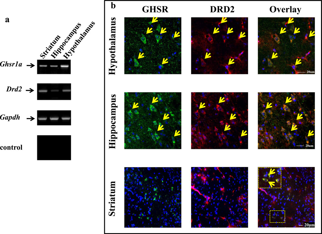
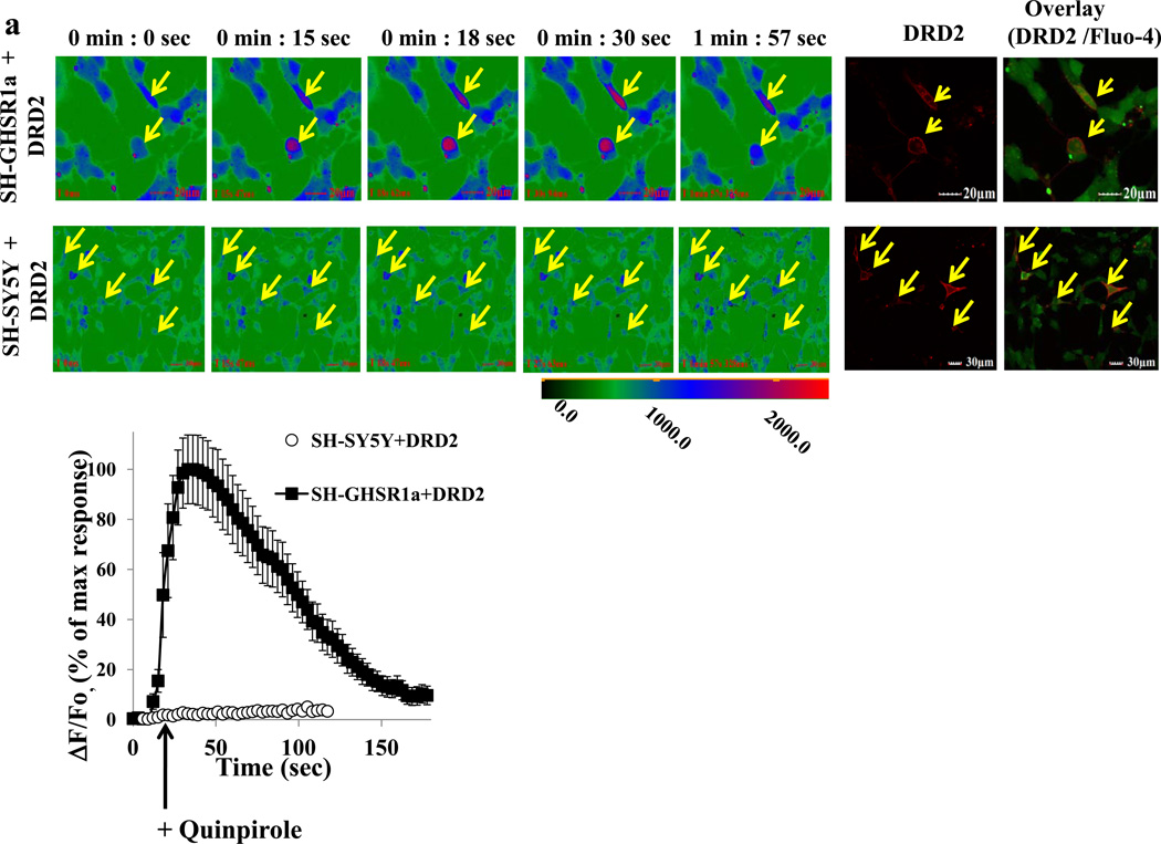
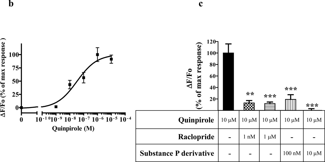
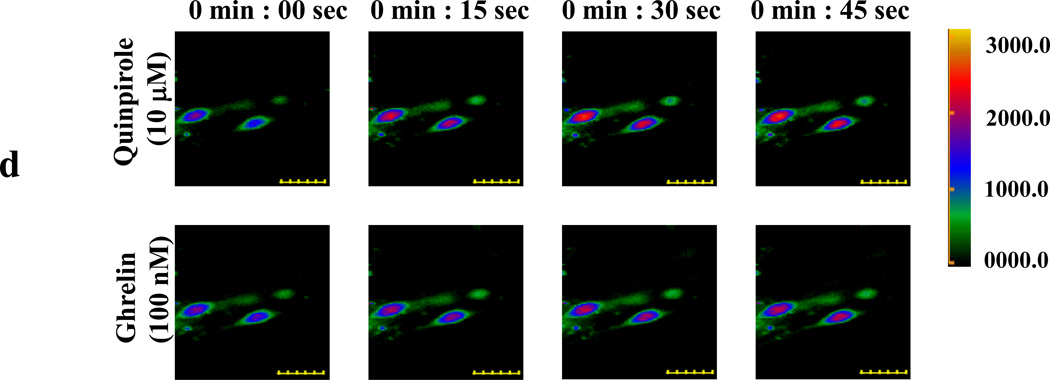
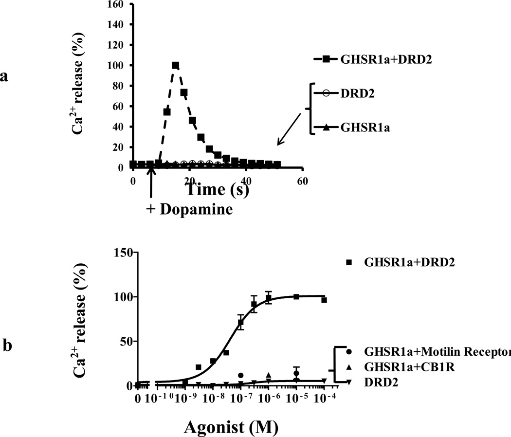
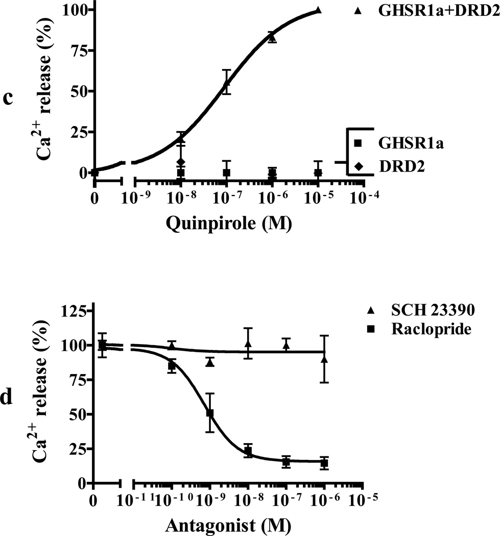
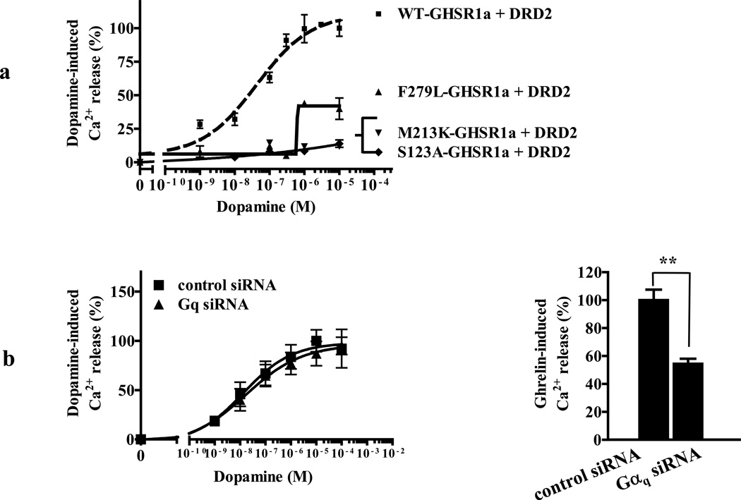
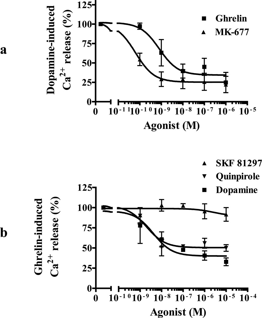
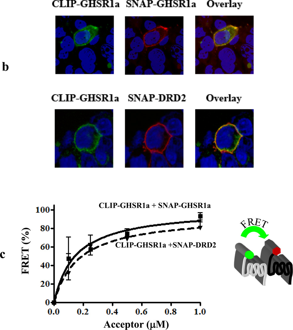
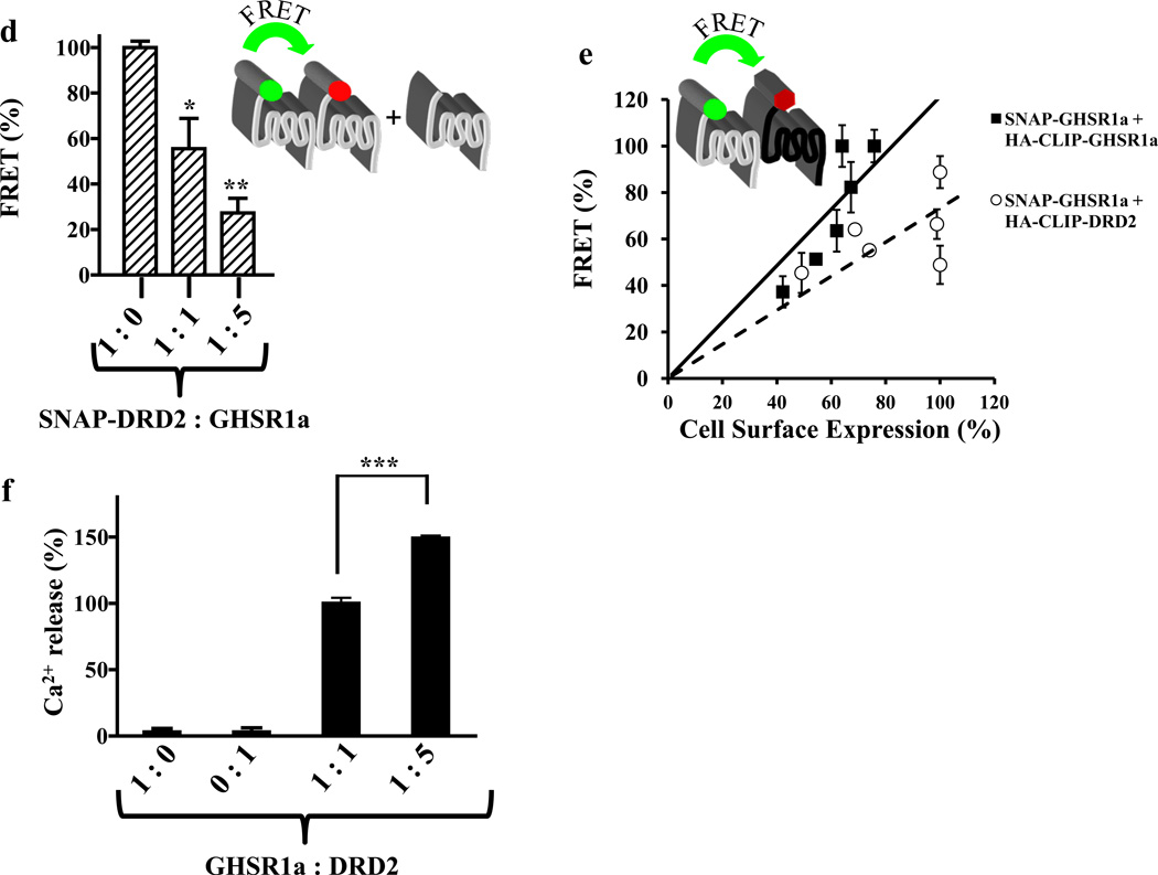
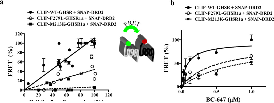
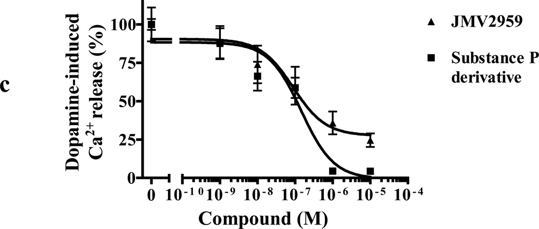
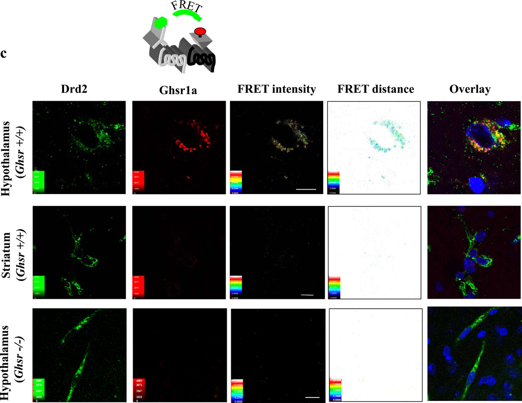
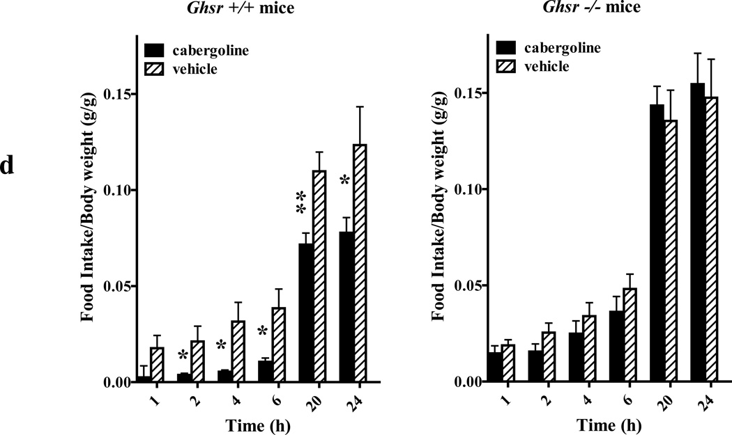
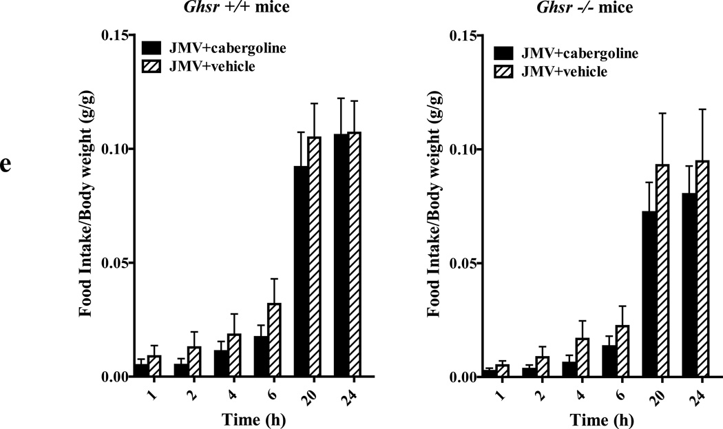
We identified subsets of neurons in the brain that coexpress the dopamine receptor subtype-2 (DRD2) and the ghrelin receptor (GHSR1a). Combination of FRET confocal microscopy and Tr-FRET established the presence of GHSR1a:DRD2 heteromers in hypothalamic neurons. To interrogate function, mice were treated with the selective DRD2 agonist cabergoline, which produced anorexia in wild-type and ghrelin⁻/⁻ mice; intriguingly, ghsr⁻/⁻ mice were refractory illustrating dependence on GHSR1a, but not ghrelin. Elucidation of mechanism showed that formation of GHSR1a:DRD2 heteromers allosterically modifies canonical DRD2 dopamine signaling resulting in Gβγ subunit-dependent mobilization of [Ca²⁺](i) independent of GHSR1a basal activity. By targeting the interaction between GHSR1a and DRD2 in wild-type mice with a highly selective GHSR1a antagonist (JMV2959) cabergoline-induced anorexia was blocked. Inhibiting dopamine signaling in subsets of neurons with a GHSR1a antagonist has profound therapeutic implications by providing enhanced selectivity because neurons expressing DRD2 alone would be unaffected.


Figures

Figure 1
Figure 1. GHSR1a and DRD2 mRNA and protein expression in mouse brain
(a) GHSR1a and DRD2 mRNA expression in mouse striatum, hippocampus and hypothalamus. No PCR products are detected in controls containing no RT; the housekeeping gapdh was used as a negative control. The sizes of PCR products are: ghsr1a, 190 bp; drd2, 286 bp; and gapdh, 428 bp. (b) Identification of neurons co-expressing GHSR1a and DRD2 in mouse brain. Brain sections from ghsr-IRES-tauGFP mice were stained with mouse DRD2 antibody and secondary Alexa647 (red); GHSR1a expressing neurons were localized by detecting GFP (green). To amplify GFP signal, sections were stained with GFP antibody and secondary Alexa488. Co-expression of GHSR1a and DRD2 is shown in overlay pictures with yellow arrows indicating individual neurons co-expressing GHSR1a and DRD2. Nuclei were stained with DAPI (blue). In striatum, box represents the area with higher magnification.
Figure 2
Figure 2. Apo-GHSR1a modifies DRD2 signaling causing quinpirole to induce mobilization of[Ca2+]i in neuronal cells
(a) SH-GHSR1a (upper panel) or parental SH-SY5Y (lower panel) neuronal cells transfected with SNAP-DRD2 were loaded with Fluo-4. Changes in [Ca2+] (shown in pseudocolor) were imaged after quinpirole (10 µM) treatment. SNAP-DRD2 expressing cells (yellow arrows) were visualized by labeling with red-fluorophore (BG-647, 2 µM). Overlay pictures represent the DRD2 expressing cells (red) loaded with Fluo-4 (green). Graphical representation of changes in [Ca2+] over time after quinpirole treatment (lower panel). Cells co-expressing GHSR1a+DRD2 (SH-GHSR1a transfected with SNAP-DRD2) show rapid Ca2+ transients; however, cells expressing only DRD2 SH-SY5Y transfected with SNAP-DRD2) show no increase in [Ca2+]. (b) Dose-dependence of quinpirole-stimulated [Ca2+]i mobilization in SH-GHSR1a cells transfected with DRD2. (c) Quinpirole-induced increase in [Ca2+]i mobilization is abolished by treatment with GHSR1a and DRD2 antagonists. Pretreatment of SH-GHSR1a cells transfected with DRD2 with raclopride or substance P derivative for 5 min attenuates quinpirole (10 µM)-induced Ca2+ signals. Data represent the mean ± s.e.m. for three independent experiments with responses from 5–10 cells analyzed for each experiment. **, P<0.01; ***, P<0.001 versus untreated control. (d) Primary neurons isolated from hypothalamus were loaded with Fluo-4. [Ca2+]i level changes (shown in pseudocolor) were imaged after quinpirole addition (10 µM, upper panels). After washing, the same cells respond to ghrelin (100 nM) treatment by increasing [Ca2+]i level (lower panels). Scale bar represents 20 µm.
Figure 3
Figure 3. Functional interactions between GHSR1a and DRD2 and involvement of Gβγ subunits in dopamine-induced Ca2+ mobilization in aequorin-HEK293 cells
(a) Activation of transfected aequorin-HEK293 cells with dopamine (10 µM) produces rapid Ca2+ transients in cells co-expressing GHSR1a+DRD2 (■) but not in cells expressing GHSR1a (▲) or DRD2 (○) alone. (b) Dose-dependent Ca2+ response to dopamine treatment in cells co-expressing GHSR1a+DRD2 (■). Dose-dependent Ca2+ responses to dopamine are not observed in cells co-expressing motilin receptor + DRD2 (●), DRD2 alone (▼) or in the presence of CB1R agonist (WIN-55212-2) in cells co-expressing GHSR1a+CB1R (▲). (c) Specificity of agonist-induced Ca2+ response. Quinpirole dose-dependently increases [Ca2+] in cells co-expressing GHSR1a+ DRD2 (▲) but not in cells expressing GHSR1a (■) or DRD2 (♦) alone. (d) In cells co-expressing GHSR1a+DRD2, dopamine-induced Ca2+ signaling is inhibited by raclopride (■) but not by the D1R specific agonist SCH 23390 (▲). (e) Pretreatment of cells with pertussis toxin (PTX, 0.25 µg/ml) inhibits dopamine-induced (100 nM) Ca2+ release in cells co-expressing GHSR1a + DRD2. (f) Pretreatment with the phospholipase C (PLC) inhibitor (U73122, 10 µM) blocks dopamine-induced (100 nM) Ca2+ release in cells co-expressing GHSR1a and DRD2. (g) In cells co-expressing GHSR1a and DRD2 the dopamine-induced (100 nM) Ca2+ signal is attenuated after pretreatment with the intracellular IP3 receptor antagonist 2-aminoethoxydiphenyl borate (2-APB). (h) Thapsigargin pretreatment (100 nM) blocks dopamine-induced (100 nM) Ca2+ release in cells co-expressing GHSR1a and DRD2. (i) Overexpression of GRK2 or the GRK2 phosphorylation mutant (GRK2-K220R)in cells co-expressing GHSR1a and DRD2 inhibit dopamine-induced (100 nM) Ca2+ mobilization. (j) The Gβγ scavenger (βARK1ct) dose-dependently decreases dopamine-induced (100 nM) Ca2+ mobilization in cells co-expressing GHSR1a + DRD2. (k) Cells co-expressing GHSR1a + DRD2 pretreated with small molecule inhibitors of Gβγ subunit signaling (M119 and M158C) and inactive control compound (M119B) for 5 min before dopamine treatment. M119B is ineffective in inhibiting dopamine-induced Ca2+ release whereas M119 and 158C inhibit the response to dopamine. The data represent the mean ± s.e.m. for three independent experiments in duplicate for each concentration point. *, P<0.05; **, P<0.01 and ***, P<0.001 versus untreated control.
Figure 4
Figure 4. Dopamine-induced Ca2+ release is independent of GHSR1a constitutive activity in cells co-expressing GHSR1a and DRD2
(a) Co-expression of GHSR1a point mutants S123A and M213K that exhibit identical constitutive activity to WT-GHSR1a, and F297L which is devoid of constitutive activity Cells co-expressing DRD2 with either S123A or M213K fail to mobilize Ca2+ in response to dopamine, whereas the GHSR1a F279L point mutant exhibits Ca2+ mobilization in response to dopamine; (b) Knockdown of Gαq subunit by siRNA does not inhibit dopamine induced Ca2+ mobilization in (left panel), whereas ghrelin-induced Ca2+ mobilization (right panel) is significantly impaired. The data represent the mean ± s.e.m. for three independent experiments in duplicate for each concentration point. **, P<0.01 versus untreated control.
Figure 5
Figure 5. Preatment of cells co-expressing GHSR1a and DRD2 with GHSR1a or DRD2 agonists results in cross-desensitization of agonist signaling
(a) Aequorin-HEK293 cells co-expressing GHSR1a + DRD2 pretreated with increasing concentrations of ghrelin (■) or MK-677 (▲) for 30 min show attenuated response to dopamine-induced (100 nM) Ca2+ mobilization. (b) Aequorin-HEK293 cells co-expressing GHSR1a + DRD2 were pretreated with increasing concentrations of dopamine (■), SKF81297 (▲) or quinpirole (▼) for 30 min and Ca2+ mobilization measured after ghrelin treatment (100 nM). The data represent the mean ± s.e.m. for three independent experiments in duplicate for each concentration point.
Figure 6
Figure 6. Detection of heteromer formation between GHSR1a and DRD2 in vitro by Tr-FRET
(a) Tr-FRET competition assays where cells were transfected with increasing ratios of SNAP-GHSR1a and untagged GHSR1a, untagged DRD2 or an untagged RXFP1. (b) Cells co-transfected with CLIP- or SNAP-tagged receptor variants, labeled specifically with either green (BC-488, 2 µM) or red fluorophore (BG-647, 2 µM) and then examined by confocal microscopy. Overlay pictures show co-localization of GHSR1a with DRD2. (c) FRET acceptor saturation assays performed using constant amount of donor fluorophore (BG-TbK, 100 nM) and increasing amounts of acceptor fluorophore (BC-647) in cells co-expressing SNAP- and CLIP-tagged receptor variants. (d) GHSR1a:DRD2 formation measured by Tr-FRET competition assays; Tr-FRET measurements on cells transfected with SNAP-DRD2 in the presence of different ratios of untagged GHSR1a. (e) Homomerization of GHSR1a and heteromerization of GHSR1a and DRD2 by Tr-FRET receptor titration. Cells were co-transfected with constant amount of SNAP-GHSR1a and increasing amounts of HA-CLIP-GHSR1a or HA-CLIP-DRD2. The HA-tagged receptor cell surface expression was determined by cell surface ELISA and Tr-FRET signal plotted as a function of cell surface expressed HA-CLIP-tagged receptor. (f) Magnitude of dopamine-induced Ca2+ release is dependent on formation of GHSR1a:DRD2 heteromers; intracellular Ca2+ mobilization induced by dopamine (10 µM) in aequorin HEK293 cells expressing different ratios of GHSR1a to DRD2. The data represent the mean ± s.e.m. for three independent experiments in duplicate for each concentration point. ** P<0.01; *** P<0.001 versus control.
Figure 7
Figure 7. GHSR1a point mutants illustrate GHSR1a:DRD2 formation is dependent on GHSR1a structure
Tr-FRET experiments comparing heteromerization of wild-type GHSR1a, M213K-GHSR1a and F279L-GHSR1a point mutants in the presence of DRD2. (a) Tr-FRET receptor titration assays were performed to assess heteromerization. Cells were co-transfected with increasing amounts of HA-CLIP-tagged receptors; the FRET signal is represented as a function of cell surface expression of HA-tagged receptors. (b) Tr-FRET acceptor titration assay to determine heteromer formation. FRET intensity signals were measured in cells co-expressing CLIP-tagged WT-GHSR1a, M213K or F279L point mutants in the presence of SNAP-DRD2 and labeled with constant amount of donor (BG-TbK, 100 nM) and increasing amount of acceptor fluorophore (BC-647). (c) Dose-dependent inhibition of dopamine-induced mobilization of [Ca2+]i in cells co-expressing GHSR1a + DRD2 by GHSR1a neutral antagonists JMV2959 and substance P derivative (d) Dose-dependent inhibition of dopamine-induced (10 µM) Ca2+ mobilization by DRD2 antagonist (raclopride, ■) and inverse agonist (sulpiride, ▲) in cells co-expressing GHSR1a + DRD2. (e) Sulpiride (■) but not raclopride (▲) inhibits ghrelin-induced (100 nM) Ca2+ mobilization in cells co-expressing GHSR1a+DRD2. The data represent the mean ± s.e.m. for three independent experiments in duplicate for each concentration point.
Figure 8
Figure 8. Detection GHSR1a:DRD2 heteromers in vivo by Tr-FRET and the dependence of the anorexigenic effect of a DRD2 agonist on GHSR1a
(a) Tr-FRET signal detection on membrane preparations from striatum and hypothalamus after labeling with red-ghrelin (100 nM, acceptor fluorophore), antibody for DRD2 and cryptate labeled secondary antibody (10 nM, donor fluorophore). Non-specific FRET signal was measured on membranes in the presence 100 nM of red-fluorophore, antibody for DRD2and cryptate labeled secondary antibody (10 nM, donor fluorophore). (b) Tr-FRET signals detected on membrane preparations of striatum and hypothalamus from ghsr+/+ and ghsr−/− mice. Membranes were labeled in the presence of 100 nM red-ghrelin (acceptor), antibody for DRD2 and 10 nM cryptate labeled secondary antibody (donor). (c) Confocal microscope FRET analysis of GHSR1a:DRD2 heteromer formation in ghsr+/+ mouse hypothalamic (upper panels) striatal (middle panels) neurons and ghsr−/− mouse hypothalamic neurons (lower panels). Brain slices were stained with 100 nM red-ghrelin (acceptor), DRD2 antibody and Cy3 labeled secondary antibody (donor). Red color indicates GHSR1a staining, green DRD2 localization. Microscopic analysis shows FRET intensity, distances separating the two receptors and co-localization of two receptors (overlay). (d) Effect of cabergoline administration on food intake in ghsr+/+ mice (n=4 for cabergoline and n=4 for vehicle; left graph) and in ghsr −/− mice (n=5 for cabergoline and n=5 for vehicle; right graph). Mice were injected i.p. with cabergoline (0.5 mg/kg) in 100 µl of physiological saline or with 100 µl saline alone (vehicle). Food intake was measured at 1, 2, 4, 6, 20 and 24 h after injections. Average body weight mass of ghsr+/+ was 31.8g and ghsr−/− was 28.1g; * P<0.05; ** P<0.01 versus control vehicle treatments. (e) The neutral GHSR1a selective antagonist JMV2959 antagonizes cabergoline induced reduction on food intake in ghsr +/+ mice (n=4 for cabergoline and n=4 for vehicle; left graph) and had no effect in ghsr −/− mice (n=5 for cabergoline and n=5 for vehicle; right graph). Mice were injected i.p. with JMV295 at 0.2 mg/kg dose, 30 min before cabergoline treatments (0.5 mg/kg). Food intake was measured at 1, 2, 4, 6, 20 and 24h after injection. (f) Effect of cabergoline administration on food intake in ghrelin+/+ mice (n=6 for cabergoline and n=4 for vehicle, left graph) and in ghrelin −/− mice (n=5 for cabergoline and n=5 for vehicle, right graph). Mice were injected i.p. with cabergoline (0.5 mg/kg) in 100 µl of physiological saline or with 100 µl saline alone (vehicle). Food intake was measured at 1, 2, 4, 6, 20 and 24 h after injections. * P<0.05 versus control vehicle treatments.

Comment in

References

    1. Albizu L, Cottet M, Kralikova M, Stoev S, Seyer R, Brabet I, Roux T, Bazin H, Bourrier E, Lamarque L, et al. Time-resolved FRET between GPCR ligands reveals oligomers in native tissues. Nat Chem Biol. 2010;6:587–594. - PMC - PubMed
    1. Bonacci TM, Mathews JL, Yuan C, Lehmann DM, Malik S, Wu D, Font JL, Bidlack JM, Smrcka AV. Differential targeting of Gbetagamma-subunit signaling with small molecules. Science. 2006;312:443–446. - PubMed
    1. Bulenger S, Marullo S, Bouvier M. Emerging role of homo- and heterodimerization in G-protein-coupled receptor biosynthesis and maturation. Trends Pharmacol Sci. 2005;26:131–137. - PubMed
    1. Button D, Brownstein M. Aequorin-expressing mammalian cell lines used to report Ca2+ mobilization. Cell Calcium. 1993;14:663–671. - PubMed
    1. Canals M, Milligan G. Constitutive activity of the cannabinoid CB1 receptor regulates the function of co-expressed Mu opioid receptors. J Biol Chem. 2008;283:11424–11434. - PubMed

Publication types

MeSH terms