Review
. 2012 Jan 27;36(1):13-21.
doi: 10.1016/j.immuni.2011.11.017.

B-1 B cell development in the fetus and adult

Affiliations
Review

B-1 B cell development in the fetus and adult

Encarnacion Montecino-Rodriguez et al. Immunity. .

Abstract

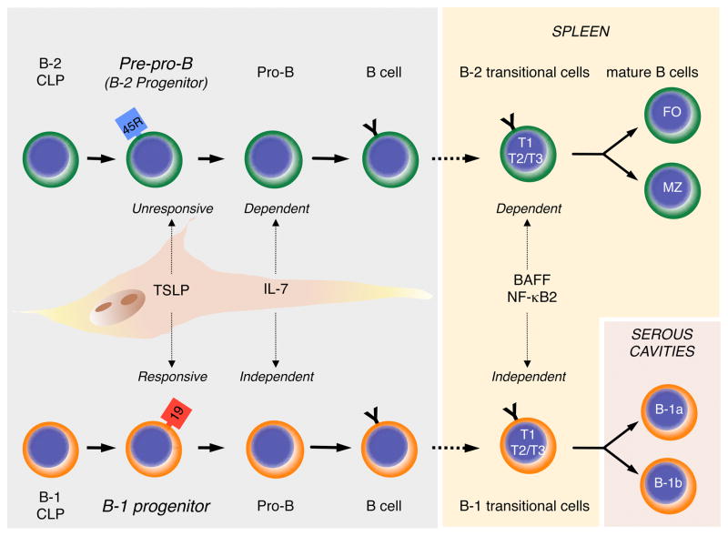
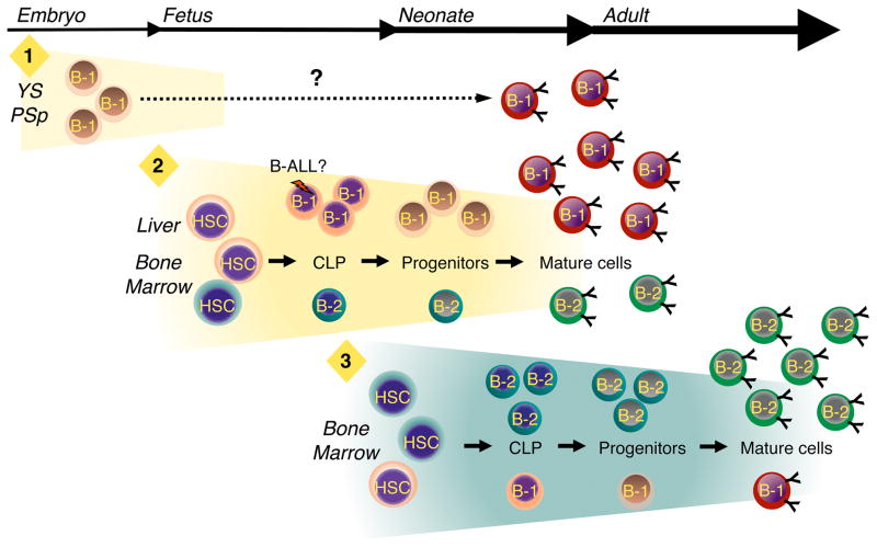
Models of hematopoiesis often depict lymphocyte production as a uniform process in which a homogenous population of hematopoietic stem cells (HSCs) generates progenitors from which all types of lymphocytes are derived. However, it is increasingly evident that these schemes are too simplistic and that the lymphoid potential of HSCs and precursors arising in the embryo, fetus, neonate, and adult is remarkably distinct. We review recent findings regarding the development of B lymphocytes, and the B-1 B cell lineage in particular, as a case in point. These studies show that B-1 and B-2 B cells involved in innate and adaptive immune responses, respectively, arise in staggered waves of development from distinct progenitors. We discuss the implications of this layered model of B cell development for understanding normal and dysregulated B lymphopoiesis.


Figures

Figure 1
Figure 1
B-1 and B-2 development. B-2 cells (Top) are produced in the bone marrow after birth. Common lymphoid progenitors (CLP) mature sequentially through pre-pro-B, pro-B, and pre-B (not shown) cell intermediates into immature sIgM+ B cells (shown as “B cell” in figure). Pre-pro-B cells are referred to as B-2 progenitors in the text. Immature sIgM+ cells migrate to the spleen where they mature through B-2 transitional 1 (T1) and Transitional 2 and 3 (T2 and T3) stages into Follicular (FO) or Marginal Zone (MZ) B cells. The stages of B-1 development are increasingly well defined, and it is now possible to propose a model of development based on the data summarized in this review (Bottom). Mature B-1 cells are generated from B-1 specified CLPs that sequentially differentiate through B-1 progenitor, pro-B, pre-B (not shown) and immature sIgM+ B-1 cell stages. The latter cells then mature in the spleen through the Transitional cell stages shown. Mature B-1 cells that migrate to serous cavities acquire the B-1a and B-1b phenotype. The figure also shows that B-1, but not B-2, progenitors are TSLP responsive, that B-2, but not B-1, development is dependent on IL-7, and that B-1 transitional cell survival and maturation is BAFF and NF-κB2 independent.
Figure 2
Figure 2
B-1 and B-2 development occur in distinct, overlapping waves. Three waves of B-1 development are proposed. The first wave (“1”) initiates in the yolk sac (YS) and para-aortic splanchnopleura (PSp) region before the emergence of definitive hematopoietic stem cells (HSC). It is not clear, as indicated by the dashed line, whether B-1 progenitors produced during this initial phase mature into B-1 cells that become part of the adult B-1 pool. If so, they may do so in YS and PSp or migrate to the fetal liver and bone marrow and complete maturation in those tissues. During the second wave (”2”), which initiates in the fetal liver and fetal bone marrow, HSCs generate B-1 progenitors from which mature B-1 cells are derived. Although B-2 development initiates during this second phase, B-1 production predominates. Whether or not B-1 and B-2 CLPs and progenitors are produced from a single type of HSC or multiple B-1 and B-2 specified stem cells exist, as indicated by the different colored HSCs, is not known. B-1 production peaks during late embryogenesis and then begins to decline just before birth. The third wave (“3”) of B cell development takes place in the bone marrow and results primarily in the production of B-2 B cells. B-1 B cells can be produced during this third wave, but the efficiency with which this occurs in comparison to wave two is substantially reduced.

Similar articles

Cited by

References

    1. Allman D, Lindsley RC, DeMuth W, Rudd K, Shinton SA, Hardy RR. Resolution of three nonproliferative immature splenic B cell subsets reveals multiple selection points during peripheral B cell maturation. J Immunol. 2001;167:6834–6840. - PubMed
    1. Allman D, Pillai S. Peripheral B cell subsets. Curr Opin Immunol. 2008;20:149–157. - PMC - PubMed
    1. Alugupalli KR, Gerstein RM. Divide and conquer: division of labor by B-1 B cells. Immunity. 2005;23:1–2. - PubMed
    1. Alugupalli KR, Leong JM, Woodland RT, Muramatsu M, Honjo T, Gerstein RM. B1b lymphocytes confer T cell-independent long-lasting immunity. Immunity. 2004;21:379–390. - PubMed
    1. Antin J, Emerson S, Martin P, Gadol N, Ault K. A major lymphoid subpopulation in human fetal spleen: phenotypic and functional studies. J Immunol. 1986;136:505–510. - PubMed

Publication types