. 2012 Jan 27;45(2):244-54.
doi: 10.1016/j.molcel.2011.11.026.

Scd6 targets eIF4G to repress translation: RGG motif proteins as a class of eIF4G-binding proteins

Affiliations

Scd6 targets eIF4G to repress translation: RGG motif proteins as a class of eIF4G-binding proteins

Purusharth Rajyaguru et al. Mol Cell. .

Abstract

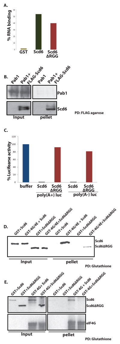
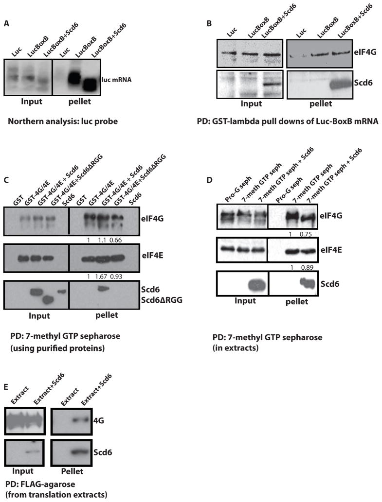
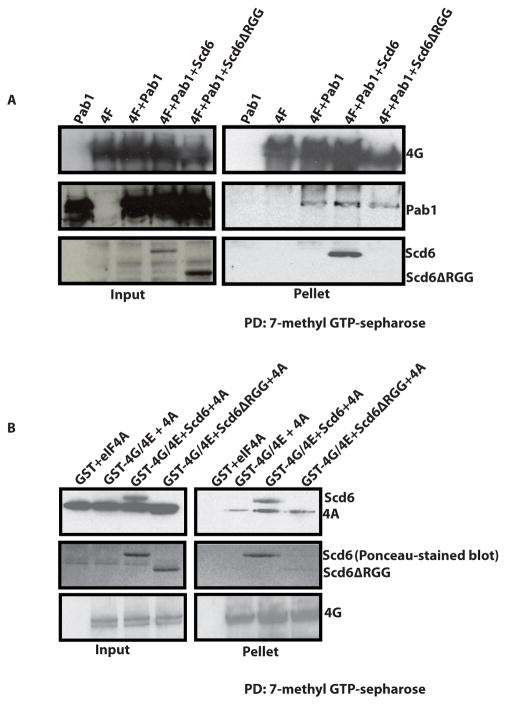
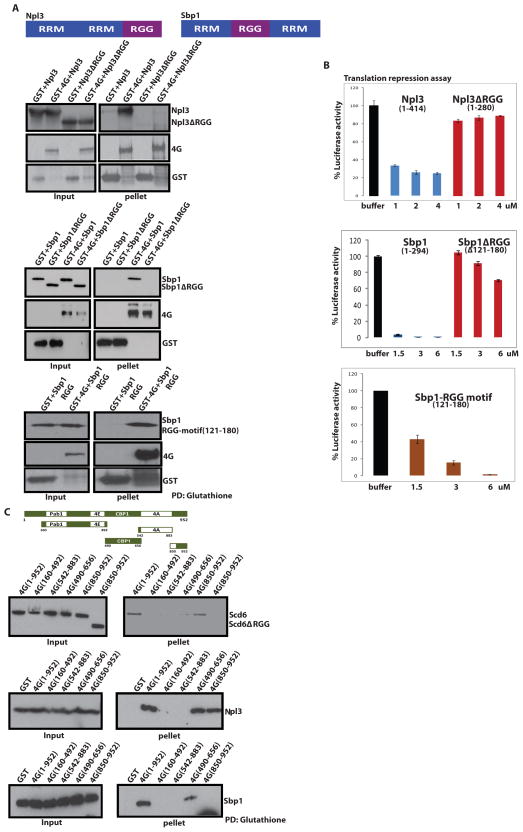
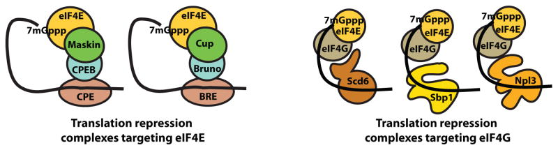
The formation of mRNPs controls the interaction of the translation and degradation machinery with individual mRNAs. The yeast Scd6 protein and its orthologs regulate translation and mRNA degradation in yeast, C. elegans, D. melanogaster, and humans by an unknown mechanism. We demonstrate that Scd6 represses translation by binding the eIF4G subunit of eIF4F in a manner dependent on its RGG domain, thereby forming an mRNP repressed for translation initiation. Strikingly, several other RGG domain-containing proteins in yeast copurify with eIF4E/G and we demonstrate that two such proteins, Npl3 and Sbp1, also directly bind eIF4G and repress translation in a manner dependent on their RGG motifs. These observations identify the mechanism of Scd6 function through its RGG motif and indicate that eIF4G plays an important role as a scaffolding protein for the recruitment of translation repressors.


Figures

Figure 1
Figure 1. Scd6 binds eIF4G
A) 32P labeled MFA2 mRNA was incubated with GST, GST-Scd6 or GST-Scd6ΔRGG followed by pull down using glutathione sepharose. Radioactivity on beads after three washes was measured by Cerenkov counting. B) Purified His-Pab1 was incubated with FLAG-Scd6 followed by FLAG pull down. Pab1 and Scd6 were detected by using polyclonal anti-Pab1 and anti-Scd6 antibodies respectively. C) Purified Scd6 and RGG mutant were tested for their effects on translation of 100 ng of poly(A−) and poly(A+) luciferase message at 6uM concentration as described previously (Nissan et al., 2010). D) Glutathione sepharose pull downs performed to look at interaction of purified GST-4G/E with recombinant Scd6 and Scd6ΔRGG mutant protein. Scd6 protein was detected with anti-FLAG antibody following manufacturer’s instructions. E) Glutathione sepharose pull downs were performed to look at interaction of GST-4G in bacterial extracts with recombinant Scd6 and Scd6ΔRGG mutant protein. 6ug of each protein was used in 200ul reaction mixture. Scd6 protein was detected with anti-FLAG antibody following manufacturer’s instructions. Typically 10% of total reaction was loaded in the ‘input’ lanes and 100% of total pulled-down material was loaded in ‘pellet’ lanes.
Figure 2
Figure 2. Scd6 forms a tri-complex with eIF4E/G on cap
A) Northern analysis of luciferase mRNA containing BoxB repeats pulled down from translation extracts with GST-lambda protein (see procedures) +/− purified His-tagged Scd6 at 6uM concentration. B) Western analysis of proteins coming along with luciferase mRNA containing BoxB using antibodies indicated. C) 7-methyl-GTP sepharose pull downs were performed using recombinant purified eIF4G/4E and Scd6 proteins (see procedures). D) 7-methyl-GTP sepharose pull downs performed from yeast translation extracts in presence of Scd6 or Scd6ΔRGG. E) FLAG-pull downs from translation reactions to pull down recombinant Scd6. 20 ul translation reactions were diluted to 200ul followed by addition of FLAG-agarose beads. Binding was performed for an hour with end to end rotation followed by three washes of beads for 10′ each. The beads were then boiled in SDS-loading buffer and analyzed by SDS-PAGE followed by western blotting. Typically 10% of total reaction was loaded in the ‘input’ lanes and 100% of total pulled-down material was loaded in ‘pellet’ lanes. Bands in C and D were quantified by using Image J program.
Figure 3
Figure 3. Scd6 does not destabilize Pab1-4G or 4A-4G interactions
A) 7-methyl-GTP sepharose pull downs were performed to look at interaction of GST-eIF4G/eIF4E complex with recombinant Pab1 in presence of Scd6 or Scd6ΔRGG protein as described in materials and methods. 8ug of GST-eIF4G/eIF4E was incubated with wt or mutant Scd6 protein for 1h at 4°C with end-to-end rotation. Following this Pab1 was added to reaction mixture and incubated for 1h with end-to-end rotation. Finally, 7-methyl-GTP sepharose was added to reaction mix and incubated for 2h. Washing was performed as described in materials and methods. eIF4G and Pab1 were detected with polyclonal antibodies. B) Glutathione pull down assay to analyze effect of purified Scd6 on eIF4A-eIF4E/G interaction. Incubation and washing conditions were same as mentioned in A. eIF4G and eIF4A were detected with polyclonal antibodies. The anti-eIF4A antibody cross-reacts with purified Scd6 (contains both His and FLAG-tag). The middle panel depicts ponceau-stained blot with clearly visible wt and mutant Scd6 proteins. This was done since eIF4A and Scd6ΔRGG run at similar position (in top panel). Typically 10% of total reaction was loaded in the ‘input’ lanes and 50 or 100% of total pulled-down material was loaded in ‘pellet’ lanes.
Figure 4
Figure 4. Scd6 localizes to and affects granules
A) Strain bearing chromosomal GFP-Edc3 was transformed with plasmid expressing Pbp1-mCherry. Cells were grown to 0.3–0.4 OD600 in appropriate minimal media with 2% glucose and then stressed in minimal medium lacking glucose for 10 minutes before observing under the microscope. B) Strain bearing chromosomal GFP-Scd6 was transformed with plasmid expressing Edc3-mCherry. Cells were grown to 0.3–0.4 OD600 in appropriate minimal media with 2% glucose and then stressed in minimal medium lacking glucose for 10 minutes before observing under the microscope. C) Strain bearing chromosomal GFP-Scd6 was transformed with Pbp1-mCherry. Cells were grown to 0.3–0.4 OD600 in appropriate minimal media with 2% glucose and then stressed in minimal medium lacking glucose for 10 minutes before observing under the microscope. D) Wild type or scd6Δ strains were transformed with vector expressing Dcp2-mCherry. Cells were grown and stressed in absence of glucose as described in A and observed under the microscope. E) Wild type or scd6Δ strains were transformed with vector expressing Dcp2-mCherry and Pab1-GFP. Cells were grown up to 0.3–0.4 OD600 stressed by adding 0.5% sodium azide (or equal of water for control cells) to the medium, incubated for 30 minutes at 30°C before observing under microscope. F) Wild type strains expressing Edc3-mCherry and Pab1-GFP were transformed with either empty vector or vector driving over-expression of SCD6 or SCD6ΔRGG from the Gal-inducible promoter. These strains were grown to 0.3–0.4 OD600 in appropriate minimal media with 2% sucrose as carbon source followed by growth in medium containing 1.8% galactose-0.2% sucrose for 2 hours. Cells were then observed under the microscope. G) eIF4G-GFP, eIF4E-GFP and eIF3b-GFP strain were transformed with empty vector or vector driving over expressing SCD6 under Gal-inducible promoter. These strains were grown to 0.3–0.4 OD600 in appropriate minimal media with 2% sucrose as carbon source followed by growth in medium containing 1.8% galactose-0.2% sucrose for 2 hours. Cells were then observed under the microscope.
Figure 5
Figure 5. Multiple RGG Domain Containing Proteins bind eIF4G and repress translation
A) Schematic representation of Sbp1 and Npl3 proteins with their domain organization. A) Top- GST pull downs with recombinant eIF4G in bacterial extracts and either full length His-Npl3 or its RGG-deletion mutants. Npl3 was detected using antisera against Npl3. A) Middle- GST pull downs with recombinant eIF4G in bacterial extracts and either full length His-Sbp1 or its RGG-deletion mutants. Sbp1 was detected using anti-His antibody. A) Bottom- GST pull downs with recombinant eIF4G in bacterial extracts and His-Sbp1 RGG-motif (121–180). RGG motif was detected using anti-His antibody B) In vitro translation assay was performed as described in Nissan et al., 2010 with the addition of recombinant full length or RGG-deleted variant of Npl3 (top panel), Sbp1 (middle panel) and Sbp1 RGG-motif (121–180) (bottom panel). Error bars represent standard error of three experiments. C) Top- Schematic representation of the 4G-deletion constructs used for mapping binding sites of Scd6, Npl3 and Sbp1. Bottom- GST pull downs of full length and subdomains of GST- tagged eIF4G with Scd6, Npl3, and Sbp1. Scd6, Npl3 and Sbp1 were detected in pull downs by antibodies against the His tag on these recombinant proteins. We observed approximately 60–70% pull downs for various 4G-fragments. Typically 10% of total reaction was loaded in the ‘input’ lanes and 100% of total pulled-down material was loaded in ‘pellet’ lanes. See also Fig. S1 which is connected to this figure.
Figure 6
Figure 6. Two paradigms for translation repression complexes utilizing components of eIF4F

References

    1. Abramoff M, Magelhaes P, Ram S. Image processing with ImageJ. Biophotonics Intl. 2004;11:36–42.
    1. Anderson P, Kedersha N. RNA granules: post-transcriptional and epigenetic modulators of gene expression. Nat Rev Mol Cell Biol. 2009;10(6):430–6. - PubMed
    1. Barbee S, Estes P, Cziko A, Hillebrand J, Luedeman R, Coller J, Johnson N, Howlett I, Geng C, Ueda R, Brand A, Newbury S, Wilhelm J, Levine R, Nakamura A, Parker R, Ramaswami M. Staufen- and FMRP-containing neuronal RNPs are structurally and functionally related to somatic P bodies. Neuron. 2006;52:997–1009. - PMC - PubMed
    1. Boag P, Nakamura A, Blackwell K. A conserved RNA-protein complex component involved in physiological germline apoptosis regulation in C. elegans. Development. 2005;132:4975–4986. - PubMed
    1. Boag P, Atalay A, Robida S, Reinke V, Blackwell K. Protection of specific maternal messenger RNAs by the P body protein CGH-1 (Dhh1/RCK) during Caenorhabditis elegans oogenesis. J Cell Biol. 2008;182(3):543–57. - PMC - PubMed

Publication types

MeSH terms