

doi: 10.1186/1755-8166-5-8.
Prenatal diagnosis of a trisomy 7/trisomy 13 mosaicism
Affiliations
- PMID: 22284936
- PMCID: PMC3293093
- DOI: 10.1186/1755-8166-5-8
Item in Clipboard
Prenatal diagnosis of a trisomy 7/trisomy 13 mosaicism
Mol Cytogenet.
.
Abstract
Double aneuploidy mosaicism of two different aneuploidy cell lines is rare. We describe for the first time a double trisomy mosaicism, involving chromosomes 7 and 13 in a fetus presenting with multiple congenital anomalies. No evidence for chimerism was found by DNA genotyping. The origin of both trisomies are consistent with isodisomy of maternal origin. Therefore, it is most likely that the double trisomy mosaicism arose from two independent events very early in embryonic development. The trisomy 7 and 13 cells were shown to be of maternal origin.
Figures
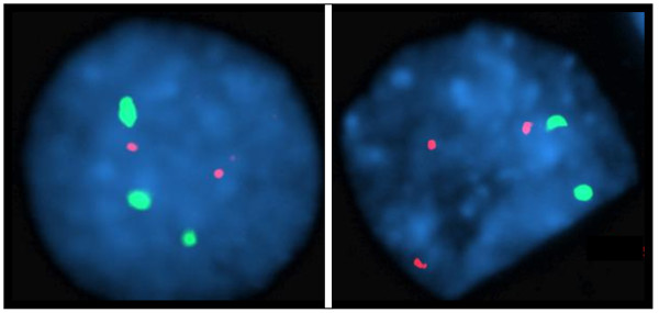
Interphase FISH results. FISH was performed with probes for chromosome 7 (Abbott-Vysis CEP 7, 7p11.1-q11.1, spectrum green) and chromosome 13 (Abbott-Vysis LSI 13 RB1, 13q14.2, spectrum orange). The left panel shows a cell with a trisomy 7 and a disomy 13. The right panel displays a cell with a trisomy 13 and a disomy 7.
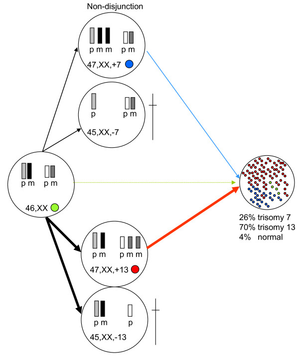
Possible mechanism for the origin of double aneuploidy mosaicism in this fetus. Schematic representation of the mechanisms that may have lead to a mosaic pattern of both trisomy 7 and trisomy 13 cells in a single fetus. Two independent non-disjunction events may have taken place in a 46, XX zygote, resulting in both a trisomy 7 cell line (26%) and a trisomy 13 cell line (70%). Both monosomic cells are not viable. In fetal tissue, 4% of the cells in fetal tissue showed a normal signal pattern with FISH.
References
-
- Abe K, Harada N, Itoh T, Hirakawa O, Niikawa N. Trisomy 13/trisomy 18 mosaicism in an infant. Clin Genet. 1996;50:300–303. - PubMed
-
- Cogulu O, Tirpan C, Ozkinay F, Gunduz C, Ozkinay C. The second case with 47, XY, + 8 [38]/45, X0. Turk J Pediatr. 2002;44:86–89. - PubMed
-
- Eiben B, Hansen S, Goebel R, Hammans W. Tissue-specific 45, X0/47, XY, +13 mosaicism in an 18-year-old woman. Hum Genet. 1989;82:391–392. - PubMed
LinkOut - more resources
Full Text Sources
