

Increased myocardial prevalence of C-reactive protein in human coronary heart disease: direct effects on microvessel density and endothelial cell survival
- PMID: 22285194
- PMCID: PMC3899797
- DOI: 10.1016/j.carpath.2011.12.003
Increased myocardial prevalence of C-reactive protein in human coronary heart disease: direct effects on microvessel density and endothelial cell survival
Abstract
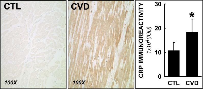
Background: Elevated plasma C-reactive protein (CRP) is a biomarker of cardiovascular diseases (CVDs), but its potential roles as a participant of the disease process are not well defined. Although early endothelial cell injury and dysfunction are recognized events in CVD, the initiating events are not well established. Here we investigated the local myocardial CRP levels and cardiac microvessel densities in control and CVD tissue samples. Using in vitro methodologies, we investigated the direct effects of CRP on human endothelial cells.
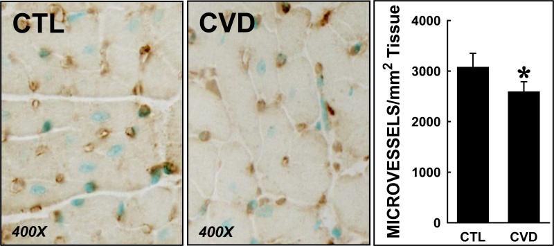
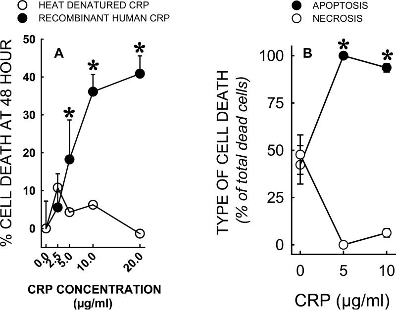
Methods: Cardiac specimens were collected at autopsy within 4 h of death and were classified as normal controls or documented evidence of CVD. The regional prevalence of CRP and the cardiac microvessels (<40 μm) were investigated using immunohistochemistry. For in vitro experiments, human umbilical vein endothelial cells were incubated with CRP. Intracellular oxidant levels were assessed using 2',7'-dichlorofluorescein diacetate fluorescence microscopy, and cell survival was concurrently determined. Effects of chemical antioxidants on endothelial cell survival were also tested.
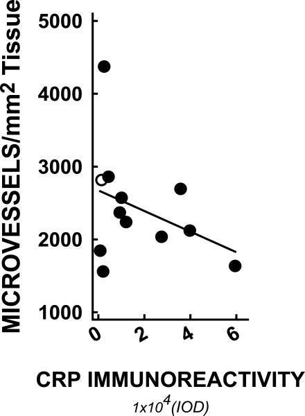
Results: Myocardial CRP levels were elevated in CVD specimens. This was associated with reduced cardiac microvessels, and this rarefaction was inversely correlated to adjacent myocardial CRP prevalence. CRP caused concentration-dependent increases in oxidant production and cell apoptosis.
Conclusions: These findings provide evidence supporting myocardial CRP as a locally produced inflammatory marker and as a potential participant in endothelial toxicity and microvascular rarefaction.
Copyright © 2012 Elsevier Inc. All rights reserved.
Figures


References
-
- Black S, Kushner I, Samols D. C-reactive Protein. J Biol Chem. 2004 Nov 19;279(47):48487–90. - PubMed
-
- Dong Q, Wright JR. Expression of C-reactive protein by alveolar macrophages. J Immunol. 1996 Jun 15;156(12):4815–20. - PubMed
-
- Ouchi N, Kihara S, Funahashi T, Nakamura T, Nishida M, Kumada M, et al. Reciprocal association of C-reactive protein with adiponectin in blood stream and adipose tissue. Circulation. 2003 Feb 11;107(5):671–4. - PubMed
-
- Ridker PM, Cook N. Clinical usefulness of very high and very low levels of C-reactive protein across the full range of Framingham Risk Scores. Circulation. 2004 Apr 27;109(16):1955–9. - PubMed
Publication types
MeSH terms
Substances
Grants and funding
LinkOut - more resources
Full Text Sources
Research Materials
Miscellaneous
