

Intrinsic axonal degeneration pathways are critical for glaucomatous damage
- PMID: 22285251
- PMCID: PMC3831512
- DOI: 10.1016/j.expneurol.2012.01.014
Intrinsic axonal degeneration pathways are critical for glaucomatous damage
Abstract
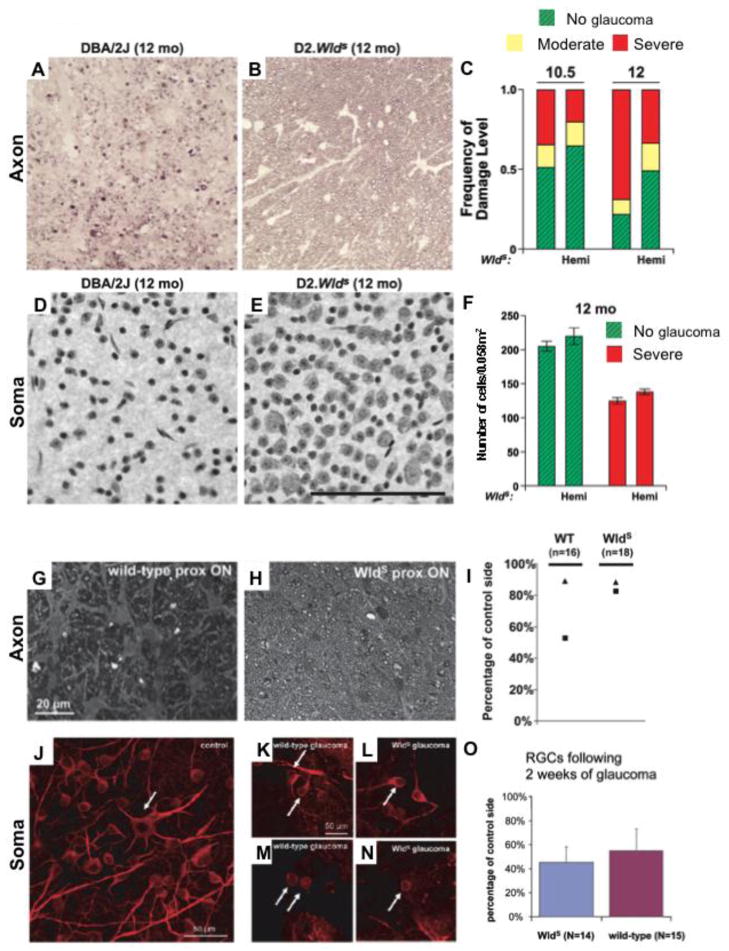
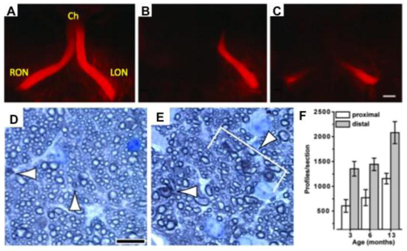
Glaucoma is a neurodegenerative disease affecting 70million people worldwide. For some time, analysis of human glaucoma and animal models suggested that RGC axonal injury in the optic nerve head (where RGC axons exit the eye) is an important early event in glaucomatous neurodegeneration. During the last decade advances in molecular biology and genome manipulation have allowed this hypothesis to be tested more critically, at least in animal models. Data indicate that RGC axon degeneration precedes soma death. Preventing soma death using mouse models that are mutant for BAX, a proapoptotic gene, is not sufficient to prevent the degeneration of RGC axons. This indicates that different degeneration processes occur in different compartments of the RGC during glaucoma. Furthermore, the Wallerian degeneration slow allele (Wld(s)) slows or prevents RGC axon degeneration in rodent models of glaucoma. These experiments and many others, now strongly support the hypothesis that axon degeneration is a critical pathological event in glaucomatous neurodegeneration. However, the events that lead from a glaucomatous insult (e.g. elevated intraocular pressure) to axon damage in glaucoma are not well defined. For developing new therapies, it will be necessary to clearly define and order the molecular events that lead from glaucomatous insults to axon degeneration.
Keywords: Astrocyte; Axon; BAX; Glaucoma; Microglia; Optic nerve; Optic nerve head; Retinal ganglion cell; Wld(s).
Copyright © 2012 Elsevier Inc. All rights reserved.
Figures
Similar articles
-
The WldS gene delays axonal but not somatic degeneration in a rat glaucoma model.Eur J Neurosci. 2008 Sep;28(6):1166-79. doi: 10.1111/j.1460-9568.2008.06426.x. Epub 2008 Sep 9. Eur J Neurosci. 2008. PMID: 18783366
-
Assessment of retinal ganglion cell damage in glaucomatous optic neuropathy: Axon transport, injury and soma loss.Exp Eye Res. 2015 Dec;141:111-24. doi: 10.1016/j.exer.2015.06.006. Epub 2015 Jun 9. Exp Eye Res. 2015. PMID: 26070986 Review.
-
Role of hypoxia-inducible factor-1α in preconditioning-induced protection of retinal ganglion cells in glaucoma.Mol Vis. 2013 Nov 23;19:2360-72. eCollection 2013. Mol Vis. 2013. PMID: 24319330 Free PMC article.
-
Under pressure: cellular and molecular responses during glaucoma, a common neurodegeneration with axonopathy.Annu Rev Neurosci. 2012;35:153-79. doi: 10.1146/annurev.neuro.051508.135728. Epub 2012 Apr 12. Annu Rev Neurosci. 2012. PMID: 22524788 Review.
-
DRP1 inhibition rescues retinal ganglion cells and their axons by preserving mitochondrial integrity in a mouse model of glaucoma.Cell Death Dis. 2015 Aug 6;6(8):e1839. doi: 10.1038/cddis.2015.180. Cell Death Dis. 2015. PMID: 26247724 Free PMC article.
Cited by
-
Identification of the first noncompetitive SARM1 inhibitors.Bioorg Med Chem. 2020 Sep 15;28(18):115644. doi: 10.1016/j.bmc.2020.115644. Epub 2020 Jul 17. Bioorg Med Chem. 2020. PMID: 32828421 Free PMC article.
-
In vivo imaging methods to assess glaucomatous optic neuropathy.Exp Eye Res. 2015 Dec;141:139-53. doi: 10.1016/j.exer.2015.06.001. Epub 2015 Jun 3. Exp Eye Res. 2015. PMID: 26048475 Free PMC article. Review.
-
Discovery and clinical translation of novel glaucoma biomarkers.Prog Retin Eye Res. 2021 Jan;80:100875. doi: 10.1016/j.preteyeres.2020.100875. Epub 2020 Jul 10. Prog Retin Eye Res. 2021. PMID: 32659431 Free PMC article. Review.
-
Can We Design a Nogo Receptor-Dependent Cellular Therapy to Target MS?Cells. 2018 Dec 20;8(1):1. doi: 10.3390/cells8010001. Cells. 2018. PMID: 30577457 Free PMC article. Review.
-
The PKG Inhibitor CN238 Affords Functional Protection of Photoreceptors and Ganglion Cells against Retinal Degeneration.Int J Mol Sci. 2023 Oct 17;24(20):15277. doi: 10.3390/ijms242015277. Int J Mol Sci. 2023. PMID: 37894958 Free PMC article.
References
-
- Abu-Amero KK, Morales J, Bosley TM. Mitochondrial abnormalities in patients with primary open-angle glaucoma. Invest Ophthalmol Vis Sci. 2006;47:2533–41. - PubMed
-
- Adalbert R, Gillingwater TH, Haley JE, Bridge K, Beirowski B, et al. A rat model of slow Wallerian degeneration (WldS) with improved preservation of neuromuscular synapses. Eur J Neurosci. 2005;21:271–7. - PubMed
-
- Anderson DR. Introductory comments on blood flow autoregulation in the optic nerve head and vascular risk factors in glaucoma. Surv Ophthalmol. 1999;43(Suppl 1):S5–9. - PubMed
-
- Anderson DR, HAQ . The Optic nerve. In: WMHJ, editor. Adler’s Physiology of the Eye. St Louis: Mosby Year Book; 1992. pp. 616–40.
Publication types
MeSH terms
Grants and funding
LinkOut - more resources
Full Text Sources
Other Literature Sources
Medical
Research Materials
