Review
. 2013 Aug:246:72-83.
doi: 10.1016/j.expneurol.2012.01.011. Epub 2012 Jan 18.

Axon degeneration in Parkinson's disease

Affiliations
Review

Axon degeneration in Parkinson's disease

Robert E Burke et al. Exp Neurol. 2013 Aug.

Abstract

Parkinson's disease (PD) is the most common neurodegenerative disease of the basal ganglia. Like other adult-onset neurodegenerative disorders, it is without a treatment that forestalls its chronic progression. Efforts to develop disease-modifying therapies to date have largely focused on the prevention of degeneration of the neuron soma, with the tacit assumption that such approaches will forestall axon degeneration as well. We herein propose that future efforts to develop neuroprotection for PD may benefit from a shift in focus to the distinct mechanisms that underlie axon degeneration. We review evidence from human post-mortem studies, functional neuroimaging, genetic causes of the disease and neurotoxin models that axon degeneration may be the earliest feature of the disease, and it may therefore be the most appropriate target for early intervention. In addition, we present evidence that the molecular mechanisms of degeneration of axons are separate and distinct from those of neuron soma. Progress is being made in understanding these mechanisms, and they provide possible new targets for therapeutic intervention. We also suggest that the potential for axon re-growth in the adult central nervous system has perhaps been underestimated, and it offers new avenues for neurorestoration. In conclusion, we propose that a new focus on the neurobiology of axons, their molecular pathways of degeneration and growth, will offer novel opportunities for neuroprotection and restoration in the treatment of PD and other neurodegenerative diseases.

Keywords: 1-methyl-4-phenyl-1,2,3,6-tetrahydropyridine; 1-methyl-4-phenylpyridinium; 6-OHDA; 6-hydroxydopamine; AAV; AD; AVs; Akt; Alzheimer's disease; Autophagy; DLB; ILB; LB; LRRK2; Lewy body; MFB; MPP(+); MPTP; PD; Parkinson's disease; SN; Striatum; Substantia nigra; TH; VMAT2; Wallerian degeneration slow; Wld(S); [(3)H]TBZOH; adeno-associated virus; autophagic vacuoles; dementia with Lewy Bodies; incidental Lewy bodies; leucine rich repeat kinase 2; mTor; medial forebrain bundle; substantia nigra; tritiated α-dihydrotetrabenazine; tyrosine hydroxylase; vesicular monoamine transporter; α-Synuclein.


Figures

Fig. 1
Fig. 1
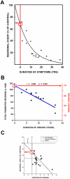
Estimates of loss of SN dopamine neurons at the time of PD symptom onset. (A) Fearnley and Lees (Fearnley and Lees, 1991) examined the number of pigmented neurons in the SN in relation to duration of symptoms, and performed a regression analysis based on an exponential decline in their number. They estimated about a 30% total loss, adjusted for age. (B) A similar estimate can be derived from the data of Ma and colleagues, based on their dissector counts of pigmented SN neurons (Ma et al., 1997). A linear regression analysis of their data with extrapolation to time of disease onset yields an estimate of about a 30% loss. (C) Greffard and colleagues performed counts of neurons in the SNpc, and determined density per unit volume. By either a linear or a negative exponential best fit analysis, they estimated a 30% loss at the time of symptom onset (Greffard et al., 2006). (Figure modified from (Cheng et al., 2010)).
Fig. 2
Fig. 2
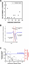
Estimates of loss of striatal dopamine terminal markers at time of symptom onset. (A) A graphical representation of the data presented in the oft-cited Bernheimer study to support the statement that there is an 80% reduction at the time of disease onset (Bernheimer et al., 1973). In the Bernheimer study, only 13 brains from patients with a diagnosis of PD were subjected to biochemical analysis. No regression analysis was performed. (B) Reiderer and Wuketich (Riederer and Wuketich, 1976) measured caudate dopamine content in two PD cohorts, one with an age of onset at 60 ± 1 years, and a second at 73 ± 1 years. Back extrapolation indicates a 68% and an 82% decrease, respectively, in caudate dopamine at the time of onset of disease in the two groups. (C) Scherman and colleagues analyzed [3H]TBZOH binding to the vesicular monoamine transporter in post-mortem caudate nucleus of 54 PD patients (Scherman et al., 1989). Polynomial regression analysis indicated a loss of 49% of binding sites at time of disease onset. (Figure modified from (Cheng et al., 2010)).
Fig. 3
Fig. 3
In patients with unilateral parkinsonism, motor signs do not yet appear when striatal dopaminergic terminal markers are depleted only 51% for 11C-DTBZ or 56% for 11C-MP (green bars), but appear on the affected side when these markers are depleted 62% and 71% respectively (red bars). (The data shown are from (Lee et al., 2000).
Fig. 4
Fig. 4
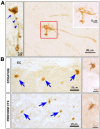
Axonopathy in hLRRK2(R1441G) BAC transgenic mice. (A) At the single axon level, staining for tyrosine hydroxylase (TH) reveals fragmentation (blue arrowheads), axonal spheroids (blue arrow), and dystrophic neurites at axon terminals (red square and inset). (B) Axonal abnormalities in the striatum and piriform cortex of the transgenic mice are also revealed by immunostaining for phosphorylated tau. Spheroids (blue arrows) and dystrophic neurites (side panels) similar to those visualized by TH staining, are observed. (Images adapted from Li et al (Li et al., 2009)).
Fig. 5
Fig. 5
Autophagy occurs in dopaminergic neuron cell bodies and axons following intra-striatal 6-OHDA injection. The presence of AVs in dopaminergic axons and cell bodies is identified by dsRed-LC3 labeling in TH-GFP mice. In the top panels, two clusters of AVs (red arrows) are identified in a GFP-labeled dopaminergic axon in the MFB at 2 days following intra-striatal 6-OHDA. AVs are also observed in dopaminergic cell bodes in the SNpc, shown in the lower panels. A single example is identified by a red arrow. (Images modified from (Cheng et al., 2011)).
Fig. 6
Fig. 6
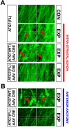
Following deletion of Atg7, axons of SNpc dopamine neurons are resistant to retrograde axonal degeneration. (A) In the absence of AAV Cre injection, Atg7fl/fl:TH-GFP mice show a loss of MFB dopaminergic axons (visualized by confocal microscopy of endogenous GFP), and the appearance of axonal spheroid pathology (red arrows) following unilateral 6-OHDA injection (indicated by the red vertical bar to the right). Atg7wt/wt:TH-GFP mice injected with AAV Cre show a similar axon loss and pathology. However, following injection of AAV Cre, Atg7fl/fl:TH-GFP mice show minimal axon loss and pathology following 6-OHDA injection. (B) Following deletion of Atg7, nigro-striatal axons in Atg7fl/fl:TH-GFP mice show less pathology, and relatively preserved number, following anterior MFB axotomy, in comparison to Atg7wt/wt:TH-GFP mice. In the Atg7wt/wt:TH-GFP mice, numerous axon spheroids are observed (red arrows). (Images modified from (Cheng et al., 2011)).

Similar articles

Cited by

References

    1. Agid Y, Agid F, Ruberg M. Biochemistry of neurotransmitters in Parkinson's disease. In: Marsden CD, Fahn S, editors. Movement Disorders 2. Butterworths; London: 1987. pp. 166–230.
    1. Avery MA, Sheehan AE, Kerr KS, Wang J, Freeman MR. Wlds requires Nmnat1 enzymatic activity and N16-VCP interactions to suppress Wallerian degeneration. J Cell Biol. 2009;184:501–513. - PMC - PubMed
    1. Barrientos SA, Martinez NW, Yoo S, Jara JS, Zamorano S, Hetz C, Twiss JL, Alvarez J, Court FA. Axonal degeneration is mediated by the mitochondrial permeability transition pore. J Neurosci. 2011;31:966–978. - PMC - PubMed
    1. Bernheimer H, Birkmayer W, Hornykiewicz O, Jellinger K, Seitelberger F. Brain dopamine and the syndromes of Parkinson and Huntington. Clinical, morphological and neurochemical correlations. J Neurol Sci. 1973;20:415–455. - PubMed
    1. Bonifati V, Rizzu P, van Baren MJ, Schaap O, Breedveld GJ, Krieger E, Dekker MC, Squitieri F, Ibanez P, Joosse M. Mutations in the DJ-1 gene associated with autosomal recessive early-onset parkinsonism. Science. 2003;299:256–259. - PubMed

Publication types